

Low-grade mucoepidermoid carcinoma mimicking benign cystic lesions in the salivary gland: A diagnostic dilemma
- PMID: 38525087
- PMCID: PMC10960343
- DOI: 10.1177/20363613241242397
Low-grade mucoepidermoid carcinoma mimicking benign cystic lesions in the salivary gland: A diagnostic dilemma
Abstract
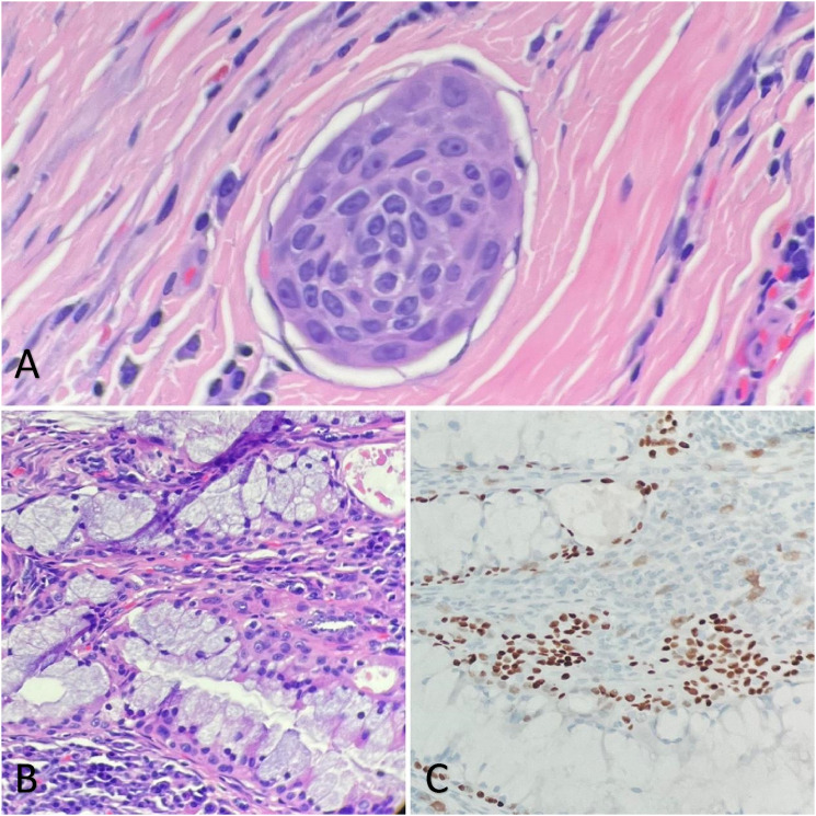
Mucoepidermoid carcinoma (MEC) is a common malignancy arising in the parotid gland. The diagnosis of MEC is typically based on its morphological features alone, characteristically containing mucocytes, intermediate cells and epidermoid cells. However, when cystic degeneration is diffuse, it is challenging to distinguish MEC from other benign cystic tumors. This is a case report of a 58-year-old Caucasian man who presented with a parotid mass. H&E sections of the mass reveal multiloculated cysts lined by bland-looking epithelium with only rare papillary architectures. The papillary proliferation contains mucocytes, and epidermoid cells highlighted by the p63 immunohistochemistry study. The diagnosis was confirmed by FISH result of positive MAML2 (11q21) rearrangement. Patient underwent parotidectomy and is disease-free 6 months post-surgery. MEC with cystic degeneration is a common diagnostic pitfall which can mimic many benign lesions in the salivary gland. We present a rare case with MEC with extensive cystic change, its molecular and pathologic findings and review the diagnostic features of MEC, its benign mimickers and useful tools for distinguishing these entities.
Keywords: Salivary gland tumor; acinic cell carcinoma; cystic degeneration; low grade mucoepidermoid carcinoma; rare tumors.
© The Author(s) 2024.
Conflict of interest statement
The author(s) declared no potential conflicts of interest with respect to the research, authorship, and/or publication of this article.
Figures


Similar articles
-
[Salivary papillary cystic low-grade mucoepidermoid carcinoma and cystadenoma: a comparison of clinicopathological and genetic features].Zhonghua Kou Qiang Yi Xue Za Zhi. 2022 Nov 9;57(11):1134-1140. doi: 10.3760/cma.j.cn112144-20220615-00327. Zhonghua Kou Qiang Yi Xue Za Zhi. 2022. PMID: 36379892 Chinese.
-
Molecular Profiling of Salivary Oncocytic Mucoepidermoid Carcinomas Helps to Resolve Differential Diagnostic Dilemma With Low-grade Oncocytic Lesions.Am J Surg Pathol. 2020 Dec;44(12):1612-1622. doi: 10.1097/PAS.0000000000001590. Am J Surg Pathol. 2020. PMID: 33002921
-
Warthin-Like Mucoepidermoid Carcinoma: A Morphological Spectrum - A Report of 3 Cases with Histological and Cytological Findings and Review of the Literature.Acta Cytol. 2022;66(3):244-252. doi: 10.1159/000521134. Epub 2022 Feb 4. Acta Cytol. 2022. PMID: 35124667 Review.
-
Case report: The diagnostic pitfall of Warthin-like mucoepidermoid carcinoma.Front Oncol. 2024 Jun 20;14:1391616. doi: 10.3389/fonc.2024.1391616. eCollection 2024. Front Oncol. 2024. PMID: 38988706 Free PMC article.
-
Warthin-like Mucoepidermoid Carcinoma of the Parotid Gland: Unusual Morphology and Diagnostic Pitfalls.Anticancer Res. 2019 Jun;39(6):3213-3217. doi: 10.21873/anticanres.13461. Anticancer Res. 2019. PMID: 31177170 Review.
Cited by
-
Polymorphous Adenocarcinoma of the Parotid Gland: A Rare Entity in Asians With a Unique Cystic Presentation.Clin Case Rep. 2025 Apr 21;13(4):e70441. doi: 10.1002/ccr3.70441. eCollection 2025 Apr. Clin Case Rep. 2025. PMID: 40264727 Free PMC article.
-
Comparative analysis of the World Health Organization Reporting System for Head and Neck Cytopathology and the Milan System for Reporting Salivary Gland Cytopathology.Cancer Cytopathol. 2025 Sep;133(9):e70041. doi: 10.1002/cncy.70041. Cancer Cytopathol. 2025. PMID: 40853710 Free PMC article.
