. 2024 Mar 7;3(3):pgae102.
doi: 10.1093/pnasnexus/pgae102. eCollection 2024 Mar.

Cryogenic electron microscopy and tomography reveal imperfect icosahedral symmetry in alphaviruses

Affiliations

Cryogenic electron microscopy and tomography reveal imperfect icosahedral symmetry in alphaviruses

David Chmielewski et al. PNAS Nexus. .

Abstract

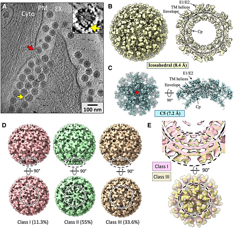
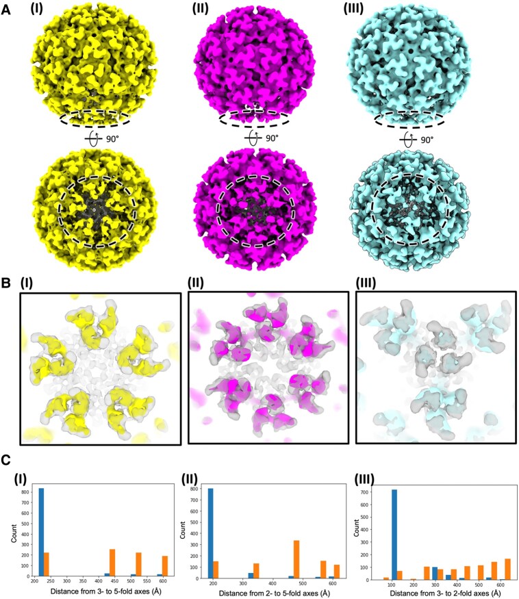
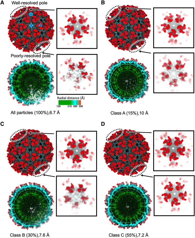
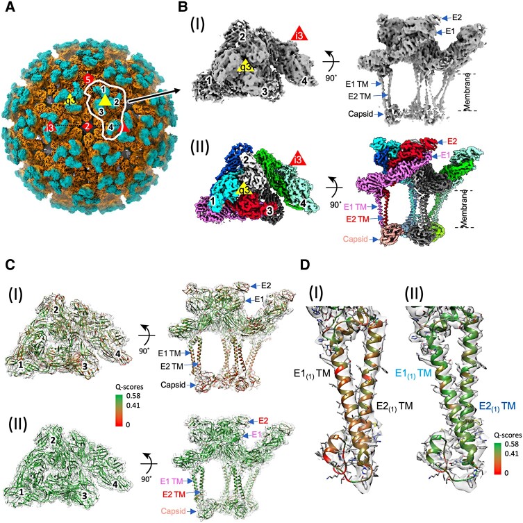
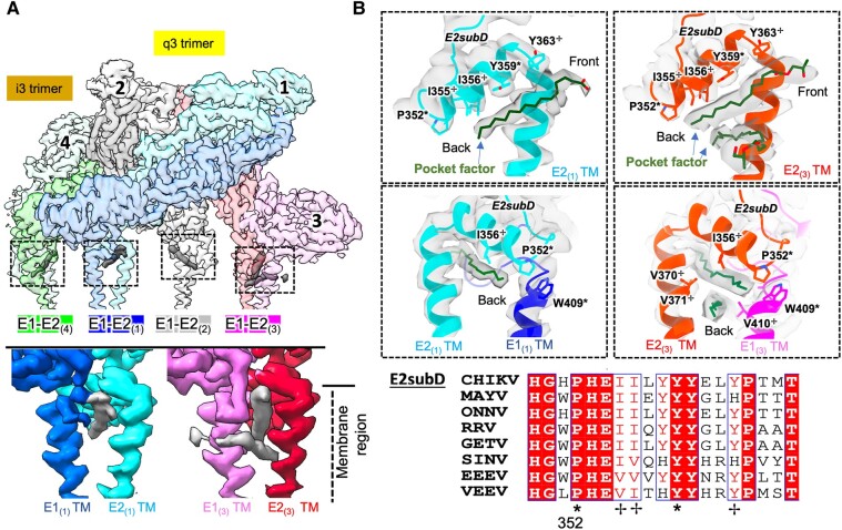
Alphaviruses are spherical, enveloped RNA viruses with two-layered icosahedral architecture. The structures of many alphaviruses have been studied using cryogenic electron microscopy (cryo-EM) reconstructions, which impose icosahedral symmetry on the viral particles. Using cryogenic electron tomography (cryo-ET), we revealed a polarized symmetry defect in the icosahedral lattice of Chikungunya virus (CHIKV) in situ, similar to the late budding particles, suggesting the inherent imperfect symmetry originates from the final pinch-off of assembled virions. We further demonstrated this imperfect symmetry is also present in in vitro purified CHIKV and Mayaro virus, another arthritogenic alphavirus. We employed a subparticle-based single-particle analysis protocol to circumvent the icosahedral imperfection and boosted the resolution of the structure of the CHIKV to ∼3 Å resolution, which revealed detailed molecular interactions between glycoprotein E1-E2 heterodimers in the transmembrane region and multiple lipid-like pocket factors located in a highly conserved hydrophobic pocket. This complementary use of in situ cryo-ET and single-particle cryo-EM approaches provides a more precise structural description of near-icosahedral viruses and valuable insights to guide the development of structure-based antiviral therapies against alphaviruses.

Keywords: alphavirus; cryo-EM; cryo-ET; imperfect icosahedral symmetry.


Figures

Fig. 1.
Fig. 1.
Subtomogram averaged structure of CHIKV virions in situ. A) Tomographic slice view of CHIKV-infected human cells with budding particles along the PM and released particles outside the cell (EX) showing imperfections in icosahedral lattice. EX, extracellular; CYTO, cytoplasm. Inset: zoom-in view of a single released virion with distortions. B) STA of released virion with imposed icosahedral symmetry at 8.4 Å resolution. Central section of density (right) with labeled structural components of virion. C) 7.2 Å STA following focused refinement around penton at a 5-fold axis, with top view (left) and side view (right). D) 3D classes of released virions, with percent of total particles assigned to each class. Poorly resolved pentamer regions displayed with dashed circle. E) Comparison of density at the poorly resolved penton (a dashed circle) between class I and class III. Zoom-in views with half-cut representation (top) and surface view (bottom) shows expansion of class III penton relative to class I.
Fig. 2.
Fig. 2.
In situ CHIKV virions with imperfect icosahedral symmetry at one pole. A) STAs of icosahedrally averaged virions released the symmetry to (I) C5 symmetry, (II) C2 symmetry, or (III) C3 symmetry. Poorly resolved regions displayed with dashed circle. B) Comparison of the density maps released to (I) C5 symmetry, (II) C2 symmetry, or (III) C3 symmetry with the map with icosahedral symmetry in semi-transparency. C) Histograms of distances from (I) a bad 3-fold axis to a bad 5-fold axis (left) vs. to a random 5-fold axis (right); (II) a bad 2-fold axis to a bad 5-fold axis (left) vs. to a random 5-fold axis (right); and (III) a bad 3-fold axis to a bad 2-fold axis (left) vs. to a random 2-fold axis (right).
Fig. 3.
Fig. 3.
Asymmetric reconstructions of biochemically purified CHIKV particles. A) Radial-colored density maps (above) with half-cut representation (below) following asymmetric (C1) reconstruction of CHIKV from all particles. Zoom-in views of the well-resolved pole and poorly resolved pole show the two respective pentamers of spikes. Following 3D classification, B) class A, C) class B, and D) class C maps are displayed in the same way as (A), including the entire particle, cross-sectional view, and two penton views with different degrees of 5-fold symmetry appearance. The percentage of particles assigned to each class after 3D classification displayed in parenthesis. All maps are displayed at the same density threshold (3σ). RNA density was masked out for visualization.
Fig. 4.
Fig. 4.
Cryo-EM structure of biochemically purified CHIKV. A) The 4.18 Å resolution radial-colored density map of CHIKV following refinement with icosahedral symmetry imposed. Penton, triangle, and diamond represent 5-, 3-, and 2-fold symmetry axes. An ASU is marked with white outline. The four E1–E2 heterodimers in the CHIKV ASU are labeled with 1–4. B) View of CHIKV ASU after reconstruction (I) icosahedral refinement at ∼4.2 Å and (II) after focused classification and refinement at ∼3.1 Å resolution. C) The ASU model fits in the map (I) before or (II) after additional processing with residues colored by Q-scores to show the improvement of map density resolvability. D) Zoom-in view of a single E1(1) and E2(1) with resolved transmembrane helices (TM) displayed with Q-scores distribution. Expected Q-score values 0.58 and 0.41 correspond to 3 and 4 Å resolution. Subscripts of E1 or E2 represent the numbering of E1–E2 dimer in an ASU in (B).
Fig. 5.
Fig. 5.
Transmembrane helices and endodomain interactions in biochemically purified CHIKV ASU. A) Multiple interactions of E1 TM region with E2 TM and Cp. Zoom-in views of residues involved in E1(1)–E2(1) TM interactions (i, ii), and E1(1)–Capsid(1) interaction (iii). B) Interaction across subunits in the q3 spike and neighboring i3 spike. E1(4)–E2(1) interaction within q3-i3 spike displayed as zoom-in views. Residues labeled with a cross or star indicate highly/completely* conserved residues across different alphaviruses in Fig. S7. Each protein molecule is colored uniquely and matches colors in Fig. 4B(II).
Fig. 6.
Fig. 6.
Lipid-like densities within the E1–E2 dimer of CHIKV. A) CHIKV ASU map highlights the positions of pocket factor densities (dashed squares). Two densities were found in the E1(3)–E2(3) dimer (bottom right) and one density in other dimers (bottom left). B) Zoom-in model views of E1–E2 hydrophobic pockets, with key residues forming interactions with the pocket factor labeled. The alkyl chain models of phospholipid fit into the extra densities in the CHIKV E1–E2 hydrophobic pocket. Residues labeled with a cross, or star are highly/completely* conserved residues across different alphaviruses. E2subD:E2 subdomain D.

Similar articles

Cited by

References

    1. Strauss JH, Strauss EG. 1994. The alphaviruses: gene expression, replication, and evolution. Microbiol Rev. 58:491–562. - PMC - PubMed
    1. de Lima Cavalcanti TYV, Pereira MR, de Paula SO, Franca RFdO. 2022. A review on chikungunya virus epidemiology, pathogenesis and current vaccine development. Viruses. 14(5):969. - PMC - PubMed
    1. Bartholomeeusen K, et al. 2023. Chikungunya fever. Nat Rev Dis Primers. 9:17. - PMC - PubMed
    1. Button JM, Qazi SA, Wang JC-Y, Mukhopadhyay S. 2020. Revisiting an old friend: new findings in alphavirus structure and assembly. Curr Opin Virol. 45:25–33. - PMC - PubMed
    1. Forsell K, Xing L, Kozlovska T, Cheng RH, Garoff H. 2000. Membrane proteins organize a symmetrical virus. EMBO J. 19:5081–5091. - PMC - PubMed