. 2024 Apr 15;13(4):bio060156.
doi: 10.1242/bio.060156. Epub 2024 Apr 5.

Distinct features of the regenerating heart uncovered through comparative single-cell profiling

Affiliations

Distinct features of the regenerating heart uncovered through comparative single-cell profiling

Clayton M Carey et al. Biol Open. .

Abstract

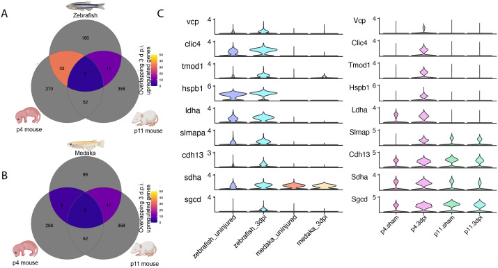
Adult humans respond to heart injury by forming a permanent scar, yet other vertebrates are capable of robust and complete cardiac regeneration. Despite progress towards characterizing the mechanisms of cardiac regeneration in fish and amphibians, the large evolutionary gulf between mammals and regenerating vertebrates complicates deciphering which cellular and molecular features truly enable regeneration. To better define these features, we compared cardiac injury responses in zebrafish and medaka, two fish species that share similar heart anatomy and common teleost ancestry but differ in regenerative capability. We used single-cell transcriptional profiling to create a time-resolved comparative cell atlas of injury responses in all major cardiac cell types across both species. With this approach, we identified several key features that distinguish cardiac injury response in the non-regenerating medaka heart. By comparing immune responses to injury, we found altered cell recruitment and a distinct pro-inflammatory gene program in medaka leukocytes, and an absence of the injury-induced interferon response seen in zebrafish. In addition, we found a lack of pro-regenerative signals, including nrg1 and retinoic acid, from medaka endothelial and epicardial cells. Finally, we identified alterations in the myocardial structure in medaka, where they lack primordial layer cardiomyocytes and fail to employ a cardioprotective gene program shared by regenerating vertebrates. Our findings reveal notable variation in injury response across nearly all major cardiac cell types in zebrafish and medaka, demonstrating how evolutionary divergence influences the hidden cellular features underpinning regenerative potential in these seemingly similar vertebrates.

Keywords: Evolution; Heart; Immunity; Interferon; Myocardium; Regeneration.


Conflict of interest statement

Competing interests The authors declare no competing or financial interests.

Figures

Fig. 1.
Fig. 1.
A single-cell atlas of cardiac injury response in zebrafish and medaka. (A) Experimental overview for collection of ventricles and single-cell sequencing. The number of independent samples and total number of quality filtered cells for each time point are indicated. (B) Representative images of Acid-Fuchsin Orange staining of collagen (blue), fibrin (red), and muscle fibers (tan) in heart sections showing cryoinjury-induced fibrin and collagen deposition in both species (arrows). Anatomical labels indicate ventricle (labeled V), atrium (labeled A), and bulbus arteriosus (labeled BA). (C) UMAP embedding of all sampled cells from each species and time point integrated into a single dataset. A total of 22 clusters were identified and colored by major cardiac cell type (cardiomyocyte, orange shades; endothelial/mural, purple shades; epicardial, green shades; leukocyte, blue shades). (D) Gene expression dot plot showing average gene expression of marker genes for cells classified as the indicated cell type. Two marker genes are displayed for each cell type. Dot sizes represent percent of cells expressing the indicated gene (pct.exp), color indicates average scaled gene expression across all cells in the indicated tissue.
Fig. 2.
Fig. 2.
Medaka lack an endogenous injury-induced interferon response. (A) Gene expression heatmap showing scaled average gene expression for 12 interferon-stimulated genes in the indicated species and tissue type at each time point. (B) RNA in situ hybridization of isg15 (interferon-responding cells), kdrl (endothelial cells), and myl7 (cardiomyocytes) in ventricle cryosections in the indicated species and time point. Scale bars: 200 µm. Anatomical labels: V, intact ventricle; BA, bulbus arteriosus; W, wound area. Images are representative of at least three individuals at each time point. (C) Gene expression feature plot for ifnphi1 across all zebrafish cardiac cell types, color scale=expression level. (D) Quantification of proportion of zebrafish endothelial cells expressing ifnphi1 at each time point.
Fig. 3.
Fig. 3.
Medaka display altered tissue-resident and injury-responsive immune cell populations. (A) UMAP embedding and sub-clustering of all leukocytes, with cells classified as T lymphocytes (TL), B lymphocytes (BL), granulocytes (GN), or macrophages (MF). (B) Gene expression dot plot of marker genes for each immune cell cluster. (C) RNA in situ hybridization of mpeg1.1 (macrophages), isg15 (interferon response), and myl7 (cardiomyocytes) in ventricle cryosections in the indicated species and time point. Scale bars: 200 µm, anatomical labels: V, intact ventricle; W, wound area. Representative images are shown from at least three animals at each time point. (D,E) Quantification of number of macrophages per mm2 in either the intact myocardium (ventricle) or wound area (wound) in zebrafish (D) or medaka (E) * indicates a P-value <0.05 using a t-test comparing with uninjured ventricle. n=3 individuals were used for macrophage quantification from each timepoint based on an average of three sections per individual. (F,G) Gene expression violin plots from all macrophages in the indicated time point and species for tnfa (F) and cd9b (G). (H) Quantification of the proportion of macrophages expressing tnfa at each time point in each species.
Fig. 4.
Fig. 4.
Zebrafish and medaka share a partially overlapping fibrotic response to injury. (A) UMAP embedding of re-clustered endothelial and mural cells classified as either endocardial endothelium (eEC), coronary endothelium (cEC), lymphatic endothelium (lEC), fibroblast-like endothelial cells (fEC), and mural cells (mural). (B) Gene expression dot plot of marker genes for each endothelial cell classification. (C) UMAP embedding of re-clustered epicardial cells classified as canonical epicardial cells (cEP), fibroblast-like epicardial cells (fEP), or zebrafish-specific epicardial cells (zEP). (D) Gene expression dot plot of marker genes for each epicardial cell classification. (E,F) Quantification of proportion of endothelial (E,F) or epicardial (G) cells classified as the indicated cell type.
Fig. 5.
Fig. 5.
Medaka epicardial and endothelial cells fail to produce many pro-regenerative signals. (A) Gene expression feature plots for the indicated pro-regenerative genes in all cells. (B) Quantification of the proportion of all epicardial cells expressing nrg1 in the indicated species and time point (C-G). Gene expression violin plots showing induction of pro-regenerative signals in the indicated species and time point comparing: (C) aldh1a2 expression in fEC and fEP cells, (D) cntf expression in fEC and eEC cells, (E) cxcl12a expression in fEP and cEP cells, (F) cxcr4a and apln expression in cEC cells, and (G) cxcl8a, vegfd, and angpt1 expression in zEP cells.
Fig. 6.
Fig. 6.
Medaka lack primordial myocardium and have few cortical layer cardiomyocytes. (A) RNA in situ hybridization of myl7 labeling myocardium in zebrafish and medaka ventricles of the indicated age. Dotted line indicates border between trabecular and cortical layers. Images are representative of at least three individuals at each time point. Scale bars: 200 µM. (B) UMAP embedding of re-clustered cardiomyocytes identified as either trabecular (tCM) or cortical (cCM). (C) Gene expression dot plot showing expression of marker genes for each CM cell cluster. (D) Proportion of cardiomyocytes in trabecular or cortical cell clusters from all single-cell samples from zebrafish and medaka. (E,F) UMAP embedding of ventricular cardiomyocytes clustered separately from uninjured (E) zebrafish or (F) medaka. (G,H) Gene expression feature plots for top marker genes for primordial cardiomyocytes in zebrafish (E) or medaka (F). (I) RNA in situ hybridization of myl7, acta2, and hey2 in uninjured zebrafish hearts. Scale bars: 200 µM. (J) RNA in situ hybridization of myl7 and acta2 in uninjured medaka heart. Scale bars: 200 µM.
Fig. 7.
Fig. 7.
Zebrafish cardiomyocytes share a cardioprotective gene signature with neonatal mouse. (A,B) Venn diagram counting overlapping genes upregulated at 3 d.p.i. in postnatal day 4 or 11 mouse cardiomyocytes with (A) zebrafish cardiomyocytes or (B) medaka cardiomyocytes. (C) Gene expression violin plots of cardioprotective genes uniquely upregulated in regenerating p4 mouse and zebrafish cardiomyocytes.

Update of

Similar articles

Cited by

References

    1. Aurora, A. B. and Olson, E. N. (2014). Immune modulation of stem cells and regeneration. Cell Stem Cell 15, 14-25. 10.1016/j.stem.2014.06.009 - DOI - PMC - PubMed
    1. Bevan, L., Lim, Z. W., Venkatesh, B., Riley, P. R., Martin, P. and Richardson, R. J. (2020). Specific macrophage populations promote both cardiac scar deposition and subsequent resolution in adult zebrafish. Cardiovasc. Res. 116, 1357-1371. 10.1093/cvr/cvz221 - DOI - PMC - PubMed
    1. Bise, T., de Preux Charles, A.-S. and Jaźwińska, A. (2019). Ciliary neurotrophic factor stimulates cardioprotection and the proliferative activity in the adult zebrafish heart. npj Regen Med. 4, 1-14. 10.1038/s41536-019-0064-9 - DOI - PMC - PubMed
    1. Brosseau, C., Colas, L., Magnan, A. and Brouard, S. (2018). CD9 Tetraspanin: a new pathway for the regulation of inflammation? Front. Immunol. 9, 2316. 10.3389/fimmu.2018.02316 - DOI - PMC - PubMed
    1. Brundel, B. J. J. M., Ke, L., Dijkhuis, A.-J., Qi, X., Shiroshita-Takeshita, A., Nattel, S., Henning, R. H. and Kampinga, H. H. (2008). Heat shock proteins as molecular targets for intervention in atrial fibrillation. Cardiovasc. Res. 78, 422-428. 10.1093/cvr/cvn060 - DOI - PubMed

Publication types

LinkOut - more resources