. 2024 Mar 25;147(1):61.
doi: 10.1007/s00401-024-02701-5.

TMEM106B coding variant is protective and deletion detrimental in a mouse model of tauopathy

Affiliations

TMEM106B coding variant is protective and deletion detrimental in a mouse model of tauopathy

George A Edwards 3rd et al. Acta Neuropathol. .

Abstract

TMEM106B is a risk modifier of multiple neurological conditions, where a single coding variant and multiple non-coding SNPs influence the balance between susceptibility and resilience. Two key questions that emerge from past work are whether the lone T185S coding variant contributes to protection, and if the presence of TMEM106B is helpful or harmful in the context of disease. Here, we address both questions while expanding the scope of TMEM106B study from TDP-43 to models of tauopathy. We generated knockout mice with constitutive deletion of TMEM106B, alongside knock-in mice encoding the T186S knock-in mutation (equivalent to the human T185S variant), and crossed both with a P301S transgenic tau model to study how these manipulations impacted disease phenotypes. We found that TMEM106B deletion accelerated cognitive decline, hind limb paralysis, tau pathology, and neurodegeneration. TMEM106B deletion also increased transcriptional correlation with human AD and the functional pathways enriched in KO:tau mice aligned with those of AD. In contrast, the coding variant protected against tau-associated cognitive decline, synaptic impairment, neurodegeneration, and paralysis without affecting tau pathology. Our findings reveal that TMEM106B is a critical safeguard against tau aggregation, and that loss of this protein has a profound effect on sequelae of tauopathy. Our study further demonstrates that the coding variant is functionally relevant and contributes to neuroprotection downstream of tau pathology to preserve cognitive function.

Keywords: Alzheimer’s disease; Cognitive resilience; Frontotemporal dementia; Mouse model; Neurodegeneration; TMEM106B; Tau; Tauopathy.


Conflict of interest statement

Declarations

Conflict of interest CZ is a paid employee of NeuroScience Associates. The remaining authors declare no competing interests.

Figures

Fig. 1
Fig. 1
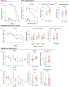
TMEM106B deletion accelerates paralysis and worsens cognitive behavior. a Onset of hind limb paralysis requiring euthanasia was measured for tau animals, excluding mice used in behavioral testing. The age at paralysis onset was significantly earlier in KO:tau than tau mice. (tau = 27, KO:tau = 43). b Spatial learning in the Morris water maze was assessed by the distance traveled to reach the escape platform. KO:tau mice performed significantly worse than tau animals on days 3 and 5 of training. c KO:tau mice recalled the escape location less well than tau animals during daily short term memory testing (c). but this difference did not persist during long-term memory testing (d). e Cognitive flexibility measured by changing the platform location each day showed that KO:tau mice performed more poorly than tau mice on trial 5. Graph shows average performance across days as a function of trial number (8 trials/day). f Short-term memory for the changing platform location was no different for any given location (left), but was worse overall in KO:tau mice when averaged across the three locations. g-h During the learning phase of radial arm water maze, KO:tau mice made more incorrect arm entries (g reference memory, left) and more repeated arm entries (h working memory, right and middle graphs). Long-term memory was also impaired in KO:tau mice compared to tau when measured as incorrect arm entries (g right), but did not reach significance for repeated arm errors (h, right). See Supplementary Table 1 for ANOVA statistics. (b-h tau = 10, KO:tau = 10) For all scatter plots: open circles = female, closed circles = male. * p < 0.05, ** p < 0.01, *** p < 0.001
Fig. 2
Fig. 2
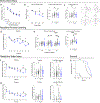
The T186S coding variant protects against cognitive decline and delays paralysis in tau mice. a No difference was noted between KI:tau and tau groups during the learning phase of Morris water maze, measured as distance to the escape platform. b Short-term memory for the escape location was better in KI:tau mice than tau on day 4 (left). When short-term memory in daily probe trials was averaged across days, the improvement in KI:tau vs tau mice was nearly significant at p = 0.065 (right). c Long-term memory for the escape location was better in KI:tau mice than tau animals. Track plots show example search paths for one animal of each genotype during the long-term probe trial (right). (a-c: WT = 18, KI = 21, tau = 20, KI:tau = 20). d Cognitive flexibility measured by changing the platform location each day showed that KI:tau mice performed better than tau mice on trials 5 and 7. Graph shows average performance across days as a function of the trial number (8 trials/day). e Short-term memory for each new platform location was better in KI:tau than tau mice by the second day of testing (platform location NW), and this difference widened with the third day of testing (platform location SE, left). On average, KI:tau mice showed better short-term memory for the changing platform locations than tau mice (right). (d, e: WT = 18, KI = 21, tau = 19, KI:tau = 19). f, g During the learning phase of radial arm water maze, the number of incorrect arm entries made by KI:tau was lower than in tau mice on trial 7 (f reference memory, right), but did not reach significance when averaged across trials (f, middle). Both genotypes made the same number of repeated arm entries during training (g, working memory, right and middle graphs). Remarkably, KI:tau mice recalled the trained escape arm better than tau animals during long-term testing, both when measured as incorrect arm entries (f, reference errors, right) and when measured as repeated arm entries (g working errors, right). (f, g: WT = 16, KI = 18, tau = 17, KI:tau = 19). h Onset of hind limb paralysis requiring euthanasia occurred significantly later in KI:tau than tau mice. tau = 23, KI:tau = 23. Significant values shown here reflect ANOVA testing with all four genotypes. For all scatter plots: open circles = female, closed circles = male. * p < 0.05, ** p < 0.01, *** p < 0.001, **** p < 0.0001. See Supplementary Table 1 for ANOVA statistics
Fig. 3
Fig. 3
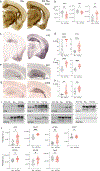
TMEM106B deletion worsens degeneration and tau pathology. a Campbell–Switzer silver staining did not identify tangles, but did reveal marked ventricular enlargement in the KO:tau animals compared to tau alone. b Silver-stained sections were used to measure the summed area of the hippocampus (HPC), overlying cortex (CTX) and adjacent lateral ventricle (LV) in tau and KO:tau mice to demonstrate a quantitative difference in the summed area of all three regions upon TMEM106B deletion. c-h Immunostaining and quantitation of AT8 (c, d), Iba1 (e, f), and CD68 (g, h) all demonstrate marked worsening of tau pathology and microgliosis upon TMEM106B deletion. Graphs in d, f, g show the percent area stained by each marker within hippocampus (HPC) and cortex (CTX). (a-h: tau = 10, KO:tau = 10). i, j Western blots for phospho-tau epitopes AT8 (S202/T205), PHF1 (S396/S404), AT100 (T212/S214), AT270 (T181), and AT180 (T231), total human tau CP27, and total tau Tau5 (i) and quantitation of band intensity (j). (i, j: WT = 2, KO = 2, tau = 6, KO:tau = 6). Open circles = female, closed circles = male. * p < 0.05, ** p < 0.01, *** p < 0.001, **** p < 0.0001
Fig. 4
Fig. 4
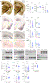
TMEM106B T186S protects against degeneration without changing tau pathology. a Campbell–Switzer silver stain hinted at ventricular preservation in T186S KI:tau mice compared with tau alone. b Summed areas of hippocampus (HPC), cortex (CTX), and lateral ventricle (LV) were measured from silver-stained sections spanning the full extent of hippocampus. Tau animals carrying the T186S variant retained more cortical tissue and showed less ventricular enlargement than tau alone. c-h Immunostaining and percent area quantitation of AT8 (c, d), Iba1 (e, f), and CD68 (g, h) were no different between genotypes in hippocampus or cortex. (a-h: tau = 10, KI:tau = 10). i, j Western blots for phospho-tau epitopes AT8 (S202/T205), PHF1 (S396/S404), AT100 (T212/S214), AT270 (T181), and AT180 (T231), total human tau CP27, and total tau with Tau5 (i) and quantitation of band intensity (j). (i, j: WT = 2, KI = 2, tau = 6, KI:tau = 6). Open circles = female, closed circles = male. * p < 0.05
Fig. 5
Fig. 5
TMEM deletion significantly alters the transcriptional signature of tauopathy. a Volcano plot showing FDR log10 p value as a function of log2 fold change in forebrain gene expression between KO and WT (left) and KO:tau vs. tau (right). Mice were 9–10 mo of age at time of harvest. 16 DEGs distinguish KO from WT. TMEM106B deletion had a far greater effect on transcriptional signature in the context of tauopathy. n = 5–7/genotype. b GSEA for the KEGG AD signature approached significant enrichment for KO:tau vs tau with an ES of 1.71 and FDR q = 0.06 (left). GSEA for the KEGG lysosome signature was significantly enriched for KO:tau vs. tau with an ES of 1.55 and FDR q = 0.034 (right)
Fig. 6
Fig. 6
TMEM deletion enhances functional overlap with AD. a Venn diagrams illustrating the number and overlap of DEGs identified by comparison of human AD vs healthy controls (HC, turquoise) against KO:tau vs. tau (red; left) b Relationship between human AD and mouse brain for DEGs identified by comparison of KO:tau vs tau. Values for human AD gene expression (relative to HC) were derived from the AMP-AD database on the X axis (log2 fold change) against the corresponding expression level for KO:tau vs WT (red) or tau vs WT (grey). Correlation (Pearson’s r value) and linear regression with human AD expression is significantly higher in the KO:tau animals than in mice expressing tau alone. c Relationship between human AD and mouse brain for human NFT-associated genes. Correlation with human NFT-associated gene expression is again increased by TMEM106B deletion. d Network clustering showing several enriched modules among the KO:tau vs tau DEGs that overlap and change in the same direction as human AD brain. Colors distinguish separate, but related, functional modules. Outlines indicate genes concordant with single cell analyses testing the effect of NFT burden in human AD brain. e Heat map illustrating DEGs used to generate functional modules in d, plotted to show the degree of change against WT (mouse, left) or HC (human, right) controls. TMEM106B deletion increases the magnitude of change towards a more AD-like expression pattern compared to tau alone. DEG expression is shown for both KO:tau and tau groups against 4 different human AD brain regions. TCX = temporal cortex, PHG = parahippocampal gyrus, STG = superior temporal gyrus, DLPFC = dorsolateral prefrontal cortex. n = 5–7 mice per genotype. **** p < 0.0001
Fig. 7
Fig. 7
TMEM106B T186S coding variant enhances hippocampal plasticity in tauopathy mice. Acute hippocampal slices from 8 mo old KI:tau and tau mice were used to measure synaptic responses in the Schaeffer collateral pathway. a Input–output (or I–V) curve plotting amplitude of the field excitatory postsynaptic potential (fEPSP) as a function of simulation intensity. Basal synaptic transmission was unchanged by TMEM106B coding variant in this setting. n = KI:tau 9 slices from 4 mice, tau 10 slices from 3 mice. b fEPSP amplitude was normalized to baseline and plotted as a function of time, before and after tetanic stimulation, to induce long-term potentiation. Potentiation was significantly greater in slices from KI:tau mice than tau. c Average normalized fEPSP amplitude measured over the last 5 min of recording (55–60 min after tetanus) highlights the difference in synaptic potentiation between genotypes. n = KI:tau 16 slices from 6 mice, tau 18 slices from 5 mice. **p < 0.01

Update of

References

    1. Allen M, Carrasquillo MM, Funk C, Heavner BD, Zou F, Younkin CS et al. (2016) Human whole genome genotype and transcriptome data for Alzheimer’s and other neurodegenerative diseases. Sci Data 3:160089. 10.1038/sdata.2016.89 - DOI - PMC - PubMed
    1. Bennett DA, Schneider JA, Arvanitakis Z, Wilson RS (2012) Overview and findings from the religious orders study. Curr Alzheimer Res 9:628–645 - PMC - PubMed
    1. Briggs DI, Defensor E, Memar Ardestani P, Yi B, Halpain M, Seabrook G et al. (2017) Role of endoplasmic reticulum stress in learning and memory Impairment and Alzheimer’s disease-Like neuropathology in the PS19 and APP(Swe) mouse models of tauopathy and amyloidosis. ENeuro. 10.1523/ENEURO.0025-17.2017 - DOI - PMC - PubMed
    1. Brody AH, Nies SH, Guan F, Smith LM, Mukherjee B, Salazar SA et al. (2022) Alzheimer risk gene product Pyk2 suppresses tau phosphorylation and phenotypic effects of tauopathy. Mol Neurodegener 17:32. 10.1186/s13024-022-00526-y - DOI - PMC - PubMed
    1. Cabron AS, Borgmeyer U, Richter J, Peisker H, Gutbrod K, Dormann P et al. (2023) Lack of a protective effect of the Tmem106b “protective SNP” in the Grn knockout mouse model for frontotemporal lobar degeneration. Acta Neuropathol Commun 11:21. 10.1186/s40478-023-01510-3 - DOI - PMC - PubMed