. 2024 Mar;143(3):455-469.
doi: 10.1007/s00439-024-02655-4. Epub 2024 Mar 25.

Heterozygous loss-of-function variants in DOCK4 cause neurodevelopmental delay and microcephaly

Affiliations

Heterozygous loss-of-function variants in DOCK4 cause neurodevelopmental delay and microcephaly

Charlotte Herbst et al. Hum Genet. 2024 Mar.

Abstract

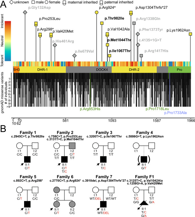
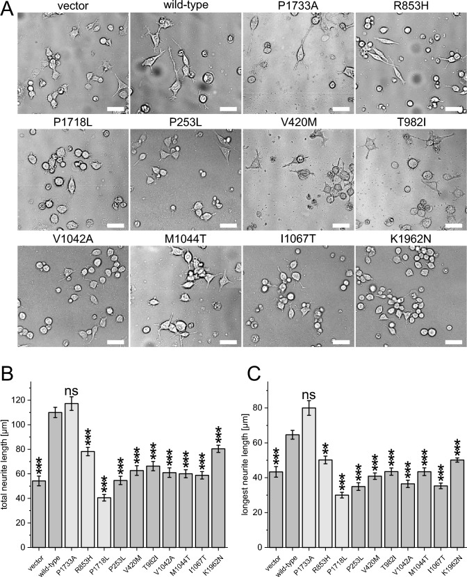
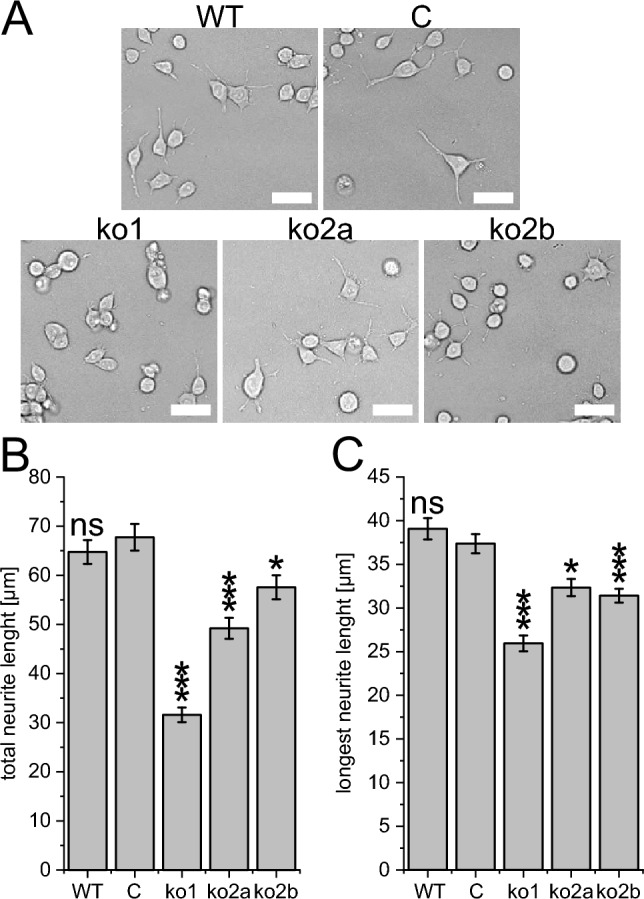
Neurons form the basic anatomical and functional structure of the nervous system, and defects in neuronal differentiation or formation of neurites are associated with various psychiatric and neurodevelopmental disorders. Dynamic changes in the cytoskeleton are essential for this process, which is, inter alia, controlled by the dedicator of cytokinesis 4 (DOCK4) through the activation of RAC1. Here, we clinically describe 7 individuals (6 males and one female) with variants in DOCK4 and overlapping phenotype of mild to severe global developmental delay. Additional symptoms include coordination or gait abnormalities, microcephaly, nonspecific brain malformations, hypotonia and seizures. Four individuals carry missense variants (three of them detected de novo) and three individuals carry null variants (two of them maternally inherited). Molecular modeling of the heterozygous missense variants suggests that the majority of them affect the globular structure of DOCK4. In vitro functional expression studies in transfected Neuro-2A cells showed that all missense variants impaired neurite outgrowth. Furthermore, Dock4 knockout Neuro-2A cells also exhibited defects in promoting neurite outgrowth. Our results, including clinical, molecular and functional data, suggest that loss-of-function variants in DOCK4 probable cause a variable spectrum of a novel neurodevelopmental disorder with microcephaly.


Conflict of interest statement

There are no competing interests.

Figures

Fig. 1
Fig. 1
Variants in DOCK4 and pedigrees. A Location of missense and null variants in DOCK4 with respect to the DOCK4 domain structure (GenBank: NM_014705.4). The x-axis represents the corresponding DOCK4 amino acid position of DOCK4. Variants reported in this study are labeled with the corresponding p-code; females are indicated as circles and males as squares. Indicated in bold are confirmed de novo variants. De novo variants that were identified in individuals with global development delay from other studies with a lacking detailed phenotypic description or inconclusive evidence are indicated in gray (see also Table S1). Missense variants in gnomAD with allele count are shown below the protein scheme. Positive controls (used for neurite outgrowth assay; p.Arg853His, p.Pro1718Leu) are labeled in green and the negative control (p.Pro1733Ala) is labeled in blue. The tolerance landscape from MetaDome is shown color-coded above the protein scheme. B Family pedigrees. Individuals with DOCK4-related disorder are indicated by filled black shapes with an arrow pointing to the individual. Open shapes represent unaffected individuals. Squares represent males, circles represent females, and triangles with a diagonal line represents termination of pregnancy. Gray-colored shapes indicate individuals with comparable milder symptoms. Of note, the phenotype of the father (I:2) of individual II:1 (family 6) is unknown. A = Generation number; B = number of that individual in that generation. Segregation results for all individuals tested are indicated with either red (presence of the DOCK4 variant) and/or black (presence of the reference allele). Abbreviations: SH3: N-terminal Src homology 3; DHR: dock homology region; DOCK4: dedicator of cytokinesis protein 4; Pro: proline-rich C-terminal end; WT: wild type
Fig. 2
Fig. 2
Structural analysis of the DOCK4 variants. A Model of the DOCK4 globular region (residues 1-1587) indicating the sites of the variants of interest in space-filled presentation. The SH3, DHR-1, DOCK4 and DHR-2 domain are colored in red, yellow, white and orange, respectively. The interaction partners ELMO and Rac-1 are shown in violet and blue. B, C Vicinity of residue 982 in the B wild-type and C p.Thr982Ile variant. In this variant, the longer isoleucine sidechain forms steric clashes (red arrow) with Leu923. D Interactions of the Ile1067 sidechain with Val1106 and Lys1109. E In the p.Ile1067Thr variant, these interactions cannot be formed by the shorter threonine sidechain (sites of missing interactions marked as dotted circles)
Fig. 3
Fig. 3
De novo and inherited missense variants in DOCK4 affect neurite outgrowth in Neuro-2A cells. A Neuro-2A cells were transfected with plasmids expressing wild-type DOCK4, the negative control p.Pro1733Ala, the positive controls p.Arg853His and p.Pro1718Leu, and the mutant variants p.Pro253Leu, p.Val420Met, p.Thr982Ile, p.Val1042Ala, p.Met1044Thr, p.Ile1067Thr, p.Lys1962Asn or vector. Transfected cells were sorted and collected by flow cytometry and then incubated in Neurobasal A media containing 2% B27 supplement (differentiation media) for 36 h. Scale bar, 50 µm. B, C Average length of total and average length of the longest neurite were measured. The results of a one-way ANOVA with the Games–Howell post hoc test (each compared to wild type) are indicated as: **: p < 0.005; ***: p < 0.0005; ns: p > 0.05. At least 70 cells/group were analyzed in each experiment. For better readability, the one letter code has been used to describe the variants in this figure
Fig. 4
Fig. 4
Knockout of Dock4 in Neuro-2A cells impairs neurite outgrowth. Neuro-2A Dock4 knockout cells by using the Alt-R CRISPR-Cas9 system utilizing one nonspecific control guide RNA (C) and two different guide RNAs (ko1 and ko2) that were used to generate three knockout clones (ko1, ko2a and ko2b). A Neuro-2A cells were incubated in Neurobasal A media containing 2% B27 supplement for 36 h for differentiation. Scale bar, 50 µm. B Average length of total and C average length of the longest neurite were measured. The results of a one-way ANOVA with the Games–Howell post hoc test (each compared to cells treated with the nonspecific control guide RNA:C) are indicated as: *: p < 0.05; ***: p < 0.0005; ns: p > 0.05. At least 70 cells/group were analyzed in each experiment

References

    1. Abraham S, Scarcia M, Bagshaw RD, McMahon K, Grant G, Harvey T, Yeo M, Esteves FOG, Thygesen HH, Jones PF, Speirs V, Hanby AM, Selby PJ, Lorger M, Dear TN, Pawson T, Marshall CJ, Mavria G. A Rac/Cdc42 exchange factor complex promotes formation of lateral filopodia and blood vessel lumen morphogenesis. Nat Commun. 2015;6:7286. doi: 10.1038/ncomms8286. - DOI - PMC - PubMed
    1. Cacheiro P, Muñoz-Fuentes V, Murray SA, Dickinson ME, Bucan M, Nutter LMJ, Peterson KA, Haselimashhadi H, Flenniken AM, Morgan H, Westerberg H, Konopka T, Hsu C-W, Christiansen A, Lanza DG, Beaudet AL, Heaney JD, Fuchs H, Gailus-Durner V, Sorg T, Prochazka J, Novosadova V, Lelliott CJ, Wardle-Jones H, Wells S, Teboul L, Cater H, Stewart M, Hough T, Wurst W, Sedlacek R, Adams DJ, Seavitt JR, Tocchini-Valentini G, Mammano F, Braun RE, McKerlie C, Herault Y, de Angelis MH, Mallon A-M, Lloyd KCK, Brown SDM, Parkinson H, Meehan TF, Smedley D. Human and mouse essentiality screens as a resource for disease gene discovery. Nat Commun. 2020;11(1):655. doi: 10.1038/s41467-020-14284-2. - DOI - PMC - PubMed
    1. Chen Q, Peto CA, Shelton GD, Mizisin A, Sawchenko PE, Schubert D. Loss of modifier of cell adhesion reveals a pathway leading to axonal degeneration. J Neurosci Official J Soc Neurosci. 2009;29(1):118–130. doi: 10.1523/JNEUROSCI.3985-08.2009. - DOI - PMC - PubMed
    1. Cioclu MC, Mosca I, Ambrosino P, Puzo D, Bayat A, Wortmann SB, Koch J, Strehlow V, Shirai K, Matsumoto N, Sanders SJ, Michaud V, Legendre M, Riva A, Striano P, Muhle H, Pendziwiat M, Lesca G, Mangano GD, Nardello R, Lemke JR, Møller RS, Soldovieri MV, Rubboli G, Taglialatela M. KCNT2-related disorders: phenotypes, functional, and pharmacological properties. Ann Neurol. 2023;94(2):332–349. doi: 10.1002/ana.26662. - DOI - PubMed
    1. Ehrhart F, Willighagen EL, Kutmon M, van Hoften M, Curfs LMG, Evelo CT. A resource to explore the discovery of rare diseases and their causative genes. Sci Data. 2021;8(1):124. doi: 10.1038/s41597-021-00905-y. - DOI - PMC - PubMed

Substances

LinkOut - more resources