

doi: 10.4103/singaporemedj.SMJ-2021-268.
Epub 2024 Mar 26.
Contrast-enhanced spectral mammography
Affiliations
- PMID: 38527307
- PMCID: PMC11060636
- DOI: 10.4103/singaporemedj.SMJ-2021-268
Item in Clipboard
Contrast-enhanced spectral mammography
Singapore Med J.
.
No abstract available
Conflict of interest statement
There are no conflicts of interest.
Figures
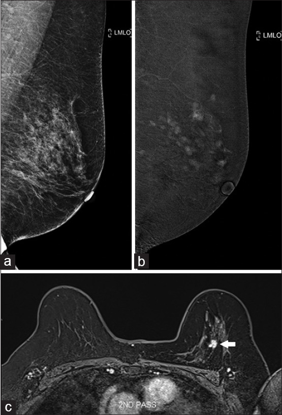
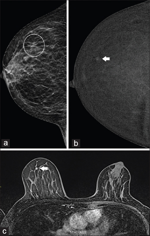
Case 1: A 69-year-old woman. (a) Full-field digital mammography shows no suspicious lesions in the left breast. (b) Contrast-enhanced spectral mammography shows multiple enhancing foci. (c) MR image confirms the presence of multiple enhancing lesions; the largest lesion (arrow) was targeted for MRI-guided vacuum-assisted biopsy, which showed low-grade ductal carcinoma in situ.
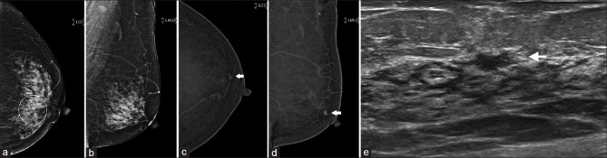
Case 2: A 69-year-old woman. (a & b) Full-field digital mammography (FFDM) shows architectural distortion in the central left breast (circle in a). Contrast-enhanced spectral mammography shows (c) no focal enhancement corresponding to the area of architectural distortion, but (b & c) a separate enhancing focus is detected in the upper outer periareolar left breast (arrows), which is not seen on FFDM. (e) US image shows an ill-defined, 8-mm hypoechoic lesion in the corresponding location (arrow); biopsy of the lesion showed intraductal carcinoma.
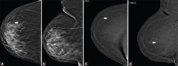
Case 3: A 75-year-old woman. (a & b) Full-field digital mammography shows a tiny nodule in the upper outer quadrant of the right breast (arrows). (c & d) Contrast-enhanced spectral mammography confirms the diagnosis. Subsequent excision biopsy revealed low-grade ductal carcinoma in situ and atypical lobular hyperplasia.
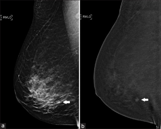
Case 4: A 62-year-old woman. (a) Full-field digital mammography shows a nodular opacity in the right lower outer quadrant (arrow). However, no sonographic correlate was found. (b) Contrast-enhanced spectral mammography shows the presence of a nodule in the right lower outer quadrant (arrow), which was proven to be fibroadenomatoid hyperplasia on stereotactic-guided biopsy.
Case 5: A 59-year-old woman. (a) Full-field digital mammography shows an asymmetry in the outer right breast (circled), visible only on craniocaudal view. (b) Contrast-enhanced spectral mammography (CESM) shows that the lesion was localised to the right upper outer quadrant (arrow). (c) Subsequent MR image shows type I enhancement kinetics, suggestive of a benign lesion (arrow), which confirmed the CESM finding.
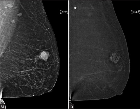
Case 6: A 62-year-old woman. (a) Full field digital mammography shows a spiculated mass seen in the left upper outer quadrant. (b) Contrast-enhanced spectral mammography for local staging shows enhancement of the mass, with no additional enhancing foci, confirming unifocal disease.
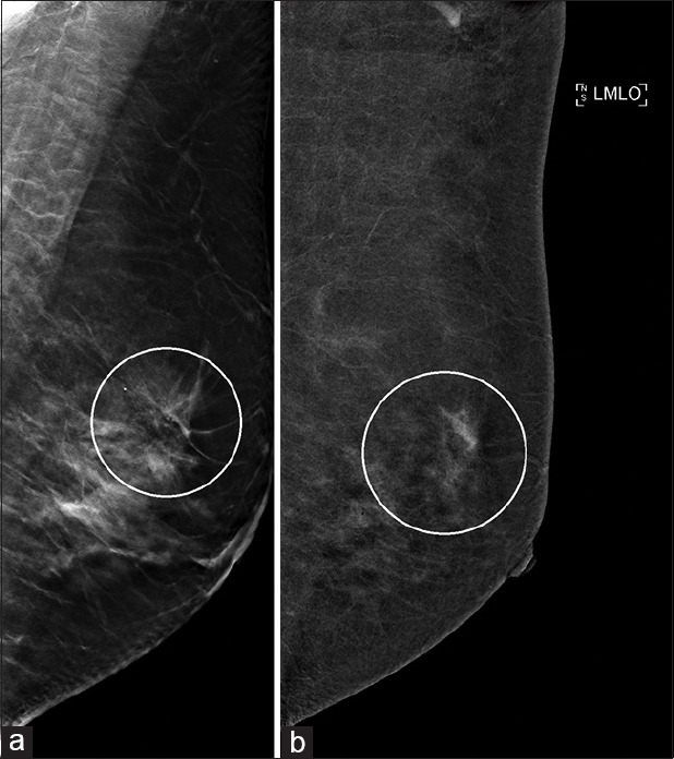
Case 7: Local staging for a 46-year-old woman with biopsy-proven right breast invasive ductal carcinoma. (a) Full-field digital mammography shows an area of architectural distortion in the left breast upper outer quadrant (circle). (b) Contrast-enhanced spectral mammography shows closely related clustered non-mass enhancement (circle). On biopsy, this was found to be a radial scar, and subsequent MRI also showed no suspicious abnormality.
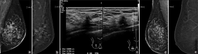
Case 8: A 57-year-old woman. (a) Full-field digital mammography (FFDM) of the left breast shows a spiculated mass in the left upper central breast, whereas (b) contrast-enhanced spectral mammography (CESM) for local staging shows extensive non-mass enhancement extending to the nipple, indicating more extensive disease than that seen on FFDM. (c) US image of the contralateral right breast shows a suspicious, taller-than-wide hypoechoic lesion; however (d & e) FFDM and CESM show no corresponding abnormality in the right breast. On complete excision, the right breast lesion showed fibrocystic change.
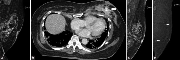
Case 9: A 59-year-old woman. (a) Full-field digital mammography (FFDM) of the left breast shows architectural distortion and extensive pleomorphic calcifications. High-grade ductal carcinoma in situ was found on core biopsy. (b) Staging CT image shows multiple enhancing foci in the left breast (arrow). (c) Post-neoadjuvant chemotherapy FFDM and (d) contrast-enhanced spectral mammography show improvement in the architectural distortion and a marked reduction in enhancing foci (arrows).
References
-
- Francescone MA, Jochelson MS, Dershaw DD, Sung JS, Hughes MC, Zheng J, et al. Low energy mammogram obtained in contrast-enhanced digital mammography (CEDM) is comparable to routine full-field digital mammography (FFDM) Eur J Radiol. 2014;83:1350–5. - PubMed
-
- Fallenberg EM, Schmitzberger FF, Amer H, Ingold-Heppner B, Balleyguier C, Diekmann F, et al. Contrast-enhanced spectral mammography vs. mammography and MRI – clinical performance in a multi-reader evaluation. Eur Radiol. 2017;27:2752–64. - PubMed
-
- Sumkin JH, Berg WA, Carter GJ, Bandos AI, Chough DM, Ganott MA, et al. Diagnostic performance of MRI, molecular breast imaging, and contrast-enhanced mammography in women with newly diagnosed breast cancer. Radiology. 2019;293:531–40. - PubMed
-
- Hobbs MM, Taylor DB, Buzynski S, Peake RE. Contrast-enhanced spectral mammography (CESM) and contrast enhanced MRI (CEMRI): Patient preferences and tolerance. J Med Imaging Radiat Oncol. 2015;59:300–5. - PubMed
MeSH terms
Substances
LinkOut - more resources
Full Text Sources
Medical
