. 2024;16(1):226-247.
doi: 10.1159/000538282. Epub 2024 Mar 25.

Type I Interferon, Induced by Adenovirus or Adenoviral Vector Infection, Regulates the Cytokine Response to Lipopolysaccharide in a Macrophage Type-Specific Manner

Affiliations

Type I Interferon, Induced by Adenovirus or Adenoviral Vector Infection, Regulates the Cytokine Response to Lipopolysaccharide in a Macrophage Type-Specific Manner

Mareike D Maler et al. J Innate Immun. 2024.

Abstract

Introduction: While TLR ligands derived from microbial flora and pathogens are important activators of the innate immune system, a variety of factors such as intracellular bacteria, viruses, and parasites can induce a state of hyperreactivity, causing a dysregulated and potentially life-threatening cytokine over-response upon TLR ligand exposure. Type I interferon (IFN-αβ) is a central mediator in the induction of hypersensitivity and is strongly expressed in splenic conventional dendritic cells (cDC) and marginal zone macrophages (MZM) when mice are infected with adenovirus. This study investigates the ability of adenoviral infection to influence the activation state of the immune system and underlines the importance of considering this state when planning the treatment of patients.

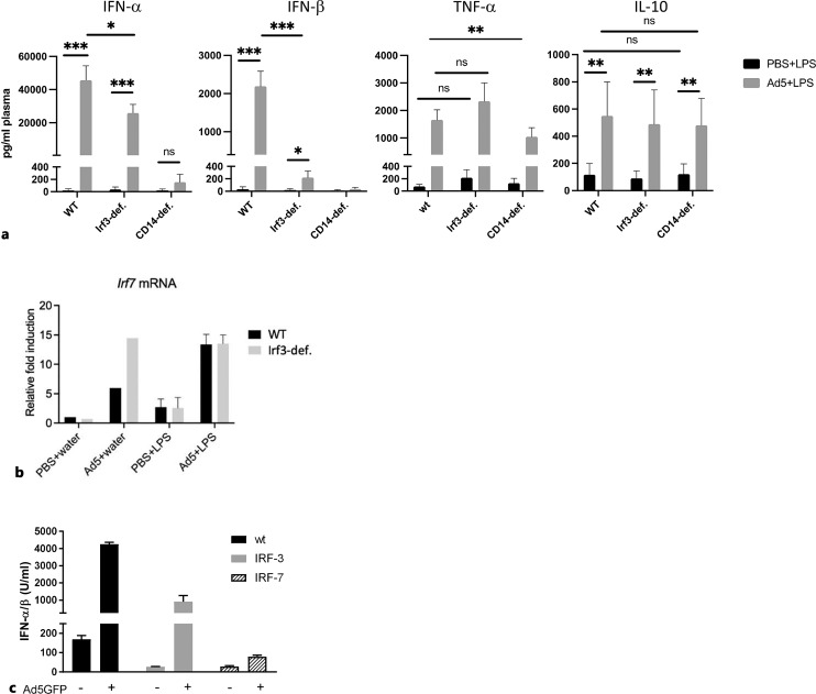
Methods: Infection with adenovirus-based vectors (Ad) or pretreatment with recombinant IFN-β was used as a model to study hypersensitivity to lipopolysaccharide (LPS) in mice, murine macrophages, and human blood samples. The TNF-α, IL-6, IFN-αβ, and IL-10 responses induced by LPS after pretreatment were measured. Mouse knockout models for MARCO, IFN-αβR, CD14, IRF3, and IRF7 were used to probe the mechanisms of the hypersensitive reaction.

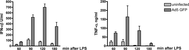
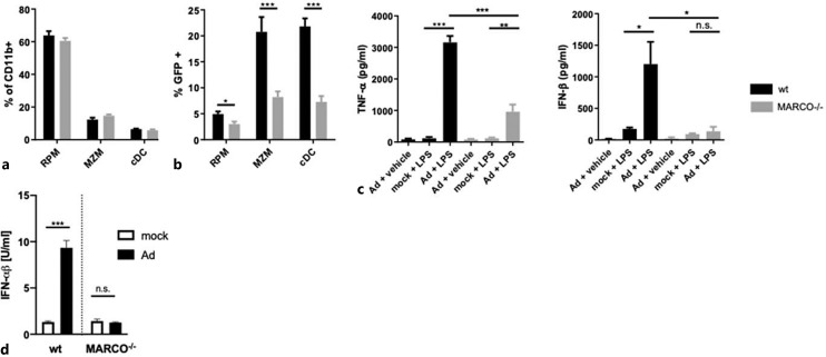
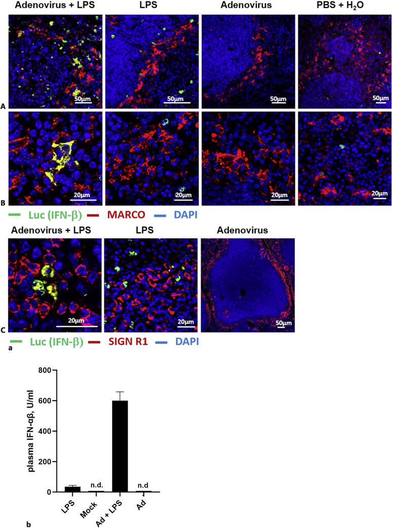
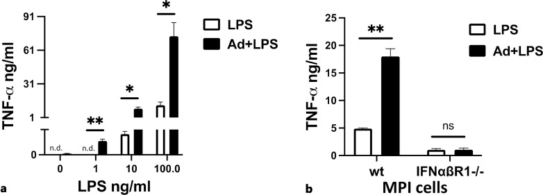
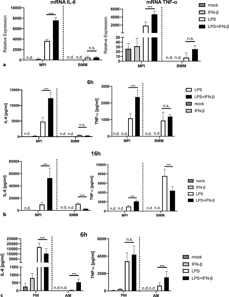
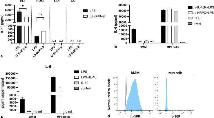
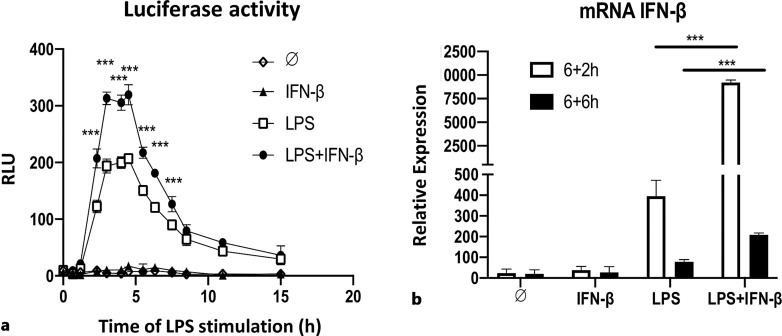
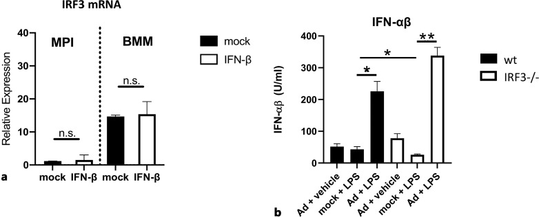
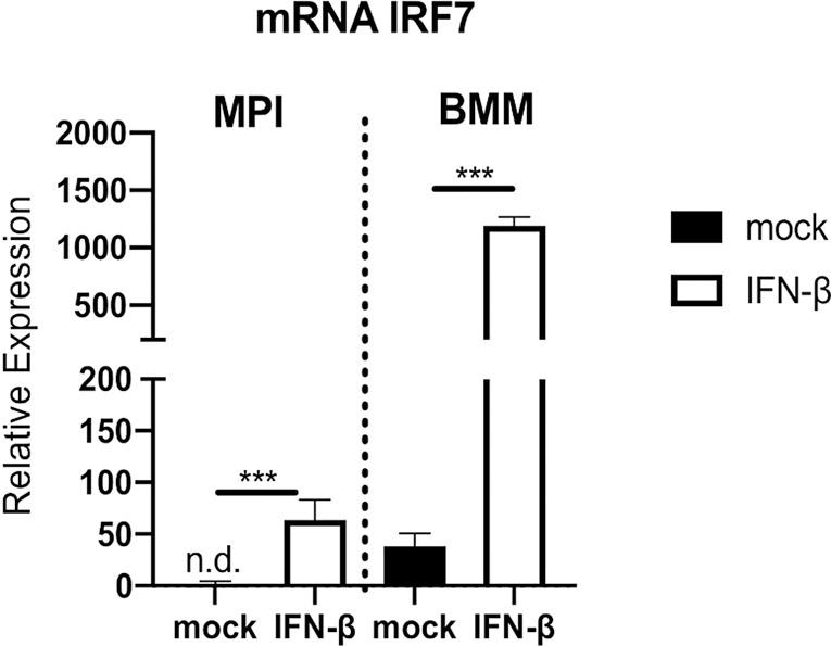
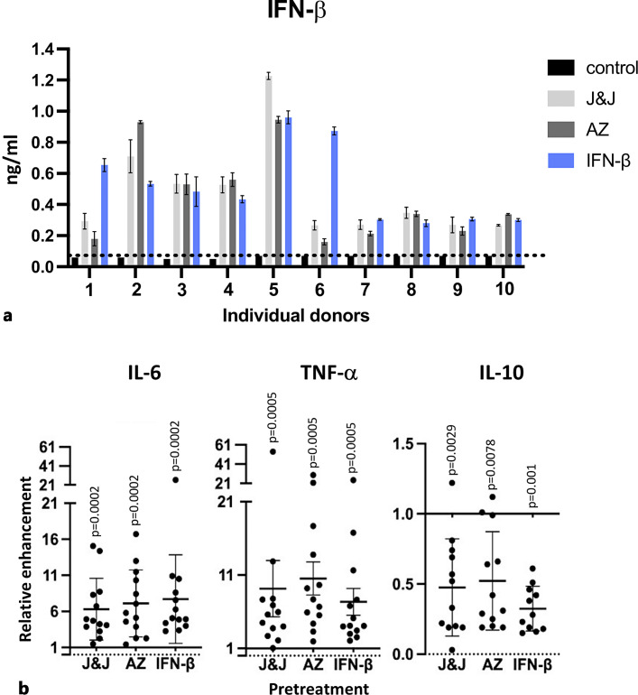
Results: We show that, similar to TNF-α and IL-6 but not IL-10, the induction of IFN-αβ by LPS increases strongly after Ad infection. This is true both in mice and in human blood samples ex vivo, suggesting that the regulatory mechanisms seen in the mouse are also present in humans. In mice, the scavenger receptor MARCO on IFN-αβ-producing cDC and splenic marginal zone macrophages is important for Ad uptake and subsequent cytokine overproduction by LPS. Interestingly, not all IFN-αβ-pretreated macrophage types exposed to LPS exhibit an enhanced TNF-α and IL-6 response. Pretreated alveolar macrophages and alveolar macrophage-like murine cell lines (MPI cells) show enhanced responses, while bone marrow-derived and peritoneal macrophages show a weaker response. This correlates with the respective absence or presence of the anti-inflammatory IL-10 response in these different macrophage types. In contrast, Ad or IFN-β pretreatment enhances the subsequent induction of IFN-αβ in all macrophage types. IRF3 is dispensable for the LPS-induced IFN-αβ overproduction in infected MPI cells and partly dispensable in infected mice, while IRF7 is required. The expression of the LPS co-receptor CD14 is important but not absolutely required for the elicitation of a TNF-α over-response to LPS in Ad-infected mice.

Conclusion: Viral infections or application of virus-based vaccines induces type I interferon and can tip the balance of the innate immune system in the direction of hyperreactivity to a subsequent exposure to TLR ligands. The adenoviral model presented here is one example of how multiple factors, both environmental and genetic, affect the physiological responses to pathogens. Being able to measure the current reactivity state of the immune system would have important benefits for infection-specific therapies and for the prevention of vaccination-elicited adverse effects.

Keywords: Adenoviral vector; Cytokines; IFN-αβ; Lipopolysaccharide; Macrophages.


Conflict of interest statement

The authors have no conflicts of interest to declare.

Figures

Fig. 1.
Fig. 1.
Enhancement of IFN-αβ and TNF-α response to LPS in adenovirus-infected mice. Groups of three C57BL/6 mice were injected with Ad or PBS and challenged 16 h later with LPS or H2O. Heparinized blood samples for cytokine measurement were collected at the indicated times after LPS treatment. IFN-αβ and TNF-α were determined by specific bioassays. A representative experiment of 2 is shown.
Fig. 2.
Fig. 2.
Requirement of scavenger receptor MARCO for the uptake of Ad by MZM and cDC and for the development of Ad-induced hypersensitivity to LPS. Splenocytes from naïve wt and MARCO−/− mice (7/strain) were obtained by mechanical disruption. a Quantification of macrophages and cDC: splenocytes of both strains were stained with anti-CD11b.PE, anti-CD11c.PE/Cy7, and anti-SIGN-R1.APC, and the CD11b+ population was further analyzed by flow cytometry with SIGN-R1/CD11c (RPM), SIGN-R1+/CD11c+ (MZM), and SIGN-R1/CD11c++ (cDC). b GFP expression after Ad infection. Splenocytes were infected with the GFP-encoding Ad for 16 h, and the GFP expression in CD11b+ cells was analyzed by flow cytometry. c TNF-α and IFN-β responses to LPS in vivo. Wt and MARCO−/− mice were injected with Ad or PBS and challenged 16 h later with LPS or H2O. Two hours after challenge, heparinized blood was collected for cytokine determination by specific ELISA (pooled results from two experiments with 3 mice/group). d IFN-αβ response to LPS in Ad-infected MPI cells. Wt and MARCO−/− MPI cells were treated with Ad or remained untreated for 16 h, washed, and stimulated with LPS for an additional 6 h. IFN-αβ was determined with a bioassay. U, relative luminescence units.
Fig. 3.
Fig. 3.
The contribution of splenic macrophages to the IFN-β response in LPS-challenged mock- and Ad-infected mice. IFN-β reporter C57BL/6 IFN-β+/Δβ-luc mice were injected with the non-fluorescent Ad2Ts1 or PBS and 16 h later challenged with LPS. Two hours after challenge, heparinized blood samples for IFN-αβ estimation or spleens for immunohistochemistry were collected and stained, as described in Materials and Methods. a Luciferase (IFN-β) expression in splenic tissue. To identify IFN-β-producing macrophages in the spleen, luciferase (Luc, green), MARCO (in A and B, red), and SIGN-R1 (in C, red) expression were visualized in paraformaldehyde-fixed, frozen tissue sections. The nuclei were stained with DAPI. A Overview at lower magnification. B, C Detailed images of the marginal zone. Representative examples are shown. b Plasma levels of IFN-αβ. The IFN-αβ in plasma was determined in a bioassay. n.d., not detectable.
Fig. 4.
Fig. 4.
Ad infection induces IFN-αβ-dependent hypersensitivity to LPS in alveolar macrophage-like mouse MPI cell lines. MPI cells in RPMI containing 10% FCS were infected with Ad and, 16 h later, stimulated with LPS. 6 h later, TNF-α in cell-free supernatants was determined by a bioassay. a TNF-α responses and over-responses are dose dependent. Infected and mock-infected wt MPI cells were stimulated with indicated amounts of LPS. b TNF-α sensitization requires the presence of the IFN-αβ receptor. Infected and mock-infected Wt and IFN-αβR−/− MPI cell lines were stimulated with 100 ng/mL LPS.
Fig. 5.
Fig. 5.
The ability of IFN-β to enhance LPS-induced TNF-α and IL-6 production is macrophage subset dependent. MPI cells, BMM, PM, and AM were pretreated with rmIFN-β and 6 h later stimulated with LPS. a mRNA expression in MPI cells and BMM. Total RNA from MPI and BMM cells was isolated 2 h after stimulation, and the cytokine expression was analyzed by qRT-PCR. Cytokine secretion by MPI cells and BMM (b) and PM and AM (c). Cytokines were determined in cell-free supernatants 6 h and 16 h after LPS addition in all cells, and MPIs and BMM, respectively, by ELISA. n.d., not detectable. Pooled results of two identical experiments carried out in triplicates are shown.
Fig. 6.
Fig. 6.
The ability to produce IL-10 and the height of IL-10R expression regulate the IL-6 response to LPS in mock- and IFN-β-pretreated macrophages. a IL-10 response to LPS: MPI’s, BMM, PC, and AM were pretreated with rmIFN-β, or remained untreated and 6 h later stimulated with LPS. The IL-10 response in culture supernatants was determined 16 h later by ELISA. IFN-β-stimulated BMM produced, on average, 110 pg IL-10/mL, while all other cell types and unstimulated control cells produced none (not shown in the figure). b IL-6 response in the presence of anti-IL-10R. BMM and MPI cells were pretreated with rmIFN-β or remained untreated and, 6 h later, stimulated with LPS in the presence or absence of 1 μg/mL anti-IL-10R or control antibody (anti-HRPO). IL-6 in culture supernatants was determined by a specific ELISA. c LPS-induced IL-6 production in the presence of exogenous IL-10. BMM and MPI cells were stimulated with LPS in the presence or absence of rmIL-10 (0.2 μg/mL). The IL-6 in culture supernatants collected 16 h later was determined by ELISA. d Cell surface IL-10R expression. The detection of IL-10R on cells was analyzed by FACS using anti-IL-10R.PE (filled blue) or isotype control (open black) antibodies. n.d., not detectable.
Fig. 7.
Fig. 7.
rIFN-β pretreatment enhances the IFN-β response to LPS in macrophages. a The time course of IFN-β production in MPI cells. IFN-β+/Δβ-luc MPI cells were treated with rmIFN-β and 6 h later stimulated with LPS. Cell-associated firefly luciferase activity was measured at the indicated time points after stimulation. RLU = relative luminescence units. b Expression of IFN-β mRNA in stimulated BMM. BMM were pretreated with rmIFN-β for 6 h and stimulated with LPS. Total cell RNA was isolated 2 and 6 h after stimulation, and IFN-β mRNA expression was analyzed by qRT-PCR.
Fig. 8.
Fig. 8.
Role of IRF3 in the induction of IFN-αβ by LPS in rmIFN-β-exposed macrophages. a IRF3 mRNA expression in IFN-β-treated MPI cells and BMM. Wt and IRF3−/− MPI cells were cultured with rmIFN-β for 6 h or remained untreated. The total cell RNA was isolated, and the mRNA expression was analyzed by qRT-PCR. b IFN-αβ response to LPS in Ad-infected wt an IRF3−/− MPI cells. Wt and IRF3−/− MPI cells were cultured with Ad or mock for 16 h, washed, and stimulated with LPS for a further 6 h. IFN-αβ in cell-free supernatants was determined by a bioassay. Supernatants of mock-treated wt and IRF3−/− cells contained no detectable amounts of IFN-αβ (not shown in the Fig.).
Fig. 9.
Fig. 9.
Treatment of macrophages with rIFN-β upregulates the of IRF7 mRNA expression. MPI cells and BMM were cultured with rmIFN-β for 2 h, and the total cell RNA was isolated. The IRF7 mRNA expression was analyzed by qRT-PCR.
Fig. 10.
Fig. 10.
Effect of IRF3 and CD14 deficiency on the response to LPS in mock- and Ad-infected mice. Wt, IRF3−/−, and CD14-deficient mice (5/group) were injected with Ad or PBS and challenged 16 h later with LPS or H2O. Heparinized blood samples for cytokine determination and spleens for RNA isolation were collected 2 h after the LPS challenge. a Cytokine production. Cytokines were determined in plasma by specific ELISA’s. b IRF7 mRNA expression. The analysis of IRF7 expression by real-time PCR was carried out using total spleen RNA. c Wt, IRF3−/−, and IRF7−/− were injected with Ad or PBS and challenged 16 h later with LPS. Heparinized blood samples were collected 2 h after LPS challenge, and IFN-αβ in plasma was determined by a bioassay.
Fig. 11.
Fig. 11.
Adenoviral vector-based SARS-CoV-2 vaccines induce type I IFN and, like exogenous IFN-β, differentially regulate LPS-induced cytokine responses in human whole blood. WB samples from healthy donors were pretreated with J&J, AZ vaccines, or hrIFN-β, or remained untreated and 24 h later stimulated or not with LPS for an additional 24 h, as described in Material and Methods section. a IFN-β determination at the time of LPS treatment. IFN-β was determined in the culture supernatants of 10 donors 24 h after the hrIFN-β pretreatment by ELISA. Supernatants of mock-pretreated WB cells contained no detectable IFN-β (control). Dotted line: detection limit. b TNF-α, IL-6, and IL-10 responses to LPS after vaccine or hrIFN-β pretreatment. The cytokine amounts were determined in whole blood supernatants from 12–13 individual donors at the end of the experiment (48 h) by ELISA. Values found in agent-pretreated, LPS-stimulated cell supernatants relative to those found in mock-pretreated and stimulated once are shown. No TNF-α, IL-6, and IL-10 was found in completely unstimulated and vaccine- or rIFN-β-only-pretreated whole blood cells (not shown).

Similar articles

Cited by

References

    1. von Jeney N, Gunther E, Jann K. Mitogenic stimulation of murine spleen cells: relation to susceptibility to Salmonella infection. Infect Immun. 1977;15(1):26–33. - PMC - PubMed
    1. Takeuchi O, Akira S. Pattern recognition receptors and inflammation. Cell. 2010;140(6):805–20. - PubMed
    1. Galanos C, Freudenberg MA, Matsuura M, Coumbos A. Hypersensitivity to endotoxin and mechanisms of host-response. Prog Clin Biol Res. 1988;272:295–308. - PubMed
    1. Freudenberg MA, Kalis C, Chvatchko Y, Merlin T, Gumenscheimer M, Galanos C. Role of interferons in LPS hypersensitivity. J Endotoxin Res. 2003;9(5):308–12. - PubMed
    1. Freudenberg MA, Tchaptchet S, Keck S, Fejer G, Huber M, Schütze N, et al. . Lipopolysaccharide sensing an important factor in the innate immune response to Gram-negative bacterial infections: benefits and hazards of LPS hypersensitivity. Immunobiology. 2008;213(3–4):193–203. - PubMed

Publication types

MeSH terms