. 2024 Jun 15:251:109918.
doi: 10.1016/j.neuropharm.2024.109918. Epub 2024 Mar 24.

A longitudinal MRI and TSPO PET-based investigation of brain region-specific neuroprotection by diazepam versus midazolam following organophosphate-induced seizures

Affiliations

A longitudinal MRI and TSPO PET-based investigation of brain region-specific neuroprotection by diazepam versus midazolam following organophosphate-induced seizures

Brad A Hobson et al. Neuropharmacology. .

Abstract

Acute poisoning with organophosphorus cholinesterase inhibitors (OPs), such as OP nerve agents and pesticides, can cause life threatening cholinergic crisis and status epilepticus (SE). Survivors often experience significant morbidity, including brain injury, acquired epilepsy, and cognitive deficits. Current medical countermeasures for acute OP poisoning include a benzodiazepine to mitigate seizures. Diazepam was long the benzodiazepine included in autoinjectors used to treat OP-induced seizures, but it is now being replaced in many guidelines by midazolam, which terminates seizures more quickly, particularly when administered intramuscularly. While a direct correlation between seizure duration and the extent of brain injury has been widely reported, there are limited data comparing the neuroprotective efficacy of diazepam versus midazolam following acute OP intoxication. To address this data gap, we used non-invasive imaging techniques to longitudinally quantify neuropathology in a rat model of acute intoxication with the OP diisopropylfluorophosphate (DFP) with and without post-exposure intervention with diazepam or midazolam. Magnetic resonance imaging (MRI) was used to monitor neuropathology and brain atrophy, while positron emission tomography (PET) with a radiotracer targeting translocator protein (TSPO) was utilized to assess neuroinflammation. Animals were scanned at 3, 7, 28, 65, 91, and 168 days post-DFP and imaging metrics were quantitated for the hippocampus, amygdala, piriform cortex, thalamus, cerebral cortex and lateral ventricles. In the DFP-intoxicated rat, neuroinflammation persisted for the duration of the study coincident with progressive atrophy and ongoing tissue remodeling. Benzodiazepines attenuated neuropathology in a region-dependent manner, but neither benzodiazepine was effective in attenuating long-term neuroinflammation as detected by TSPO PET. Diffusion MRI and TSPO PET metrics were highly correlated with seizure severity, and early MRI and PET metrics were positively correlated with long-term brain atrophy. Collectively, these results suggest that anti-seizure therapy alone is insufficient to prevent long-lasting neuroinflammation and tissue remodeling.

Keywords: Benzodiazepines; Diisopropylfluorophosphate; In vivo imaging; Neuroinflammation; Rat; Status epilepticus.


Conflict of interest statement

Declaration of competing interest The authors declare the following financial interests/personal relationships which may be considered as potential competing interests: Pamela J Lein reports financial support was provided by the National Institute of Neurological Disorders and Stroke CounterACT program. Brad A. Hobson reports financial support was provided by a T32 training program awarded to the University of California, Davis by the National Institute of General Medical Sciences and by a scholarship from the ARCS Foundation of Northern California.

Figures

Fig. 1.
Fig. 1.. Schematic of the study design.
(A) Schematic depicting the dosing paradigm used to induce status epilepticus while minimizing death. (B) Schematic depicting the experimental timeline for imaging. Animals were imaging longitudinally from 3 to 65 days post-DFP exposure with the exception of two animals in the DFP group that died prior to imaging at the 7 day time point. These animals were replaced with two time-matched DFP intoxicated animals at subsequent time points. Subsets of these animals were imaged at 95 and 168 days. Data in brackets represent the achieved scans at each time after accounting for animal death. (C) Table detailing animal numbers and compounds administered to each experimental group (vehicle control group = VEH).
Fig. 2.
Fig. 2.. Acute DFP intoxication produces lesions and signs of brain atrophy evident in T2w images that are attenuated by intervention with DZP or MDZ.
Rows are representative axial (−3.5 mm bregma) T2w images of the left hemisphere of a single animal from each experimental group. A vehicle control animal (row 1) shows homogeneous image intensity in brain parenchyma that is consistent across follow-up scans. By comparison, the DFP animal (row 2) displays hyperintense lesions in the hippocampus (arrowhead right) and piriform cortex (arrowhead left). At later time points, this animal is characterized by progressive hippocampal atrophy (arrowhead down right), profound ventricular enlargement (arrowhead down left), and hypointense lesions within the thalamus (arrowhead up). Intervention with DZP (row 3) or MDZ (row 4) appeared to prevent hippocampal hyperintensity, but not hyperintensity within the piriform cortex nor hypointensity within the thalamus. Note: not all lesions are marked by arrowheads in the array of images.
Fig. 3.
Fig. 3.. Quantification of lateral ventricular and hippocampal volumes following acute DFP intoxication demonstrate ongoing brain atrophy that is reduced by DZP and MDZ intervention.
Geometric mean ratios (GMR) depict changes in regional volumes over time relative to a comparator group. Columns represent specific comparisons between experimental groups; rows represent distinct brain regions compared at each imaging time point. Given the rodent skull maintains approximately a fixed volume in adulthood, enlargement of the lateral ventricles represents a recession of brain tissue as CSF increases to fill the space available. Data are presented as the GMR (dot) with 95% confidence interval (vertical bars) from days 3 through 168 post-exposure); the horizontal line corresponds to a GMR of 1.0 (Days 3–65, n = 8–10 animals per group; days 91 and 165; n = 3–7 animals per group). Confidence intervals that do not include 1.00, and survived correction for multiple comparisons (FDR), are colored blue and indicate a significant difference between the two groups being compared at p < 0.05.
Fig. 4.
Fig. 4.. Acute DFP intoxication produces aberrant tissue diffusion characteristics that are brain region-specific and not resolved by intervention with DZP or MDZ.
Rows are representative axial (−3.5 mm bregma) parametric maps of the ADC calculated from diffusion-weighted MRI of a single animal from each experimental group. Comparison of a VEH control animal (row 1, VEH) to animals that received DFP, with or without benzodiazepine intervention (rows 2, 3, and 4), reveals persistent lesions in the hippocampus (arrowhead down), thalamus (arrowhead up), and piriform cortex (arrowhead left) characterized by areas of increased ADC heterogeneity. Diffusion-weighted MRI also reveals hippocampal atrophy and ventricular enlargement following DFP intoxication with saline intervention. Note: not all lesions are marked by arrowheads in the array of images.
Fig. 5.
Fig. 5.. Spatiotemporal quantification of ADC heterogeneity demonstrates persistent region-specific damage following acute DFP intoxication, with or without benzodiazepine intervention.
Columns represent specific experimental groups; rows represent distinct brain regions at each imaging time point. Data are presented as the geometric mean ratio with 95% confidence interval (Day 3–65, n = 8–10 animals per group; days 91 and 165: n = 3–7 animals per group). Confidence intervals that do not include 1.00 and survived correction for multiple comparisons are colored blue and indicate a significant difference between groups at p < 0.05.
Fig. 6.
Fig. 6.. Persistent neuroinflammation in the thalamus and piriform cortex resulting from acute DFP intoxication is unaffected by diazepam or midazolam intervention.
Longitudinal PET SUV maps of a single animal from each experimental group. Data are overlaid on T2w images from the same animal (Fig. 2). The [18F]PBR111 uptake observed in the vehicle animal is largely due to non-specific binding along major blood vessels and outside the brain. By comparison, the DFP animal displays significant radiotracer uptake in the hippocampus (arrowhead down), thalamus (arrowhead up), and piriform cortex (arrowhead left) that persists for the duration of the study. Diazepam or midazolam intervention attenuates, but does not prevent, apparent radiotracer uptake in the thalamus and piriform cortex. Midazolam intervention appears to reduce radiotracer uptake in the hippocampus of this particular animal. In all scans there is minor signal penetration from outside the brain along the skull and jaw bones due to [18F] binding in bone following defluorination of the radiotracer. Note: not all inflammatory lesions are marked by arrowheads in the array of images.
Fig. 7.
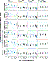
Fig. 7.. Chronic neuroinflammation following acute DFP intoxication persists in multiple regions even with midazolam or diazepam intervention.
Regional [18F]PBR111 uptake data quantified by standard uptake value (SUV) analysis normalized to the SUV of a cerebellar reference region (SUVR). Columns represent specific experimental groups; rows represent distinct brain regions at each imaging time point. Data are presented as the geometric mean ratio (dot) with 95% confidence interval (vertical bars); the horizontal line corresponds to a GMR of 1.0 (Days 3–65, n = 8–10 animals per group; days 91 and 165; n = 3–7 animals per group). Confidence intervals that do not include 1.00, and survived correction for multiple comparisons (FDR), are colored blue and indicate a significant difference between the two groups being compared at p < 0.05.

References

    1. Abdel-Rahman A, Shetty AK, Abou-Donia MB, 2002. Acute exposure to sarin increases blood brain barrier permeability and induces neuropathological changes in the rat brain: dose-response relationships. Neuroscience 113, 721–741. - PubMed
    1. Ackerman JJ, Neil JJ, 2010. The use of MR-detectable reporter molecules and ions to evaluate diffusion in normal and ischemic brain. NMR Biomed. 23, 725–733. - PMC - PubMed
    1. Altay O, Suzuki H, Hasegawa Y, Ostrowski RP, Tang J, Zhang JH, 2014. Isoflurane on brain inflammation. Neurobiol. Dis. 62, 365–371. - PMC - PubMed
    1. Andrew PM, Lein PJ, 2021. Neuroinflammation as a therapeutic target for mitigating the long-term consequences of acute organophosphate intoxication. Front. Pharmacol. 12, 674325. - PMC - PubMed
    1. Apland JP, Aroniadou-Anderjaska V, Figueiredo TH, Rossetti F, Miller SL, Braga MF, 2014. The limitations of diazepam as a treatment for nerve agent-induced seizures and neuropathology in rats: comparison with UBP302. J Pharmacol Exp Ther 351, 359–372. - PMC - PubMed

MeSH terms