. 2024 Jan-Mar;65(1):35-44.
doi: 10.47162/RJME.65.1.05.

Epidemiological and histopathological aspects of ocular melanomas in Northeastern Romania

Affiliations

Epidemiological and histopathological aspects of ocular melanomas in Northeastern Romania

Claudia Florida Costea et al. Rom J Morphol Embryol. 2024 Jan-Mar.

Abstract

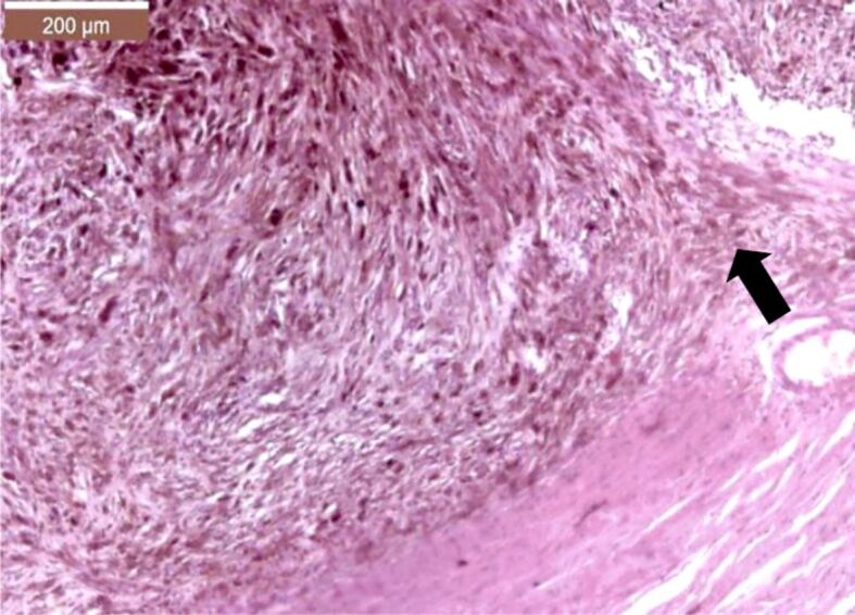
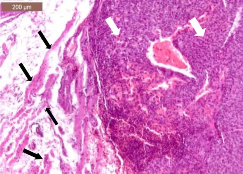
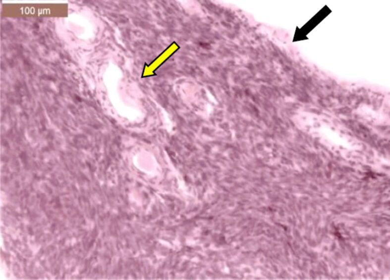
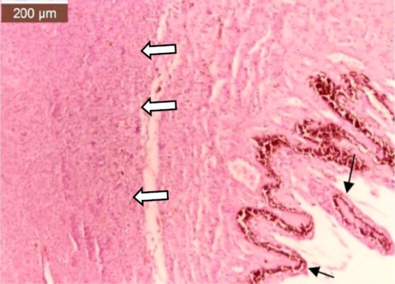
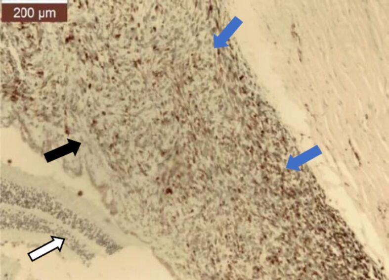
Ocular melanoma is a rare but complex disease in current medical practice. Our retrospective study spans over a period of 28 years and analyzed uveal and conjunctival melanomas that were consecutively admitted, diagnosed, and treated in the 2nd Ophthalmology Clinic of Prof. Dr. Nicolae Oblu Emergency Clinical Hospital, Iaşi, Romania. The patients were selected from the records of the Department of Pathology of our Hospital, being diagnosed by standard histopathological techniques. The aim of this study was to summarize the epidemiological and pathological aspects of uveal and conjunctival melanomas in Northeastern region of Romania. In our study, we did not notice a predilection of uveal and conjunctival melanoma to one particular gender. The most common histological subtypes of ocular melanomas were the heavily pigmented spindle cell subtype, followed by the epithelioid subtype. Our patients sought medical help in a timely manner, before the systemic invasion of the disease could develop.


Conflict of interest statement

The authors declare that they have no conflict of interests.

Figures

Figure 1
Figure 1
Moderately pigmented, choroidal spindle cell melanoma infiltrating in the surrounding sclera (black arrow). Photomicrograph exhibits a densely cellular tumor, composed of bundles of cells with spindle nuclei, with moderate atypia. Many tumor cells show an abundant amount of brown, fine granular pigment (melanin) in the cytoplasm. The tumor infiltrates the inner surface of the adjacent sclera [Hematoxylin–Eosin (HE) staining, ×100]
Figure 2
Figure 2
Poorly pigmented, choroidal mixed melanoma infiltrating in nearby sclera (black arrow). Photomicrograph shows a densely cellular tumor, composed of sheets of epithelioid-appearing cell mixed with cells with spindle-shaped nuclei with moderate atypia; from place to place, tumor cells present a reduced amount of brown, fine granular pigment (melanin) in the cytoplasm; the tumor infiltrates the inner surface of the adjacent sclera (HE staining, ×200)
Figure 3
Figure 3
Heavily pigmented choroidal mixed melanoma (blue arrow) infiltrating the surrounding sclera (black arrow) and around its nerve fibers (yellow arrow). Photomicrograph showing a densely cellular tumor, composed of sheets of epithelioid cell mixed with cells with spindle-shaped nuclei with moderate atypia; from place to place, tumor cells present a reduced amount of brown, fine granular pigment (melanin) in the cytoplasm; the tumor infiltrates into the inner surface of the adjacent sclera and around the nerve fibers from this level (HE staining, ×100)
Figure 4
Figure 4
Nonpigmented choroidal melanoma infiltrating in the orbit. Photomicrograph showing a densely cellular tumor composed of sheets of neoplastic melanocytes (white arrows) infiltrating between striated muscle fibers (black arrows) from the orbit (HE staining, ×100)
Figure 5
Figure 5
The distribution of different anatomical structures invaded by ocular melanomas (n=36)
Figure 6
Figure 6
Location of ocular melanomas according to the gender of the patients (n=36). F: Female; M: Male
Figure 7
Figure 7
Distribution of ocular melanomas according to location in the eyes (left–right) (n=36). LE: Left eye; RE: Right eye
Figure 8
Figure 8
Distribution of pathological subtypes of ocular melanomas according to the gender of the patients (n=36)
Figure 9
Figure 9
Heavily pigmented conjunctival epithelioid cells melanoma. Photomicrograph showing on the left the conjunctival epithelium (black solid arrow) and on the right, just below the conjunctiva, a densely cellular tumor (black arrowhead), whose cells have cytoplasm abundantly loaded with a fine granular brown pigment (melanin) also covering the nuclei (HE staining, ×100)
Figure 10
Figure 10
Heavily pigmented choroidal epithelioid cells melanoma. Photomicrograph showing a densely cellular tumor, composed of sheets of loosely cohesive, voluminous, epithelioid melanocytes with abundant cytoplasm, containing abundant brown, finely granular pigment (melanin), almost completely covering the nucleus (white arrows) (HE staining, ×400).
Figure 11
Figure 11
Moderately pigmented iris melanoma. Photomicrograph showing on the lower left the ciliary body (white arrow) and on the upper right the presence of a tumor (black arrow) which replaces the iris structure and is moderately cellular, being composed of atypical melanocytes; in the cytoplasm of some of the tumor cells there is a fine granular brown pigment (melanin) (blue arrow) (van Gieson staining, ×100)
Figure 12
Figure 12
Same case, with higher magnification. Photomicrograph showing the densely cellular tumor, consisting of sheets of atypical, dscohesive melanocyte. In some areas there are neoplastic cells with granular brown pigment (melanin) in the cytoplasm (blue arrows) (van Gieson staining, ×200)
Figure 13
Figure 13
Hypopigmented conjunctival spindle cells melanoma of the forniceal conjunctiva. Photomicrograph showing on the right nonkeratinized stratified columnar forniceal conjunctival epithelium (black arrow) and pseudoglands of Henle (yellow arrow) included in a densely cellular tumor composed of bundles of spindle-shaped cells. Rare tumor cells show a relatively small amount of fine granular brown pigment (melanin) in the cytoplasm (HE staining, ×200)
Figure 14
Figure 14
Moderately pigmented choroidal melanoma infiltrating the surrounding sclera and around its nerve fibers. Photomicrograph showing a densely cellular tumor composed of sheets of neoplastic melanocytes (black arrow) infiltrating the inner surface of the adjacent sclera and around the nerve fibers at its level (white arrows) (van Gieson staining, ×200)
Figure 15
Figure 15
Lightly pigmented conjunctival melanoma. Photomicrograph showing the conjunctival epithelium (black arrows) on the right, which covered a densely cellular tumor (white arrows) consisting of atypical melanocytes, some of them with granular brown pigment (melanin) in the cytoplasm (HE staining, ×100)
Figure 16
Figure 16
Same case, with higher magnification. Lightly pigmented conjunctival melanoma. Photomicrograph showing the tumor (black arrow) composed of atypical melanocytes. Some of the neoplastic cells have in their cytoplasm a fine granular brown pigment (melanin) (HE staining, ×200)
Figure 17
Figure 17
Lightly pigmented choroidal spindle cells melanoma. Photomicrograph showing a densely cellular tumor composed of bundles of spindle-shaped, moderately atypical melanocytes (HE staining, ×200)
Figure 18
Figure 18
Nonpigmented choroidal epithelioid cells melanoma. Photomicrograph showing a densely cellular tumor made up of sheets of loosely cohesive, voluminous, epithelioid tumor cells (white arrows), with abundant eosinophilic cytoplasm and pleomorphic nucleus (HE staining, ×400)
Figure 19
Figure 19
Nonpigmented ciliary body melanoma. Photomicrograph showing on the lower right the ciliary body (black arrows) and on the right a tumor (white arrows) at the base of the ciliary body infiltrating into the choroid; the tumor is densely cellular and consists of atypical melanocytes (HE staining, ×100)
Figure 20
Figure 20
Photomicrograph showing the retina detaching from choroid, as the choroid was the origin of a medium pigmented melanoma. The tumor consists of sheets of poorly cohesive atypical melanocytes, most of them with coarser granules of melanin pigment in their cytoplasm, even covering the nucleus. On the left there is the sclera that is devoid of tumor cells (black arrows) (HE staining, ×50)
Figure 21
Figure 21
Photomicrograph showing a lightly pigmented choroidal spindle cells melanoma. The retina (white arrow) was detached from the choroid (black arrow), as it was the origin of a densely cellular tumor, consisting of spindle tumor cells arranged in bundles (blue arrows). The tumor cells have a relatively small amount of fine granular brown pigment (melanin) in the cytoplasm (van Gieson staining, ×100)

References

    1. Kaliki S, Shields CL. Uveal melanoma: relatively rare but deadly cancer. Eye (Lond) 2017;31(2):241–257. - PMC - PubMed
    1. Brouwer NJ, Verdijk RM, Heegaard S, Marinkovic M, Esmaeli B, Jager MJ. Conjunctival melanoma: new insights in tumour genetics and immunology, leading to new therapeutic options. Prog Retin Eye Res. 2022;86:100971–100971. - PubMed
    1. Pandiani C, Béranger GE, Leclerc J, Ballotti R, Bertolotto C. Focus on cutaneous and uveal melanoma specificities. Genes Dev. 2017;31(8):724–743. - PMC - PubMed
    1. Patrocínio LG, Lourenço C, Prado Silva, Borges Barra, Patrocínio JA. Primary malignant orbit melanoma. Braz J Otorhinolaryngol. 2006;72(5):716–716. - PMC - PubMed
    1. Patel DR, Patel BC. Ocular melanoma. StatPearls [Internet] 2024 Jan - PubMed