. 2024 Mar 25;14(1):7025.
doi: 10.1038/s41598-024-57624-8.

Comparative response to PDT with methyl-aminolevulinate and temoporfin in cutaneous and oral squamous cell carcinoma cells

Affiliations

Comparative response to PDT with methyl-aminolevulinate and temoporfin in cutaneous and oral squamous cell carcinoma cells

J Nicolás-Morala et al. Sci Rep. .

Abstract

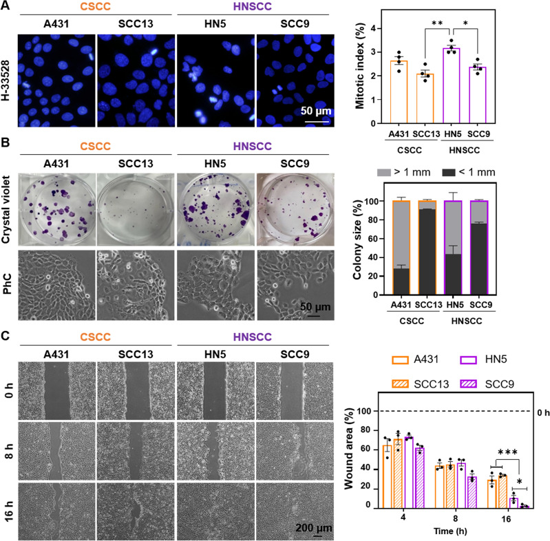
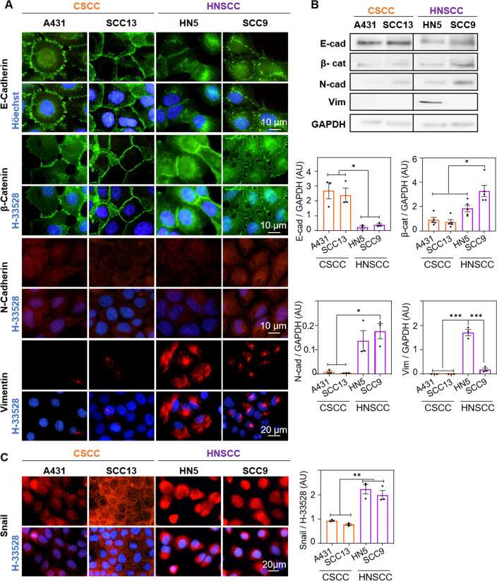
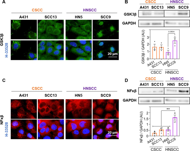
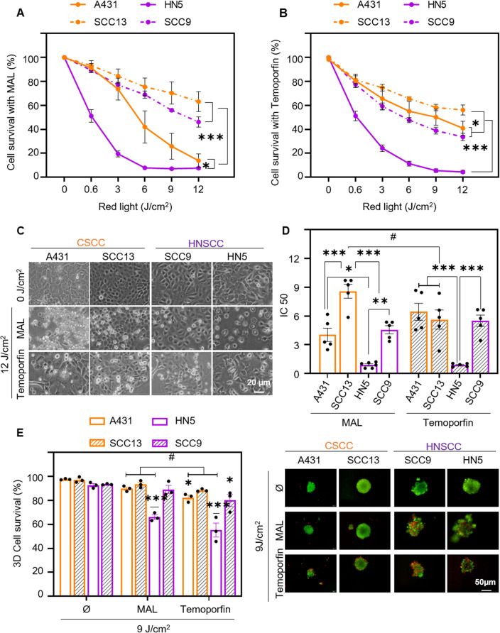
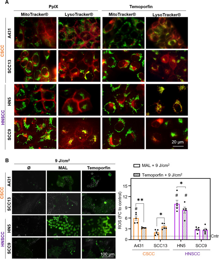
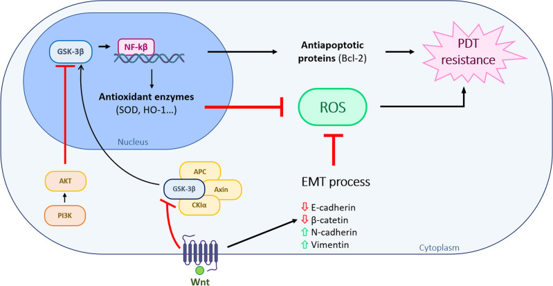
Cutaneous and Head and Neck squamous cell carcinoma (CSCC, HNSCC) are among the most prevalent cancers. Both types of cancer can be treated with photodynamic therapy (PDT) by using the photosensitizer Temoporfin in HNSCC and the prodrug methyl-aminolevulinate (MAL) in CSCC. However, PDT is not always effective. Therefore, it is mandatory to correctly approach the therapy according to the characteristics of the tumour cells. For this reason, we have used cell lines of CSCC (A431 and SCC13) and HNSCC (HN5 and SCC9). The results obtained indicated that the better response to MAL-PDT was related to its localization in the plasma membrane (A431 and HN5 cells). However, with Temoporfin all cell lines showed lysosome localization, even the most sensitive ones (HN5). The expression of mesenchymal markers and migratory capacity was greater in HNSCC lines compared to CSCC, but no correlation with PDT response was observed. The translocation to the nucleus of β-catenin and GSK3β and the activation of NF-κβ is related to the poor response to PDT in the HNSCC lines. Therefore, we propose that intracellular localization of GSK3β could be a good marker of response to PDT in HNSCC. Although the molecular mechanism of response to PDT needs further elucidation, this work shows that the most MAL-resistant line of CSCC is more sensitive to Temoporfin.


Conflict of interest statement

The authors declare no competing interests.

Figures

Figure 1
Figure 1
Cell proliferation and migration in CSCC and HNSCC. (A) Nuclei (blue) were stained with Höechst-33258 and observed by fluorescence microscopy under ultraviolet excitation light. Mitotic index of cell lines was estimated by the number of dividing cells/total cell number. At least 500 cells of each cell type were estimated. Photographs (left) and quantification (right), n = 4. (B) 100 cells per well were seeded, after 14 days of growth, the colonies formed were fixed and stained with crystal violet. Colonies were classified according to their size into < 1 mm and > 1 mm. Photographs (left) and quantification (right), n = 3. (C) Wound closure evolution after inserts removal at 0 h. Photographs were taken at 0, 8 and 16 h (left panel) and quantification of the cell-free area (right panel), n = 3. Values are represented as the mean ± SEM (*p < 0.05, **p < 0.01, ***p < 0.001).
Figure 2
Figure 2
EMT markers in CSCC and HNSCC. (A) Localization of E-cadherin, β-catenin, N-cadherin and vimentin assessed by indirect immunofluorescence. Nuclei are counterstained with Höechst-33258 (blue), n = 3. (B) Quantification of E-cadherin, β-catenin, N-cadherin and vimentin expression by Western Blot. A representative expression band and the densitometry of these bands relative to the loading control (GAPDH) are shown, n = 3. (C) Localization of Snail (red) determined by indirect immunofluorescence. Nuclei are counterstained with Höechst-33258 (blue) (left panel) and Snail expression by quantification of fluorescence intensity (right panel), n = 3. Values were represented as mean ± SEM (*p < 0.05, **p < 0.01, ***p < 0.001).
Figure 3
Figure 3
GSK3β and NF-κβ expression. (A) Localization of GSK3β (green) determined by indirect immunofluorescence. Nuclei are counterstained with Höechst-33258 (blue), n = 3. (B) Quantification of GSK3β expression by Western blot. A representative band and densitometry of the bands relative to the loading control (GAPDH) are shown, n = 3. (C) Localization of NF-κβ (red) determined by indirect immunofluorescence. Nuclei are counterstained with Höechst-33258 (blue), n = 3. (D) Expression and quantification of NF-κβ by Western blot. Representative band and densitometry of these relative to the loading control (GAPDH) are shown, n = 3. Values are represented as mean ± SEM (**p < 0.01).
Figure 4
Figure 4
Photodynamic therapy with methyl-aminolevulinate and Temoporfin. Cell survival was determined by MTT assay 24 h after incubation with 0.5 mM MAL for 5 h (A) or 24 h with 25 nM Temoporfin (B) and subsequent irradiation with red light (0 to 12 J/cm2). The results of the MTT assay are relativized to the values of absorbance at 542 nm obtained for untreated cells (indicated as control), n = 5. (C) Cell morphology after PDT (5 h of MAL or 24 h Temoporfin incubation followed by 12 J/cm2 dose) and observed by phase contrast microscopy 24 h after irradiation. (D) Half maximal inhibitory concentration (IC50) is represented for both treatments in each cell line, n = 5. (E) Cell survival after PDT (0.5 mM MAL, 9 J/cm2 or 25 nM Temoporfin, 9 J/cm2) in spheroids. Quantification of cell survival was determined by staining with acridine orange and propidium iodide and estimating the green (live) cells with respect to red (dead) cells, n = 3. Values were represented as mean ± SEM (*p < 0.05, **p < 0.01, ***p < 0.001, #p < 0.05 MAL vs Temoporfin).
Figure 5
Figure 5
Subcellular localization of photosensitizer and ROS production. (A) Cells were incubated with MAL (0.5 mM for 24 h) and with Temoporfin (25 nM for 24 h) and the localization of PS (red fluorescence) determined by fluorescence microscopy. Green fluorescence caused by MitoTracker® (mitochondria) or LysoTracker® (lysosomes) probes, n = 3. (B) ROS production detected by the DHF-DA fluorescent probe after PDT with MAL or Temoporfin and red light (9 J/cm2). Cells were incubated MAL (0.5 mM for 24 h) and with Temoporfin (25 nM for 24 h), and in the last hour DHF-DA was added, reaching a final concentration of 6 μM. The fluorescence signal was observed by using fluorescence microscopy (λexc = 436 nm). Intracellular fluorescence intensity was measured by ImageJ, n = 5. Values were represented as mean ± SEM (*p < 0.05, **p < 0.01, ***p < 0.001, #p < 0.05 different cells between same treatment).
Figure 6
Figure 6
Schematic relation between EMT and the GSK3β/NF-κβ pathway and PDT resistance. Abnormal expression of Wnt/β-catenin pathway or loss of phosphatidylinositol 3-kinase (PI3K)-Akt signalling is related to the nuclear localization of GSK3β. GSK3β nuclear translocation constitutes an upstream regulator of nuclear NF-kB, functioning as a transcription activating diverse antiapoptotic and antioxidant target enzymes. One of the mechanisms through which EMT can induce PDT resistance is the ability to provoke low quantities of ROS. This reduction in ROS production can be also produced by other signalling pathways as the activation of pro-survival signals as a result of NF-kB transcriptional activities.

Similar articles

References

    1. Sung H, et al. Global cancer statistics 2020: GLOBOCAN estimates of incidence and mortality worldwide for 36 cancers in 185 countries. CA Cancer J. Clin. 2021;71:209–249. doi: 10.3322/caac.21660. - DOI - PubMed
    1. Dotto GP, Rustgi AK. Squamous cell cancers: a unified perspective on biology and genetics. Cancer Cell. 2016;29:622–637. doi: 10.1016/j.ccell.2016.04.004. - DOI - PMC - PubMed
    1. Johnson DE, et al. Head and neck squamous cell carcinoma. Nat. Rev. Dis. Prim. 2020;6:92. doi: 10.1038/s41572-020-00224-3. - DOI - PMC - PubMed
    1. Alsahafi E, et al. Clinical update on head and neck cancer: Molecular biology and ongoing challenges. Cell Death Dis. 2019;10:540. doi: 10.1038/s41419-019-1769-9. - DOI - PMC - PubMed
    1. Corchado-Cobos R, García-Sancha N, González-Sarmiento R, Pérez-Losada J, Cañueto J. Cutaneous squamous cell carcinoma: From biology to therapy. Int. J. Mol. Sci. 2020;21:2956. doi: 10.3390/ijms21082956. - DOI - PMC - PubMed

MeSH terms