

Plate augmentation and hybrid bone grafting are effective treatments for atrophic nonunion of the femur with the original intramedullary nail retained in situ
- PMID: 38528078
- PMCID: PMC10963732
- DOI: 10.1038/s41598-024-57809-1
Plate augmentation and hybrid bone grafting are effective treatments for atrophic nonunion of the femur with the original intramedullary nail retained in situ
Abstract
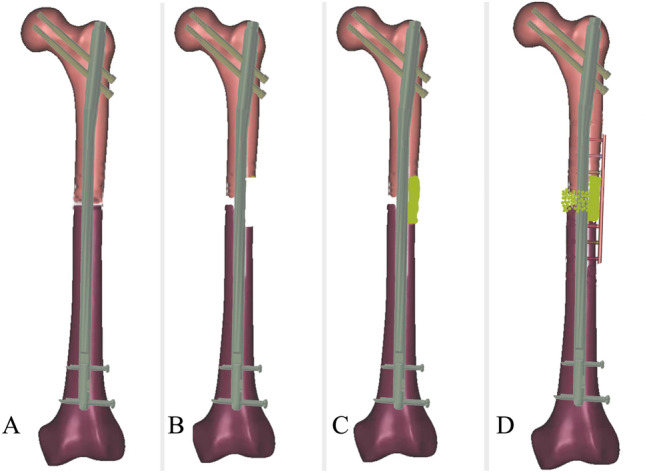
The purpose of this study is to evaluate the efficacy of plate augmentation and hybrid bone grafting for treating atrophic nonunion of the femur with original intramedullary nail retained in situ.In this study, 36 patients with atrophic nonunion of the femur who underwent surgery using the technique of plate augmentation and a hybrid bone grafting while retaining the original intramedullary nail in situ in Xi'an Honghui Hospital from January 2019 to December 2021 were enrolled. 28 patients who met the inclusion and exclusion criteria were ultimately included in the study. These 28 patients, consisting of 20 males and 8 females with a mean age of 38 years, were evaluated based on factors such as operation time, intraoperative blood loss, the average hospitalization days. Additionally, the results and function of these patients were evaluated by union time, Wu's scores of limb function and incidence of serious complications.All 28 patients achieved bone union at the 12 month follow-up, with an average follow-up time of 14.6 ± 4.2 months.The average operation time was 68.3 ± 11.2 min, and the average intraoperative blood loss was 140 ± 22.6 ml. Patients were hospitalized for an average of 5.8 ± 1.1 days. Full clinical and radiological bone union was achieved on average at 5.1 ± 1.9 months. The mean value of Wu's scores at the 12 month follow-up was significantly higher than before the operation. Limb function was excellent in 27 patients and good in one patient at the 12 month follow-up. However, five patients experienced the lower limb vein thrombosis, including one deep vein thrombosis and four lower limb intermuscular vein thromboses. One patient had a superficial infections of the surgical incision site, while three patients reported pain and numbness where their iliac bone graft was extracted at the 12 month follow-up. The technique of plate augmentation and hybrid bone grafting, combined with retaining the original intramedullary nail in situ has been shown to be a safe, effective, simply and standardizable practice for treating atrophic femoral nonunion with an intact original IMN fixation.
Keywords: Atrophic nonunion; Femur; Hybrid bone grafting; Intramedullary nail; Plate augmentation.
© 2024. The Author(s).
Conflict of interest statement
The authors declare no competing interests.
Figures


Similar articles
-
[Augmenting locking plate with autologous bone graft for the treatment of nonunion of long bone fracture in the lower extremity with retaining of the original intramedullary nail].Zhongguo Gu Shang. 2023 Dec 25;36(12):1191-5. doi: 10.12200/j.issn.1003-0034.2023.12.016. Zhongguo Gu Shang. 2023. PMID: 38130231 Chinese.
-
[A comparative study of two internal fixation techniques for femoral nonunion after intramedullary nails].Zhongguo Gu Shang. 2025 Apr 25;38(4):378-83. doi: 10.12200/j.issn.1003-0034.20240657. Zhongguo Gu Shang. 2025. PMID: 40296599 Chinese.
-
Successful management of lower limb nonunion using locking plates and bone graft with retention of intramedullary nail.Medicine (Baltimore). 2019 Mar;98(13):e14949. doi: 10.1097/MD.0000000000014949. Medicine (Baltimore). 2019. PMID: 30921194 Free PMC article.
-
[Research progress of augmentation plate for femoral shaft nonunion after intramedullary nail fixation].Zhongguo Xiu Fu Chong Jian Wai Ke Za Zhi. 2019 Dec 15;33(12):1467-1473. doi: 10.7507/1002-1892.201903073. Zhongguo Xiu Fu Chong Jian Wai Ke Za Zhi. 2019. PMID: 31823542 Free PMC article. Review. Chinese.
-
Plate Augmentation in Aseptic Femoral Shaft Nonunion after Intramedullary Nailing: A Literature Review.Bioengineering (Basel). 2022 Oct 16;9(10):560. doi: 10.3390/bioengineering9100560. Bioengineering (Basel). 2022. PMID: 36290528 Free PMC article. Review.
References
-
- Lai PJ, Hsu YH, Chou YC, Yeh WL, Ueng S, Yu YH. Augmentative antirotational plating provided a significantly higher union rate than exchanging reamed nailing in treatment for femoral shaft aseptic atrophic non-union—retrospective cohort study. BMC Musculoskelet. Disord. 2019;20(1):127. doi: 10.1186/s12891-019-2514-3. - DOI - PMC - PubMed
MeSH terms
Grants and funding
LinkOut - more resources
Full Text Sources
Miscellaneous
