. 2024 Mar 25;23(1):63.
doi: 10.1186/s12943-024-01976-2.

rs822336 binding to C/EBPβ and NFIC modulates induction of PD-L1 expression and predicts anti-PD-1/PD-L1 therapy in advanced NSCLC

Affiliations

rs822336 binding to C/EBPβ and NFIC modulates induction of PD-L1 expression and predicts anti-PD-1/PD-L1 therapy in advanced NSCLC

Giovanna Polcaro et al. Mol Cancer. .

Abstract

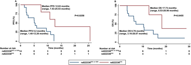
Efficient predictive biomarkers are needed for immune checkpoint inhibitor (ICI)-based immunotherapy in non-small cell lung cancer (NSCLC). Testing the predictive value of single nucleotide polymorphisms (SNPs) in programmed cell death 1 (PD-1) or its ligand 1 (PD-L1) has shown contrasting results. Here, we aim to validate the predictive value of PD-L1 SNPs in advanced NSCLC patients treated with ICIs as well as to define the molecular mechanisms underlying the role of the identified SNP candidate. rs822336 efficiently predicted response to anti-PD-1/PD-L1 immunotherapy in advanced non-oncogene addicted NSCLC patients as compared to rs2282055 and rs4143815. rs822336 mapped to the promoter/enhancer region of PD-L1, differentially affecting the induction of PD-L1 expression in human NSCLC cell lines as well as their susceptibility to HLA class I antigen matched PBMCs incubated with anti-PD-1 monoclonal antibody nivolumab. The induction of PD-L1 expression by rs822336 was mediated by a competitive allele-specificity binding of two identified transcription factors: C/EBPβ and NFIC. As a result, silencing of C/EBPβ and NFIC differentially regulated the induction of PD-L1 expression in human NSCLC cell lines carrying different rs822336 genotypes. Analysis by binding microarray further validated the competitive allele-specificity binding of C/EBPβ and NFIC to PD-L1 promoter/enhancer region based on rs822336 genotype in human NSCLC cell lines. These findings have high clinical relevance since identify rs822336 and induction of PD-L1 expression as novel biomarkers for predicting anti-PD-1/PD-L1-based immunotherapy in advanced NSCLC patients.

Keywords: Immunotherapy; NSCLC; PD-1; PD-L1; Predictive biomarker; SNP; rs822336.


Conflict of interest statement

The authors declare no competing interests.

Figures

Fig. 1
Fig. 1
Association between rs822336 and clinical outcomes in advanced NSCLC patients treated with anti-PD-1/PD-L1 therapy. PFS (A) and OS (B) of NSCLC patients treated with anti-PD-1/PD-L1 therapy were stratified based on PD-L1 rs822336 genotypes. PFS and OS were compared using the Kaplan-Meier method. Differences in patients’ survival were analyzed using a log-rang test. P < 0.05 was considered statistically significant
Fig. 2
Fig. 2
Association between rs822336 and clinical outcomes in advanced NSCLC patients treated with pembrolizumab plus platinum-based chemotherapy. PFS (A) and OS (B) of NSCLC patients treated with anti-PD-1 pembrolizumab plus platinum-based chemotherapy were stratified based on PD-L1 rs822336 genotypes. PFS and OS were compared using the Kaplan-Meier method. Differences in patients’ survival were analyzed using a log-rang test. P < 0.05 was considered statistically significant
Fig. 3
Fig. 3
Characterization of human EGFRmut and EGFRwt NSCLC cell lines. HCC827, H1975, PC-9, H460, A549, H1299, H1703 and H1437 cells were seeded into 6-well plates at the density of 2 × 106 per well. Following a 24 h incubation at 37 °C in a 5% CO2 atmosphere, cells were harvested. DNA was extracted and genotyped for rs822336 PD-L1 SNP utilizing PCR
Fig. 4
Fig. 4
Modulation by IFN-ɣ of PD-L1 expression in NSCLC cell lines carrying different rs822336 genotype. EGFRmut HCC827G/G, H1975G/G, PC-9G/G and EGFRwt H460G/G, A549G/G, H1299C/C, H1703C/C and H1437C/C cells were seeded into 6-well plates at a density of 2 × 106 cells per well and incubated with IFN-ɣ (100ng/ml). Untreated cells were used as a control. Following a 48 h incubation at 37 °C in a 5% CO2 atmosphere, cells were harvested and cell surface stained with Allophycocyanin (APC) anti-PD-L1 IgG2b Ab. APC anti-mouse IgG2b was used as a specificity control. Data, expressed as mean fluorescence intensity (MFI), are representative of the results obtained in three independent experiments. No changes in MFI were observed in specificity control following IFN-ɣ incubation (data not shown)
Fig. 5
Fig. 5
Modulation by rs822336 allele-specificity of the in vitro activity of anti-PD-1 nivolumab on NSCLC cells co-cultured with HLA-matched PBMCs. EGFRmut HCC827G/G, H1975G/G and EGFRwt H460G/G, A549G/G, H1299C/C and H1437C/C cells were seeded into 24-well plates at a density of 2 × 105 cells per well. Following a 24 h incubation at 37 °C in a 5% CO2 atmosphere, cells were co-cultured with HLA-matched PBMCs in a 1:10 ratio and incubated with nivolumab (10 µg/ml). HLA-A*11, HLA-A*01, HLA-A*24, HLA-A*25, HLA-B*40 and HLA-A*03 haplotypes were used for matching EGFRmut HCC827G/G, H1975G/G and EGFRwt H460G/G, A549G/G, H1299C/C and H1437C/C cells, respectively, with PBMCs. HLA-matched PBMCs were activated utilizing an anti-CD3 (1 µg/mL) and an anti-CD28 (1 µg/mL) T Cell TransAct (T- Act). Non-activated HLA-matched PBMCs were used as controls. Purified human IgG4 was used as a control for nivolumab. (A) Following a 48 h incubation, cancer cell viability was determined by cell counting kit-8 (CCK-8) assay. Data are expressed as mean percentages of the viability of nivolumab treated cells ± SD as compared to cells incubated with purified human IgG4. The mean percentage of cell viability and SD were calculated from three independent experiments; each of them was performed in triplicate. (B) Following a 48 h incubation, apoptosis induction was determined by Annexin V/PI assay by flow cytometry. Data are expressed as mean percentages of apoptotic cell population of nivolumab treated cells ± SD as compared to cells incubated with purified human IgG4. The mean percentage of apoptotic cells and SD were calculated from three independent experiments; each of them was performed in triplicate. (C-E) Following a 48 h incubation, LDH (C), IFN-γ (D) and TNFα (E) levels in the medium harvested from cultures of HLA-matched PBMCs with cancer cells were measured by LDH assay kit, ELISA Max Deluxe Set Human IFN-γ kit and human TNFα ELISA, respectively. Data are expressed as means of LDH, IFN-γ and TNFα levels ± SD of the results obtained in three independent experiments; each of them performed in triplicate. (ns: not significant; *P ≤ 0.05; ***P ≤ 0.001)
Fig. 6
Fig. 6
Prediction of TF binding to rs822336 based on allele-specificity. The output of the analysis contains the TFs [database accession number] predicted with a dissimilarity margin less or equal than 15% to the target sequence +/−25 bp to rs822336 G (A) and C (B) genotype. Nucleotide sequence of the potential binding site as well as the position of each TFBS are illustrated. Tables report a detail of the serum response factor (SRF) binding site prediction and the random expectation (RE) values for different levels of sequence-matrix similarity. The rate of dissimilarity between the putative and consensus sequences for TFs is indicated; RE indicates expected occurrences of the match in a random sequence of the same length as the query sequence according to the dissimilarity index based on equi–probability for the four nucleotides (RE equally) and nucleotide frequencies in the query sequence (RE query). NFI/CTF [T00094] also known as Nuclear Factor I C (NFIC)
Fig. 7
Fig. 7
Validation of TFs involved in the regulation of PD-L1 based on rs822336 allele-specificity. H1975G/G and H1299C/C cells were seeded into T75 flasks at a density of 5 × 106 cells. Following a 48 h incubation at 37 °C in a 5% CO2 atmosphere, DNA-pull down assay was performed with immobilized wt/mut oligos incubated with nuclear extracts on 4 distinct sample group combinations. Nuclear extract of H1299C/C cells was incubated with the mut oligo; nuclear extract of H1299C/C cells was incubated with the wt oligo; nuclear extract of H1975G/G cells was incubated with the mut oligo; nuclear extract of H1975G/G cells was incubated with wt oligo. Putative TFs of the rs822336 region of PD-L1 based on its allele-specificity were detected by LC-MS/MS. (A) Venn diagram represents the overlap of 315 TFs detected over the 4 analysed sample groups. (B) Pie chart shows the gene ontology (GO) classification of TFs detected over the 4 analysed sample groups. (C) Heat map summarises the average amount measured based on the observed label-free quantitation (LFQ) intensities for each of the 315 proteins detected in the 4 analysed sample groups. (D) Volcano plot shows a proteomic based comparison between H1299C/C+mut and H1975G/G+wt. Reported points indicate proteins that display both large magnitude fold-changes (x axis) and high statistical significance (y axis). Corresponding points to C/EBPβ and NFIC are highlighted
Fig. 8
Fig. 8
Regulation by C/EBPβ and NFIC silencing of PD-L1 expression in NSCLC cell lines carrying different rs822336 genotype under basal conditions or following IFN-ɣ incubation. EGFRmut H1975G/G and EGFRwt H460G/G cells (upper panel) as well as EGFRwt H1299C/C and EGFRwt H1437C/C cells (lower panel), transduced with C/EBPβ- and NFIC-specific siRNAs or siRNA-controls, were seeded into 6-well plates at a density of 2 × 106 cells per well and incubated with IFN-ɣ (100ng/ml). Untreated cells were used as a control. Following a 48 h incubation at 37 °C in a 5% CO2 atmosphere, cells were harvested and lysed. Cell lysates were analyzed by western blot with NFIC, C/EBPβ and PD-L1-specific Abs. Representative results are shown. The levels of NFIC and C/EBPβ, normalized to GAPDH and relative to untreated siRNA-control as well as levels of PD-L1, normalized to GAPDH and relative to treated siRNA-control, are plotted on the right, and expressed as mean ± SD of the results obtained in three independent experiments
Fig. 9
Fig. 9
Graphical representation of the influence of rs822336 genotype on PD-L1 induction and increased sensitivity of NSCLC cells to anti-PD-1/PD-L1 based therapy

References

    1. Siegel R, Ma J, Zou Z, Jemal A. Cancer statistics, 2014. CA Cancer J Clin. 2014;64:9–29. doi: 10.3322/caac.21208. - DOI - PubMed
    1. Scagliotti GV, Parikh P, von Pawel J, Biesma B, Vansteenkiste J, Manegold C, et al. Phase III study comparing cisplatin plus gemcitabine with cisplatin plus pemetrexed in chemotherapy-naive patients with advanced-stage non-small-cell lung cancer. J Clin Oncol. 2008;26:3543–51. doi: 10.1200/JCO.2007.15.0375. - DOI - PubMed
    1. Jänne PA, Riely GJ, Gadgeel SM, Heist RS, Ou S-HI, Pacheco JM, et al. Adagrasib in Non-small-cell Lung Cancer harboring a KRASG12C mutation. N Engl J Med. 2022;387:120–31. doi: 10.1056/NEJMoa2204619. - DOI - PubMed
    1. Peters S, Camidge DR, Shaw AT, Gadgeel S, Ahn JS, Kim D-W, et al. Alectinib versus Crizotinib in untreated ALK-Positive non-small-cell Lung Cancer. N Engl J Med. 2017;377:829–38. doi: 10.1056/NEJMoa1704795. - DOI - PubMed
    1. Wolf J, Seto T, Han J-Y, Reguart N, Garon EB, Groen HJM, et al. Capmatinib in MET exon 14-Mutated or MET-Amplified non-small-cell Lung Cancer. N Engl J Med. 2020;383:944–57. doi: 10.1056/NEJMoa2002787. - DOI - PubMed

Publication types

MeSH terms