Evaluation and management of patellar instability
- PMID: 38529132
- PMCID: PMC10929281
- DOI: 10.21037/aoj-2020-02
Evaluation and management of patellar instability
Abstract
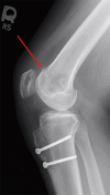
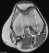
Patellar instability is a common clinical problem that primarily affects the adolescent and young adult population. The demographic and anatomic risk factors that predispose patients to patellar instability are multifactorial and include young age, female sex, trochlear dysplasia, elevated tibial tubercle to trochlear groove distance (TT-TG), patella alta, femoral and tibial malalignment, ligamentous laxity, and lack of neuromuscular control. There have been substantial efforts to predict which patients who sustain a first-time dislocation will go on to incur additional dislocations. This is particularly important because with each dislocation event, there is a significant risk of injury to the patellofemoral joint including both medial patellofemoral ligament (MPFL) stretch or rupture and damage to the cartilage which can range from simple fissures to full-thickness cartilage defects and osteochondral fractures. Prediction models have demonstrated that amongst first time dislocators, young patients with trochlear dysplasia are at the highest risk for redislocation. The current standard of care for treatment of first-time dislocators without a loose body or osteochondral fracture is nonoperative management. However, recently there has been a focus on implementing a risk-stratified approach to the surgical indications for a first-time dislocator as the high-risk population might be better treated with early surgical stabilization to prevent or reduce their risk of recurrent dislocation and its associated morbidity. Likewise, for patients with recurrent dislocations, it remains to be determined whether an isolated MPFL reconstruction is sufficient for high-risk patients with several poor prognostic risk factors or if bony realignment procedures should be implemented concurrently.
Keywords: Patella instability; medial patellofemoral ligament reconstruction (MPFL reconstruction); patellar dislocation; tibial tubercle osteotomy (TTO); trochlear dysplasia.
2022 Annals of Joint. All rights reserved.
Conflict of interest statement
Conflicts of Interest: All authors have completed the ICMJE uniform disclosure form (available at http://dx.doi.org/10.21037/aoj-2020-02). The series “Sports Related Injuries of the Female Athlete” was commissioned by the editorial office without any funding or sponsorship. BESS reports personal fees from Arthrex, outside the submitted work. In addition, a member of her family receives royalties from Arthrex, outside of the submitted work. The authors have no other conflicts of interest to declare.
Figures


References
Publication types
LinkOut - more resources
Full Text Sources
Miscellaneous
