Review
. 2022 Apr 15:7:16.
doi: 10.21037/aoj-20-118. eCollection 2022.

Anatomy of lateral meniscus

Affiliations
Review

Anatomy of lateral meniscus

Simone Perelli et al. Ann Jt. .

Abstract

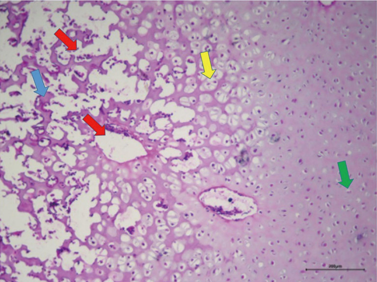
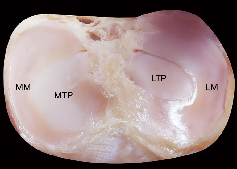
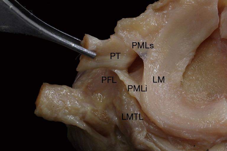
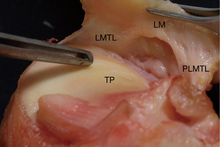
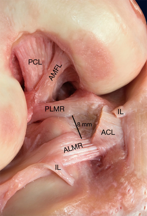
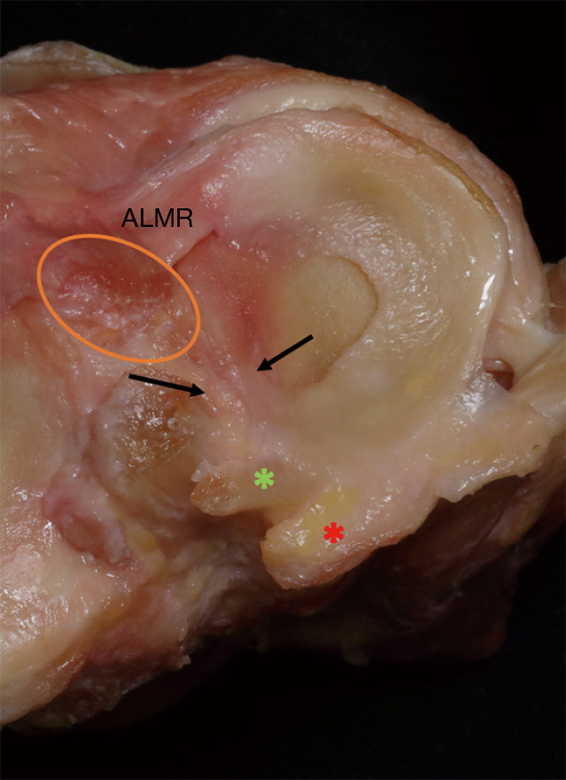
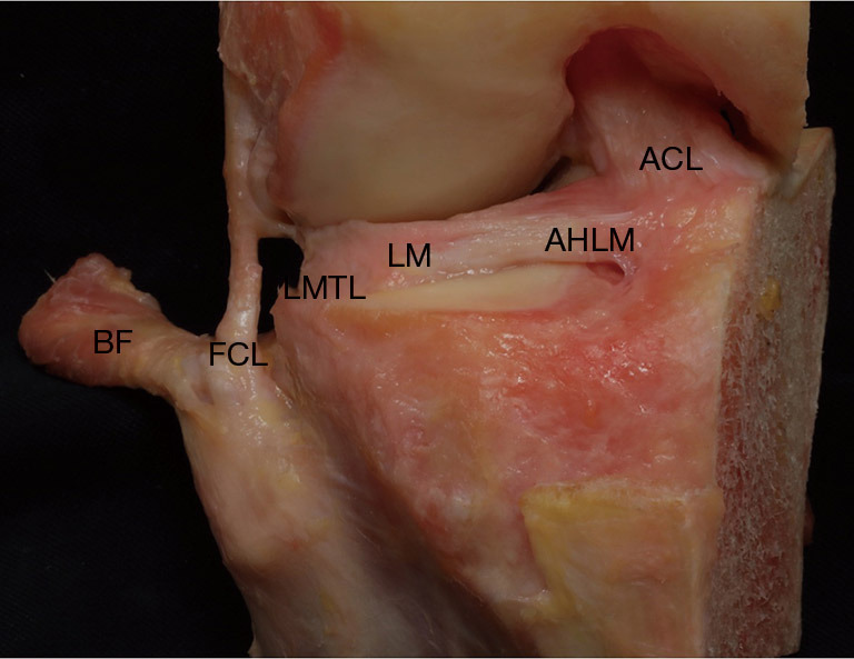
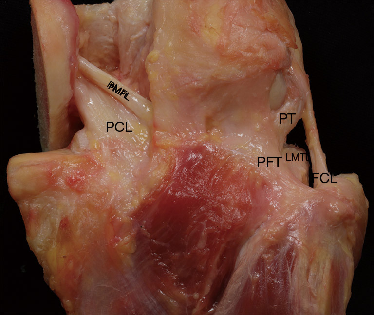
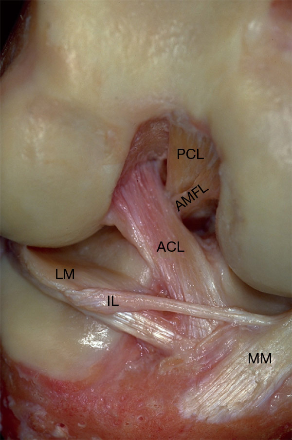
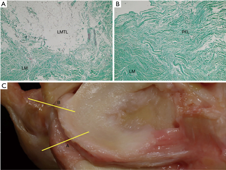
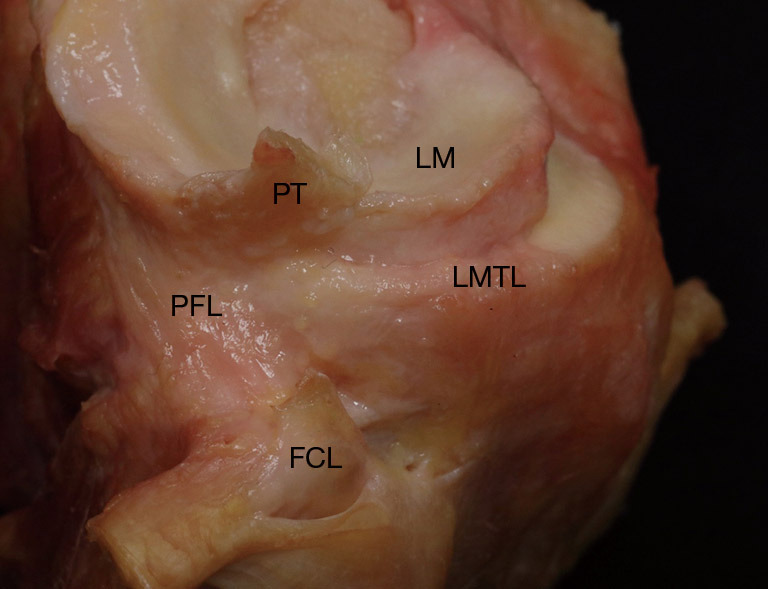
The anatomy of the lateral meniscus underlies the understanding of its unique biomechanics. Moreover, the knowledge of its microscopic structure, its vascularization and its ligament insertions can make us understand the rationale for its surgical treatment. It is well known as the respect of the anatomy leads to better results in reconstructive surgery. Knowing the differences in the shape and in the areas of insertion of the meniscal roots can be useful in case of reinserting a root or when performing a meniscal transplant. Learning about the capsular insertions, the anchoring ligaments and the areas of greatest mobility of the lateral meniscus is useful during meniscal repair and replacement surgery. This information can let us choose the most appropriate technique and the best device to face any kind of meniscal lesion. In this article, we will consider both the micro and the macro meniscal structure in order to be able to give a description as complete as possible of this fundamental structure. We will consider the interrelation of the meniscus with the neighboring anatomical structures with which it contributes to the biomechanical control of the joint. It is important to understand the interrelation with both anterior and posterior cruciate ligament (PCL) given that frequently a combined meniscal and ligamentous reconstruction is necessary.

Keywords: Lateral meniscus; anatomy; meniscal ligament; meniscal root.


Conflict of interest statement

Conflicts of Interest: All authors have completed the ICMJE uniform disclosure form (available at https://aoj.amegroups.com/article/view/10.21037/aoj-20-118/coif). The series “The Lateral Meniscus” was commissioned by the editorial office without any funding or sponsorship. JCM received payment for lectures from Smith & Nephew. The authors have no other conflicts of interest to declare.

Figures

Figure 1
Figure 1
Transversal histological section of a human knee lateral meniscus, hematoxylin and eosin stains, scale 200 µm, 10×. Red arrows show blood vessels (red arrows) between the circumferential acidophilic collagen fibers (blue arrow) of the external third. In the central zone the radial fibers are shown with the chondrocyte-like cells (yellow arrow). Finally, the circumferential fibers of the inner third are found between the fibrochondrocytes (green arrow). H&E. Scale 200 µm, 10×.
Figure 2
Figure 2
Axial view of the tibial plateau. The lateral meniscus (LM) has a more circular-shape in comparison to the medial meniscus (MM). The LM cover a wider area of the lateral tibial plateau (LTP). MTP, medial tibial plateau.
Figure 3
Figure 3
Anterior axial view with the superior popliteomeniscal ligament (PMLs) and inferior popliteomeniscal ligament (PMLi) identified. PMLs mark the posterior and anterior borders of the popliteus hiatus. FCL, fibular collateral ligament; PT, popliteus tendon; LMTL, lateral meniscotibial ligament; PFL, popliteofibular ligament.
Figure 4
Figure 4
Submeniscal view of a lateral meniscus (LM) showing the posteolateral meniscotibial ligament (PLMTL) and the lateral meniscotibial ligament (LMTL). In between there are no structures that anchor the lateral meniscus to the tibia. TP, tibial plateau.
Figure 5
Figure 5
Anterolateral view of a right knee with the anterior cruciate ligament (ACL) and the intermeniscal ligament (IL) sectioned off. The anterior meniscofemoral ligament (AMFL) inserts anterior and distal from the femoral footprint of the posterior cruciate ligament (PCL). The anterolateral (ALMR) and posterolateral (PLMR) meniscal roots have a very close insertion (8 mm distance) with some connections between PLMR and the ACL.
Figure 6
Figure 6
Axial view of a lateral meniscus. The orange circle marks the insertion area of the anterior cruciate ligament: an overlap with the anterolateral meniscal root (ALMR) is observed. The red asterisk shows the meniscal insertion of the posterior meniscofemoral ligament. The green asterisk shows the meniscal insertion of the anterior meniscofemoral ligament. The black arrows indicate the meniscoligament ligament between the posterolateral meniscal root and the posterior part of the anterior cruciate ligament.
Figure 7
Figure 7
Anterior view of a lateral meniscus. The anterior horn of lateral meniscus (AHLM) braided with the tibial footprint of the anterior cruciate ligament (ACL). The more anterior attachment of the lateral meniscotibial ligament (LMTL), thin and loose. BF, biceps femoral tendon; FCL, fibular collateral ligament; LM, lateral meniscus.
Figure 8
Figure 8
Posterior view showing the popliteo-fibular ligament (PFL), more tense and thicker than the lateral meniscotibial ligament (LMTL). The posterior meniscofemoral ligament (PMFL) runs from the posterior horn of the lateral meniscus to the medial femoral condyle. Its connections with the tibial insertion of the posterior cruciate ligament (PCL) are shown. FCL, fibular collateral ligament; PT, popliteus tendon.
Figure 9
Figure 9
Anteromedial view of a right knee showing a type I intermeniscal ligament (IL) with attachments to the anterior horn of the medial meniscus (MM) and anterior margin of the lateral meniscus (LM). ACL, anterior cruciate ligament; PCL, posterior cruciate ligament; AMFL, anterior meniscofemoral ligament.
Figure 10
Figure 10
Histological sections of a lateral meniscus. (A) Histological section of the transition between the lateral meniscus body (LM) and the lateral meniscotibial ligament (LMTL). HE staining, original magnification 100×. (B) Histological section of the transition between the lateral meniscus body and the popliteofibular ligament (PFL). Less cells are visible in the LM rather than in the LMTL. (C) Lateral meniscus. The yellow lines show where the two histological sections have been taken.
Figure 11
Figure 11
Anatomical dissection of a right knee. Sagittal view showing the peripheral insertions of the lateral meniscal body (LM), once the popliteal tendon (PT) and the fibular collateral ligament (FCL) are sectioned. The entire meniscus-tibio-popliteo-fibular complex is observed, with the LMTL positioned anterior to the fibular collateral ligament (FCL) and the more posterior popliteo-fibular ligament (PFL).

Similar articles

Cited by

References

    1. Markes AR, Hodax JD, Ma CB. Meniscus Form and Function. Clin Sports Med 2020;39:1-12. 10.1016/j.csm.2019.08.007 - DOI - PubMed
    1. Bryceland JK, Powell AJ, Nunn T. Knee Menisci. Cartilage 2017;8:99-104. 10.1177/1947603516654945 - DOI - PMC - PubMed
    1. Fox AJ, Bedi A, Rodeo SA. The basic science of human knee menisci: structure, composition, and function. Sports Health 2012;4:340-51. 10.1177/1941738111429419 - DOI - PMC - PubMed
    1. Petersen W, Tillmann B. Age-related blood and lymph supply of the knee menisci: A cadaver study. Acta Orthop Scand 1995;66:308-12. 10.3109/17453679508995550 - DOI - PubMed
    1. Gray JC. Neural and vascular anatomy of the menisci of the human knee. J Orthop Sports Phys Ther 1999;29:23-30. 10.2519/jospt.1999.29.1.23 - DOI - PubMed