. 2024 Mar 11:15:1358219.
doi: 10.3389/fimmu.2024.1358219. eCollection 2024.

Exploring type I interferon pathway: virulent vs. attenuated strain of African swine fever virus revealing a novel function carried by MGF505-4R

Affiliations

Exploring type I interferon pathway: virulent vs. attenuated strain of African swine fever virus revealing a novel function carried by MGF505-4R

Juliette Dupré et al. Front Immunol. .

Abstract

African swine fever virus represents a significant reemerging threat to livestock populations, as its incidence and geographic distribution have surged over the past decade in Europe, Asia, and Caribbean, resulting in substantial socio-economic burdens and adverse effects on animal health and welfare. In a previous report, we described the protective properties of our newly thermo-attenuated strain (ASFV-989) in pigs against an experimental infection of its parental Georgia 2007/1 virulent strain. In this new study, our objective was to characterize the molecular mechanisms underlying the attenuation of ASFV-989. We first compared the activation of type I interferon pathway in response to ASFV-989 and Georgia 2007/1 infections, employing both in vivo and in vitro models. Expression of IFN-α was significantly increased in porcine alveolar macrophages infected with ASFV-989 while pigs infected with Georgia 2007/1 showed higher IFN-α than those infected by ASFV-989. We also used a medium-throughput transcriptomic approach to study the expression of viral genes by both strains, and identified several patterns of gene expression. Subsequently, we investigated whether proteins encoded by the eight genes deleted in ASFV-989 contribute to the modulation of the type I interferon signaling pathway. Using different strategies, we showed that MGF505-4R interfered with the induction of IFN-α/β pathway, likely through interaction with TRAF3. Altogether, our data reveal key differences between ASFV-989 and Georgia 2007/1 in their ability to control IFN-α/β signaling and provide molecular mechanisms underlying the role of MGF505-4R as a virulence factor.

Keywords: African Swine Fever Virus; IFN; TRAF3; viral immune evasion; virulence factor.


Conflict of interest statement

The authors declare that the research was conducted in the absence of any commercial or financial relationships that could be construed as a potential conflict of interest.

Figures

Figure 1
Figure 1
Assessment of IFN-α levels in pigs infected with Georgia 2007/1 and ASFV-989. SPF pigs were i.m. infected with 1 x 10³ HAD50 of the Georgia 2007/1 (black squares, n = 6) and ASFV-989 strains (white squares, n = 17). (A) At the indicated days post-infection (D3 and D5), blood samples were collected and ASFV viremia (Log10 eq HAD50/mL) was measured using a qPCR test (33) that detect both Georgia 2007/1 and ASFV-989 strains. (B) The protein levels of IFN-α were quantified in the sera by ELISA and expressed as units/ml (IU/ml). Correlations were determined by comparing P72 levels at D3 and the IFN-α levels at day D5 (C, Georgia 2007/1 and D, ASFV-989. **, p < 0.005, ***, p < 0.0005 and “ns” for non significant.
Figure 2
Figure 2
The attenuated ASFV-989 strain showed increased replication and elevated IFN-α and IFN-β in porcine alveolar macrophages. PAMs were infected with Georgia 2007/1 or ASFV-989 at a MOI of 2. Black and white squares correspond to Georgia 2007/1 and ASFV-989, respectively, while white triangles correspond to non-infected PAMs (D, E). (A) At the indicated times, the supernatants were collected and used to evaluate the virus titers. (B) The expression level of the P30 viral gene was measured by RT-qPCR and normalized to that of GAPDH. Data are presented as a fold increase relative to PAMs infected with Georgia 2007/1 at 0h p.i. (C) Expressions levels of 33 viral genes were measured and shown as a fold increase relative to the Georgia 2007/1 at 4h p.i. The expression levels of IFN-α (D) and IFN-β (E) were evaluated by RT-qPCR. *, p < 0.05, **, p < 0.005 and ***, p < 0.0005. Data are representative of three independent experiments.
Figure 3
Figure 3
Activation of the IFN-β and ISRE promoters in cells expressing ASFV proteins. (A) Schematic representation of the ASFV genes at the site of the deletion. The numbers indicate amino acids (aa) positions. (B) HEK-293T cells were co-transfected with IFN-β-pGL3 and pRL-CMV reference plasmids, poly(dA:dT) and pCI-neo-3×FLAG expression vectors encoding 3×FLAG alone or fused to the indicated ASFV ORFs. After 48h, the relative luciferase activity was determined. (C) Same experiment as (B) except that transfected cells were stimulated after 24h with 1000 IU/ml of IFN-β and expression of the luciferase reporter construct controlled by ISRE repeats (pISRE-Luc) was quantified 24h later. (D) Same experiment as (B) but cells were co-transfected with full-length (FL) or the indicated fragments of MGF505-4R. All experiments were achieved in triplicate. Data represent means ± SD and are representative of three independent experiments. *, p < 0.05, **, p < 0.005 and ***, p < 0.0005.
Figure 4
Figure 4
Effects of MGF505-4R on IFN-β promoter upon activation with different components of the IFN-α/β induction pathway. HEK-293T cells were co-transfected with IFN-β-pGL3 and pRL-CMV reference plasmids and pCI-neo-3×FLAG expression vectors encoding 3×FLAG alone or fused to MGF505-4RFL, MGF505-4R1-108 or MGF505-4R109-506. The IFN-β promoter was activated by overexpressing NΔRIG-I (A), MAVS (B), TBK1 (C) or IRF3-5D (D). All experiments were achieved in triplicate. Data represent means ± SD and are representative of three independent experiments. *, p < 0.05, **, p < 0.005 and ***, p < 0.0005.
Figure 5
Figure 5
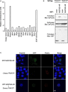
MGF505-4R interaction with TRAF3. (A) HEK-293T cells were co-transfected with pDESTN2H-N1 encoding the fragment N1 of the NanoLuciferase (aa1-aa65) fused to MGF505-4R and pDESTN2H-N2 encoding the fragment N2 of the NanoLuciferase (aa66-aa171) alone or fused to the indicated swine cellular protein. STAT1/STAT2 interaction was used as positive control. In this condition, STAT1 and STAT2 were expressed in pDESTN2H-N1 and pDESTN2H-N2, respectively. After 48h, cells were lysed, the bioluminescence was measured and data are presented as a fold increase relative to the condition where pDESTN2H-N1-MGF505-4R and pDESTN2H-N2 empty vector were co-transfected. (B) HEK-293T cells were transfected with expression vectors encoding GST alone or fused to MG505-4R and tested for the interaction with either human or swine TRAF3. Total cell lysates were prepared 48h post-transfection (cell lysate; middle panel), and co-purifications of indicated cellular proteins were assayed by pull-down using glutathione-sepharose beads (pull-down; upper panel). GST-tagged MG505-4R was detected by immunoblotting using anti-GST antibody (pull-down; lower panel), while TRAF3 was detected with an anti-3xFLAG antibody. (C) HeLa cells were co-transfected with pCherry-C1 and pEGFP-C1 plasmids encoding human TRAF3 and MG505-4R, respectively. 24 h after, cells were fixed and labeled with the dye Hoechst 33258 to stain nuclei. Intracellular localization of Hoechst-stained nuclei (blue), TRAF3 (red) and MG505-4R (green) were visualized by confocal fluorescence microscopy (×40 magnification). Scale bars represent 10 µM.
Figure 6
Figure 6
Schematic representation of the induction of IFN-α/β signaling upon the recognition of ds viral DNA.

References

    1. Guinat C, Gogin A, Blome S, Keil G, Pollin R, Pfeiffer DU, et al. . Transmission routes of African swine fever virus to domestic pigs: current knowledge and future research directions. Vet Rec. (2016) 178:262−7. doi: 10.1136/vr.103593 - DOI - PMC - PubMed
    1. Davies K, Goatley LC, Guinat C, Netherton CL, Gubbins S, Dixon LK, et al. . Survival of african swine fever virus in excretions from pigs experimentally infected with the Georgia 2007/1 isolate. Transbound Emerg Dis. (2017) 64:425−31. doi: 10.1111/tbed.2017.64.issue-2 - DOI - PMC - PubMed
    1. Olesen AS, Lohse L, Boklund A, Halasa T, Gallardo C, Pejsak Z, et al. . Transmission of African swine fever virus from infected pigs by direct contact and aerosol routes. Vet Microbiol. (2017) 211:92−102. doi: 10.1016/j.vetmic.2017.10.004 - DOI - PubMed
    1. Boinas FS, Wilson AJ, Hutchings GH, Martins C, Dixon LJ. The Persistence of African Swine Fever Virus in Field-Infected Ornithodoros erraticus during the ASF Endemic Period in Portugal. PloS One. (2011) 6:e20383. doi: 10.1371/journal.pone.0020383 - DOI - PMC - PubMed
    1. Diaz AV, Netherton CL, Dixon LK, Wilson AJ. African swine fever virus strain Georgia 2007/1 in ornithodoros erraticus ticks. Emerg Infect Dis. (2012) 18:1026−8. doi: 10.3201/eid1806.111728 - DOI - PMC - PubMed

Publication types

Substances