. 2024 Apr 12;9(94):eadn1452.
doi: 10.1126/sciimmunol.adn1452. Epub 2024 Apr 12.

The palmitoylation of gasdermin D directs its membrane translocation and pore formation during pyroptosis

Affiliations

The palmitoylation of gasdermin D directs its membrane translocation and pore formation during pyroptosis

Arumugam Balasubramanian et al. Sci Immunol. .

Abstract

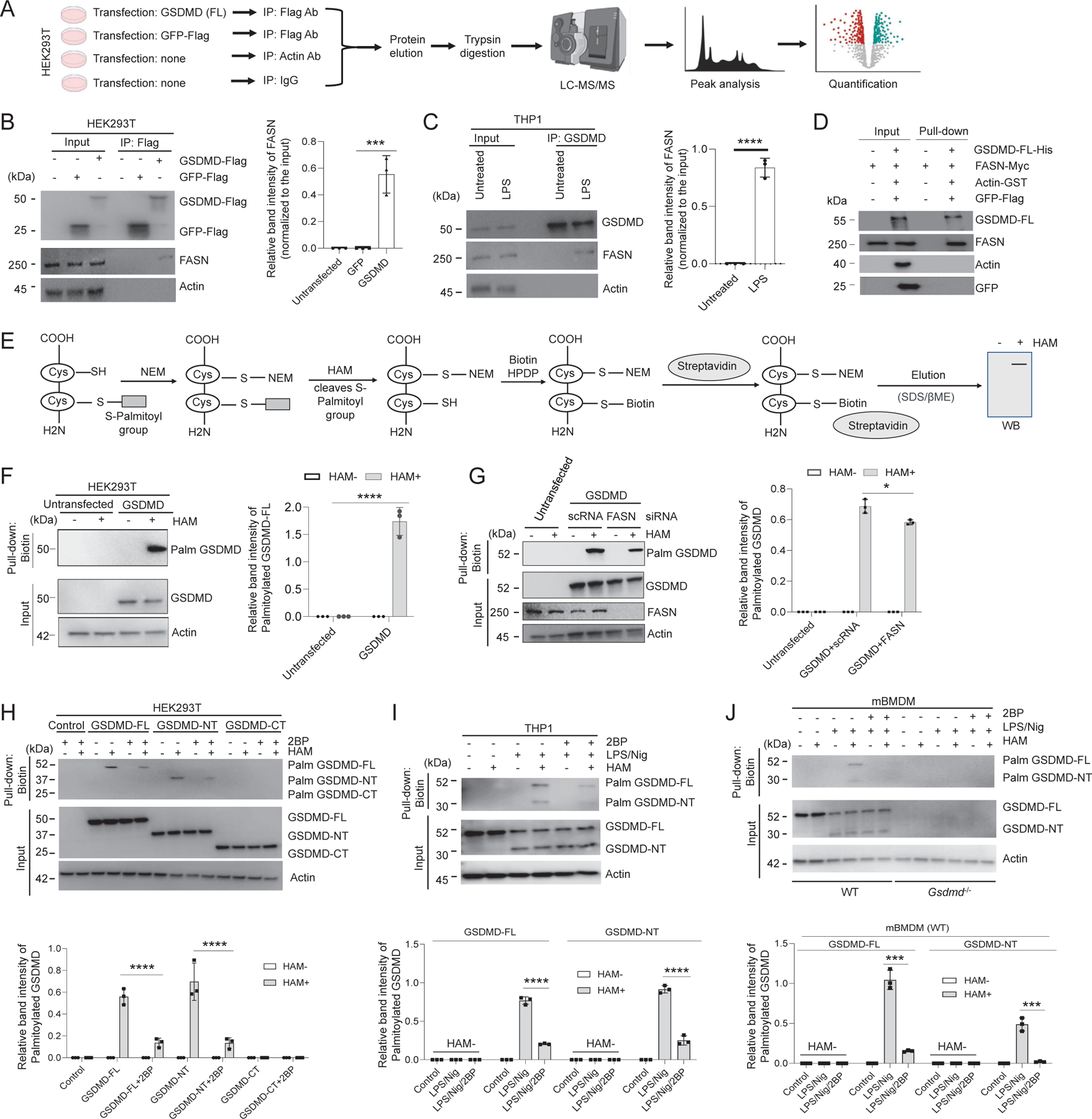
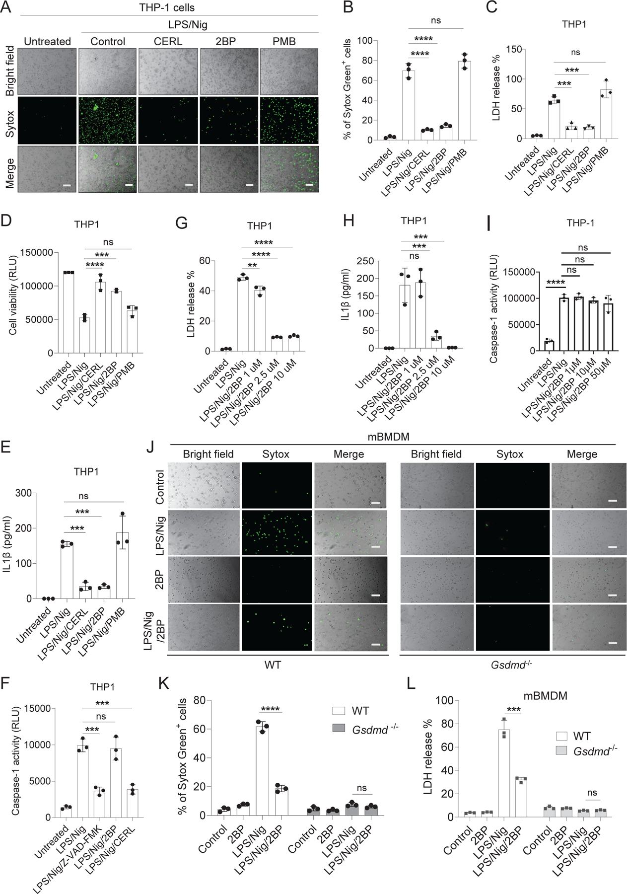
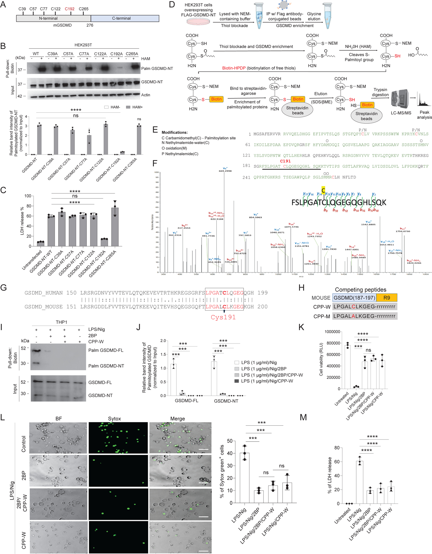
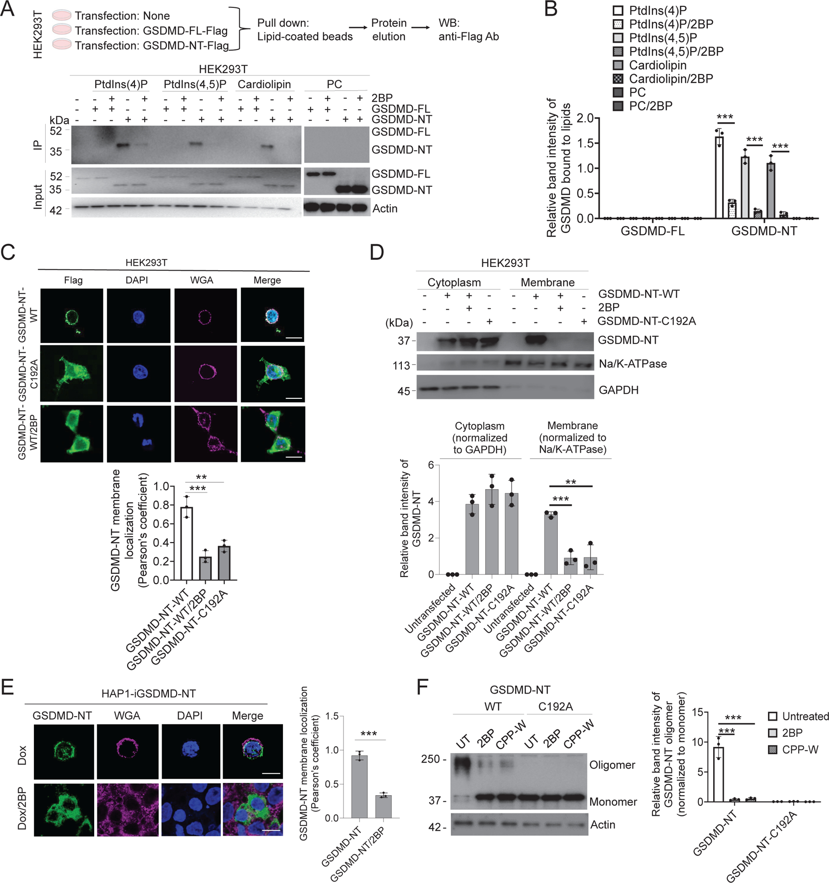
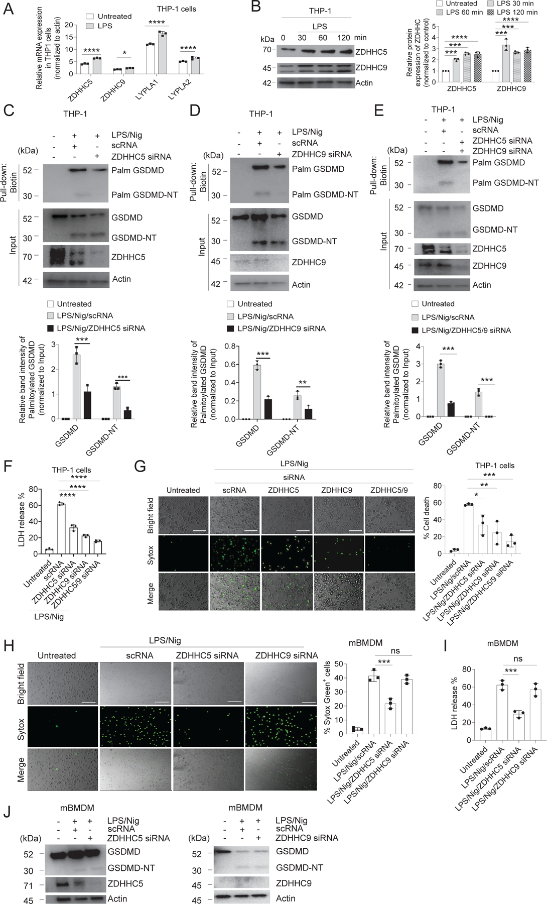
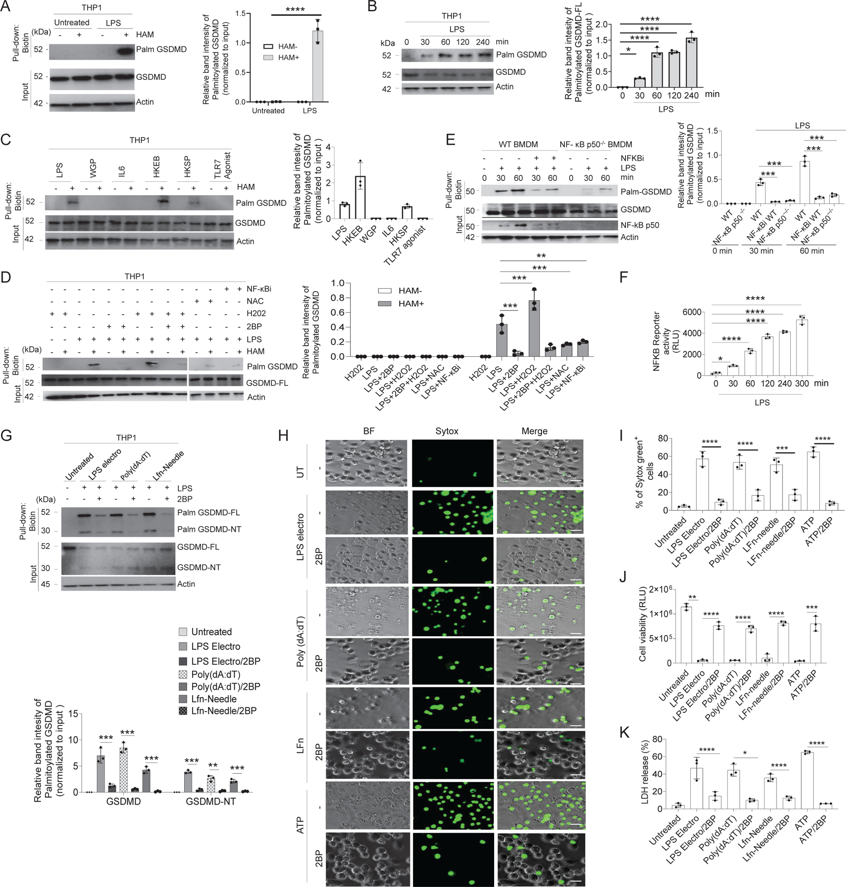
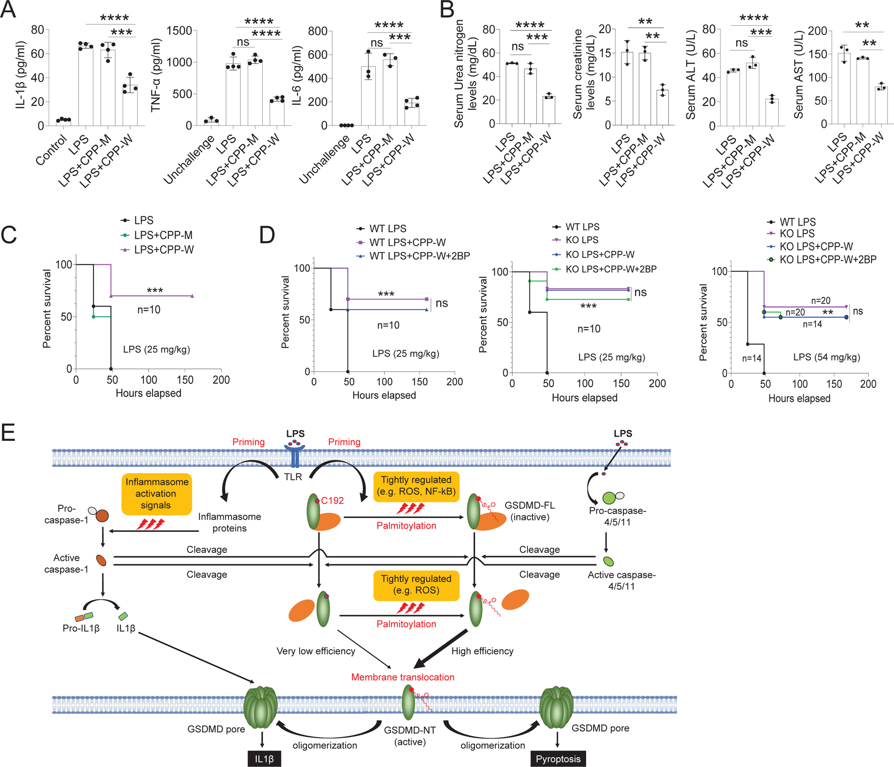
Plasma membrane perforation elicited by caspase cleavage of the gasdermin D (GSDMD) N-terminal domain (GSDMD-NT) triggers pyroptosis. The mechanisms underlying GSDMD membrane translocation and pore formation are not fully understood. Here, using a proteomic approach, we identified fatty acid synthase (FASN) as a GSDMD-binding partner. S-palmitoylation of GSDMD at Cys191/Cys192 (human/mouse), catalyzed by palmitoyl acyltransferases ZDHHC5 and ZDHHC9 and facilitated by reactive oxygen species (ROS), directly mediated membrane translocation of GSDMD-NT but not full-length GSDMD (GSDMD-FL). Palmitoylation of GSDMD-FL could be induced before inflammasome activation by stimuli such as lipopolysaccharide (LPS), consequently serving as an essential molecular event in macrophage priming. Inhibition of GSDMD palmitoylation suppressed macrophage pyroptosis and IL-1β release, mitigated organ damage, and enhanced the survival of septic mice. Thus, GSDMD-NT palmitoylation is a key regulatory mechanism controlling GSDMD membrane localization and activation, which may offer an additional target for modulating immune activity in infectious and inflammatory diseases.


Conflict of interest statement

Competing interests: J.C.K. consults and holds equity in Corner Therapeutics, Larkspur Biosciences, MindImmune Therapeutics, and Neumora Therapeutics. H.W. is a cofounder of Ventus Therapeutics. All other authors declare no competing financial interests.

Figures

Fig. 1.
Fig. 1.. GSDMD interacts with FASN and both full-length (GSDMD-FL) and cleaved GSDMD-NT are palmitoylated.
(A) Schematic of the approach and strategy used to identify GSDMD-interacting proteins by mass spectrometry. (B) FASN-GSDMD interaction in HEK293T cells expressing hGSDMD-FL-FLAG. Right: quantification of relative FASN protein bound with GSDMD-FL. n=3 biological replicates. (C) FASN–GSDMD interaction in PMA-differentiated THP-1. Right: quantification of endogenous FASN bound with GSDMD protein. n=3 biological replicates. (D) Interaction between c-Myc-tagged FASN and His-tagged hGSDMD-FL. Immunoblot analyses were performed using anti-GSDMD and other indicated antibodies. n=3 biological replicates. (E) Schematic of the steps in the acyl-biotin exchange (ABE) palmitoylation assay. (F) GSDMD palmitoylation in HEK293T cells. n=3 biologically independent experiments. (G) FLAG-hGSDMD-FL was coexpressed with control or FASN siRNA and subjected to the ABE palmitoylation assay and immunoblot analyses. n=3 biological replicates. (H) Palmitoylation of GSDMD-FL, GSDMD-N, or GSDMD-C in the presence or absence of 2BP. n=3 biological replicates. (I) GSDMD palmitoylation in PMA-differentiated macrophage-like THP-1 cells. Cells were treated with or without LPS for 3 hours, followed by 2BP treatment for 30 min and then nigericin treatment for 35 min. Immunoblot analyses were performed with anti-GSDMD antibodies. n=3 biological replicates. (J) GSDMD palmitoylation in primary mouse bone marrow-derived macrophages (mBMDM). Cells were cultured in the presence of LPS (1 μg/ml) (3 hours), followed by 2BP 10 μM (30 min) and treatment with nigericin (20 μM) for 1 hour. n=3 biological replicates. (B, C, F, G, H, I, and J) Data are mean ± SEM. Immunoblots shown are representative of three independent experiments. *P<0.05, **P<0.01, ***P<0.001, ****P<0.0001. Statistical analyses were performed using one-way ANOVA with Bonferroni’s multiple comparison test (B), unpaired Student’s t test (C), or two-way ANOVA with Bonferroni’s multiple comparison test (F-J).
Fig. 2.
Fig. 2.. Inhibition of palmitoylation impairs pyroptosis in macrophages.
(A) Representative brightfield and fluorescence images of THP-1 cells primed with LPS (3 hours) followed by treatment with palmitoylation inhibitors (cerulenin, 50 μM; 2-bromopalmitate, 10 μM) and depalmitoylation inhibitor (palmostatin B, 50 μM) for 30 min before stimulation with nigericin for 35 min. Pyroptotic pore formation was assessed with the SYTOX Green dye binding assay using ImageJ software. Scale bars: 100 μm. n=3 biological replicates. (B) Quantification of relative SYTOX uptake in THP-1 cells. (C) LDH release from THP-1 cells. n=3 biological replicates. (D) Relative cell viability was calculated using the RealTime-Glo MT Cell Viability Assay. n=3 biological replicates. (E) IL-1β secretion by THP-1 cells was measured by ELISA. n=3 biological replicates. (F) The relative abundance of active caspase in THP-1 cell culture supernatants was quantified with Caspase-1 Glo reagent (Promega). n=3 biological replicates. (G) THP-1 cells treated with or without LPS/Nig in the presence or absence of 2BP were treated at different concentrations. The resulting supernatants were quantified for LDH release. n=3 biological replicates. (H) IL-1β release from THP-1 cells. n=3 biological replicates. (I) The effect of 2BP treatment on caspase-1 activity. n=3 biological replicates. (J) mBMDM from WT and Gsdmd−/− mice were primed with or without LPS for 3 hours in the presence or absence of 2BP for 30 min before stimulation with nigericin for 1 hour. Pyroptotic pore formation was assessed with the SYTOX Green assay, and fluorescence images were captured and quantified using ImageJ. Scale bars: 100 μm. n=3 biological replicates. (K) Quantification of relative SYTOX uptake in mBMDMs. (L) LDH release from WT and Gsdmd−/− cell. n=3 biological replicates. (B to L) All data are representative of three independent experiments. Data are means ± SEM. **P<0.01, ***P<0.001, ****P<0.0001. Statistical analyses were performed using one-way (B to I) or two-way (K and L) ANOVA with Bonferroni’s multiple comparison test.
Fig. 3.
Fig. 3.. GSDMD palmitoylation occurs on Cys192.
(A) A schematic of the number of cysteine residues present in mouse GSDMD-NT. Highlighted in red is a potential cysteine site in GSDMD-NT that is palmitoylated. (B) Palmitoylation of WT GSDMD-NT or mutant GSDMD-NT (Cys39A, Cys57A, Cys77A, Cys122A, Cys192A, Cys265A) in HEK293T cells. Immunoblot analyses were performed with anti-FLAG HRP antibody. Palmitoylated proteins were quantified and normalized to corresponding input. n=3 biological replicates. (C) LDH release. n=3 biological replicates. (D) Schematic of the approach and strategy used to identify GSDMD palmitoylation by mass spectrometry. (E) Posttranslational modifications identified in GSDMD-NT protein. Protein sequences covered by MS analyses are marked in green. (F) MS/MS CID fragmentation spectrum of the peptide FSLPGATCLQGEGQGHLSQK (m/z 1058.02013, [M+2H]2+) containing carbamidomethylation at C8 (Cys192). (G to M) A competitive peptide, CPP-W, specifically inhibits GSDMD palmitoylation and suppresses macrophage pyroptosis. (G) Protein sequence alignment of human and mouse GSDMD using EMBOSS Matcher. (H) Cell-permeable, GSDMD-competing peptides. R9 represents poly-arginine. Lower case amino acid symbol (r) represents the D-form of arginine. (I) GSDMD palmitoylation in PMA differentiated THP-1 cells primed with LPS (1 μg/ml) for 2 hours at 37°C, followed by 2BP (50 μM) or 2BP plus CPP-W (50 μM) treatment for 30 min. Nigericin (10 μM, 30 min) was then added to trigger inflammasome activation. n=3 biological replicates. (J) Quantification of palmitoylated GSDMD normalized to input. (K) Relative cell viability was calculated using the RealTime-Glo MT Cell Viability Assay. n=3 biological replicates. (L) Pyroptotic pore formation was assessed with the SYTOX Green assay, and fluorescence images were captured and quantified using ImageJ. Scale bars: 50 μm. n=3 biological replicates. (M) LDH release. n=3 biological replicates. (B, C, J, K, L, and M) All data are representative of three independent experiments. Data are means ± SEM. **P<0.01, ***P<0.001, ****P<0.0001. Statistical analyses were performed using one-way (C, K, L, and M) or two-way (B and J) ANOVA with Bonferroni’s multiple comparison test.
Fig. 4.
Fig. 4.. Palmitoylation of GSDMD-NT is required for its membrane translocation and oligomerization.
(A) Schematic approach and strategy used to detect GSDMD-lipid binding. HEK293T cells were transfected with either human GSDMD-FL-FLAG or GSDMD-NT-FLAG and cultured for 24 hours in the presence or absence of 2BP (50 μM). Following cell lysis, the lysates were co-precipitated with agarose beads coated with specific lipids. Immunoblot analyses were subsequently performed using anti-FLAG HRP antibodies. n=3 biological replicates. (B) Quantification of the relative amount of GSDMD bound to the lipid-coated beads. n=3 biological replicates. (C) Representative Subcellular localization of GSDMD-NT in HEK293T cells. Scale bars: 10 μm. Green: GSDMD; blue: nuclei (DAPI); and magenta: plasma membrane (wheat germ agglutinin (WGA). Results are presented as normalized Pearson’s correlation. At least 20 cells were randomly picked and analyzed for each sample. n=3 biological replicates. (D) Distribution of GSDMD-NT and mutant in subcellular fractions of HEK293T cells subjected to immunoblot analysis. n=3 biological replicates. (E) Subcellular localization of GSDMD-NT in Dox-induced GSDMD-NT cells treated with or without 2BP. Scale bars: 10 μm. Green: GSDMD; magenta: plasma membrane (WGA); and blue: nuclei (DAPI). Images shown are representative of at least ten random fields from three independent experiments. n=3 biological replicates. (F) Inhibition of palmitoylation impaired GSDMD oligomerization. Right: quantification of relative oligomer normalized with monomer. n=3 biological replicates. (B to F) All data are representative of three independent experiments. Data are mean ± SEM. **P<0.01, ***P<0.001. Statistical analyses were performed using one-way ANOVA with Bonferroni’s multiple comparison test (C and D), unpaired Student’s t test (E), or two-way ANOVA with Bonferroni’s multiple comparison test (B and F).
Fig. 5.
Fig. 5.. ZDHHC5 and ZDHHC9-mediated palmitoylation is required for LPS/Nig-triggered pyroptosis in macrophages.
(A) Expression of palmitoylation-related genes in THP-1 cells. Real-time PCR analysis was conducted using THP-1 cells primed with LPS for 3 hours. mRNA expression of ZDHHC was quantified and normalized to actin. n=3 biological replicates. (B) LPS-induced expression of ZDHHC5 and ZDHHC9 protein in THP-1 cells. PMA differentiated THP-1 cells were treated with LPS (100 ng/ml) for indicated time. Right, quantification of relative expression of ZDHHC protein using ImageJ. n=3 biological replicates. (C to E) THP-1 cells were transiently transfected with control or siRNA targeting (C) ZDHHC5, (D) ZDHHC9, and (E) ZDHHC5/9 followed by priming with LPS for 3 hours and treatment with nigericin for 35 min. The ABE assay and immunoblot analysis were then performed with anti-GSDMD antibodies. Bar graph depicts the quantification of palmitoylated GSDMD protein normalized to relative input. n=3 biological replicates. (F) LDH release. n=3 biological replicates. (G) Fluorescence images of SYTOX Green uptake in THP-1 cells expressing control and ZDHHC siRNA. Scale bars: 100 μm. Right: quantification of relative SYTOX uptake in THP-1 cells. n=3 biological replicates. (H to J) ZDHHC5 palmitoyl enzyme mediates pyroptotic cell death in primary mouse macrophages. (H) Fluorescence images of SYTOX Green uptake in mBMDMs transfected with control or ZDHHC siRNA. Scale bars: 100 μm. Right, quantification of SYTOX uptake. n=3 biological replicates. (I) LDH release from mBMDMs. n=3 biological replicates. (J) mBMDMs transfected with control or Zdhhc siRNAs were treated with or without LPS/Nig. Cells were lysed and subjected to immunoblot analysis. Immunoblot images are representative of three independent repeats. n=3 biological replicates. (A to H) All data are representative of three independent experiments. Data are mean ± SEM. *P<0.05, **P<0.01, ***P<0.001, **** P<0.0001. Statistical analyses were performed using one-way (F to H) or two-way (A to E) ANOVA with Bonferroni’s multiple comparison test.
Fig. 6.
Fig. 6.. GSDMD palmitoylation is tightly regulated.
(A) GSDMD palmitoylation in PMA differentiated THP-1 cells stimulated with LPS (100 ng/ml) for 2 hours. n=3 biological replicates. (B) Time course of LPS-induced GSDMD palmitoylation. n=3 biological replicates. (C) GSDMD palmitoylation in PMA-differentiated THP-1 cells stimulated with LPS (1 μg/ml), whole glucan particles (WGP) (100 μg/ml), IL-6 (600 pg/ml), heat-killed E. coli (HKEB) (105 cells/ml), heat-killed S. pneumonia (HKSP) (107 cells/ml), or a TLR7 agonist (imidazoquinoline) (2.5 μg/ml) for 2 hours at 37°C. n=3 biological replicates. (D) GSDMD palmitoylation in PMA-differentiated THP-1 cells treated with or without LPS for 3 hours in the presence or absence of H2O2 (625 μM), 2BP (10 μM), NAC (3 mM), or JSH-23 (NF-κBi) (50 μM). n=3 biological replicates. (E) GSDMD palmitoylation in mouse primary bone marrow-derived WT and NF-κB p50−/− macrophages treated with LPS (100 ng/ml) in the presence or absence of NF-κBi (20 μM). n=3 biological replicates. (F) Time course of LPS-induced of NF-κB activation in PMA differentiated THP-1-NF-κB-Luc cells. n=3 biological replicates. (G to K) 2BP suppresses macrophage pyroptosis triggered by various inflammasomes. PMA-differentiated THP-1 cells were primed with LPS (1 μg/ml) for 2 hours at 37°C. The AIM2 inflammasome was activated using precomplexed Poly(dA:dT)/LyoVec (1 μg/ml) and NLRC4/NAIP inflammasome was activated using Lfn-Needle (Needle-Tox) (4 ng/ml). Non-canonical inflammasome (caspase-11) activation was induced by LPS electroporation (1 μg/ml). All subsequent assays were conducted 24 hours thereafter. (G) GSDMD palmitoylation. n=3 biological replicates. (H) Pyroptotic pore formation assessed with the SYTOX Green assay. Scale bars: 50 μM. n=3 biological replicates. (I) Quantification of relative SYTOX uptake. n=3 biological replicates. (J) Relative cell viability calculated using the RealTime-Glo MT Cell Viability Assay. n=3 biological replicates. (K) LDH release. n=3 biological replicates. (A to K) All data are representative of three independent experiments. Data are mean ± SEM. *P<0.05, **P<0.01, ***P<0.001, **** P<0.0001. Statistical analyses were performed using one-way (B, E, F, and I to K) or two-way (A, D, and G) ANOVA with Bonferroni’s multiple comparison test.
Fig. 7.
Fig. 7.. Specific inhibition of GSDMD palmitoylation with CPP-W alleviates the severity of sepsis.
(A) Peritoneal fluid IL-1β, TNF-α, and IL-6 levels in sepsis induced by LPS (20 mg per kilogram of body weight) (n=3–4 mice per group). ***P<0.001, ****P<0.0001, one-way ANOVA followed by Bonferroni’s multiple comparison test. (B) Circulating AST, ALT, BUN, and creatine levels in the serum. n=3 mice per group. **P<0.01, ***P<0.001, ****P<0.0001, one-way ANOVA followed by Bonferroni’s multiple comparison test. (C) Survival rates were calculated using Kaplan–Meier survival curves. n=10 mice per group. The experiment was performed independently three times and the data were pooled and analyzed together. **P<0.01, ***P<0.001, log-rank (Mantel–Cox) test. (D) Survival rates were calculated using Kaplan–Meier survival curves. n=10–20 mice per group. The experiment was performed independently three times, and the data were pooled and analyzed together.**P<0.01, ***P<0.001, log-rank (Mantel–Cox) test. (E) Graphical illustration of GSDMD palmitoylation and its role in macrophage pyroptosis.

Update of

Similar articles

Cited by

References

    1. Jorgensen I, Miao EA, Pyroptotic cell death defends against intracellular pathogens. Immunol Rev 265, 130–142 (2015). - PMC - PubMed
    1. Man SM, Karki R, Kanneganti TD, Molecular mechanisms and functions of pyroptosis, inflammatory caspases and inflammasomes in infectious diseases. Immunol Rev 277, 61–75 (2017). - PMC - PubMed
    1. Shi J, Zhao Y, Wang K, Shi X, Wang Y, Huang H, Zhuang Y, Cai T, Wang F, Shao F, Cleavage of GSDMD by inflammatory caspases determines pyroptotic cell death. Nature 526, 660–665 (2015). - PubMed
    1. Kayagaki N, Stowe IB, Lee BL, O’Rourke K, Anderson K, Warming S, Cuellar T, Haley B, Roose-Girma M, Phung QT, Liu PS, Lill JR, Li H, Wu J, Kummerfeld S, Zhang J, Lee WP, Snipas SJ, Salvesen GS, Morris LX, Fitzgerald L, Zhang Y, Bertram EM, Goodnow CC, Dixit VM, Caspase-11 cleaves gasdermin D for non-canonical inflammasome signalling. Nature 526, 666–671 (2015). - PubMed
    1. He WT, Wan H, Hu L, Chen P, Wang X, Huang Z, Yang ZH, Zhong CQ, Han J, Gasdermin D is an executor of pyroptosis and required for interleukin-1beta secretion. Cell Res 25, 1285–1298 (2015). - PMC - PubMed

Publication types