. 2024 May 6;221(5):e20231200.
doi: 10.1084/jem.20231200. Epub 2024 Mar 26.

Functional diversity of NLRP3 gain-of-function mutants associated with CAPS autoinflammation

Affiliations

Functional diversity of NLRP3 gain-of-function mutants associated with CAPS autoinflammation

Camille Cosson et al. J Exp Med. .

Abstract

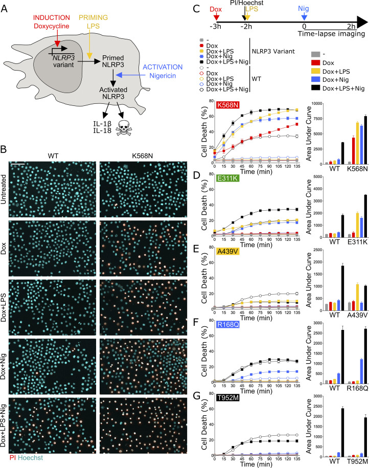
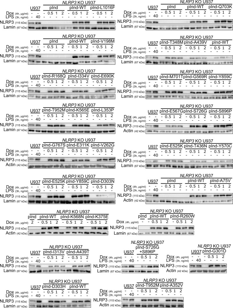
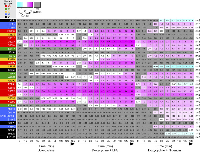
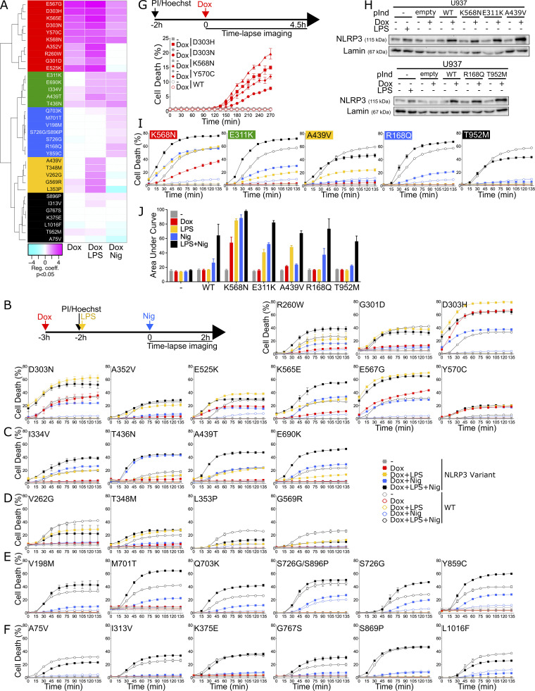
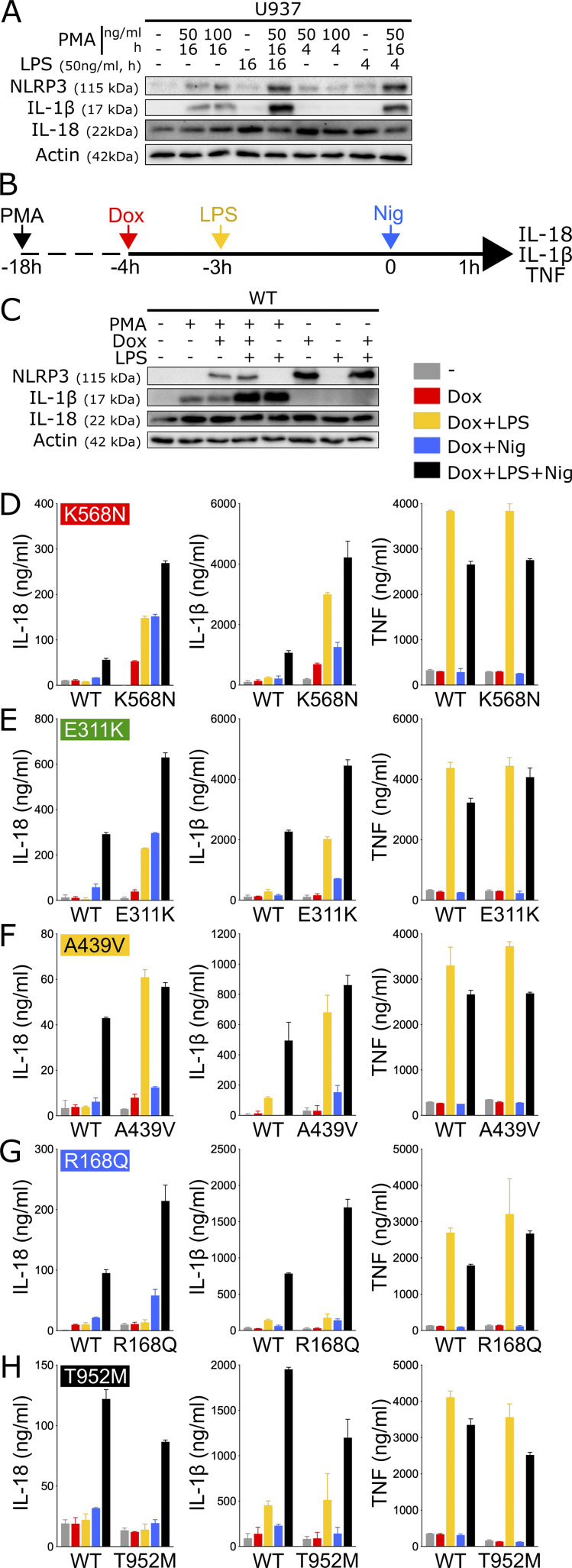
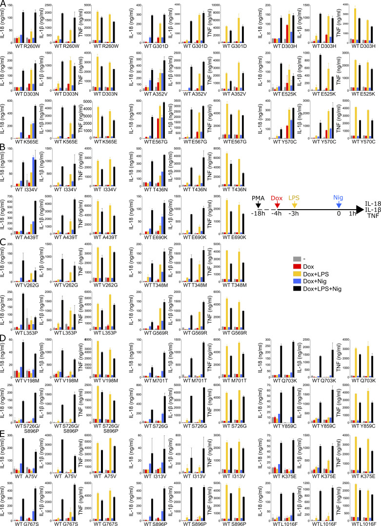
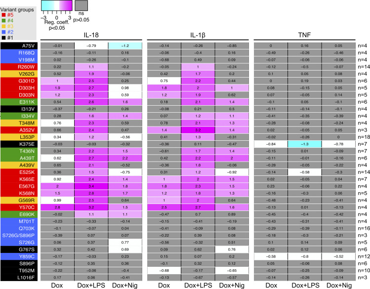
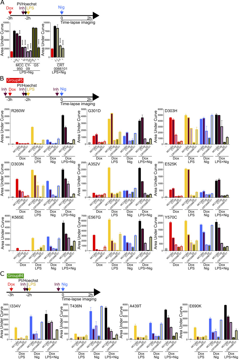
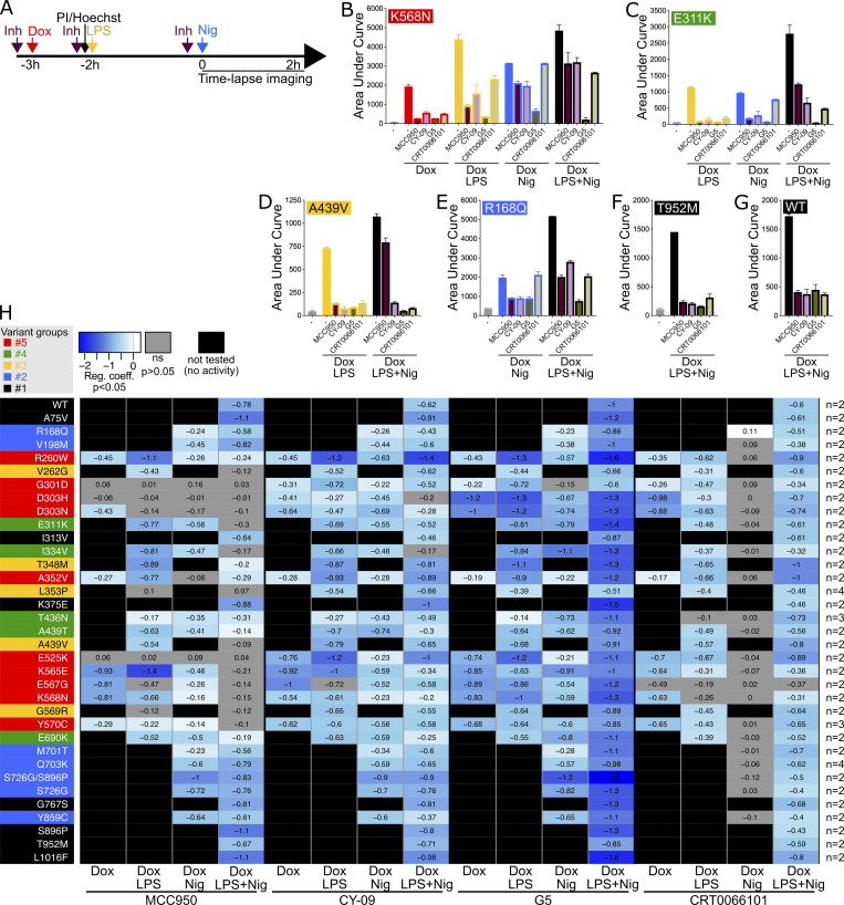
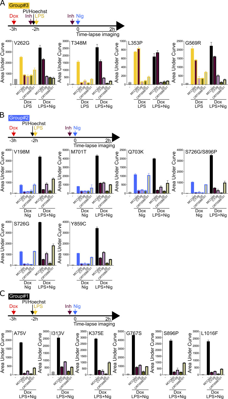
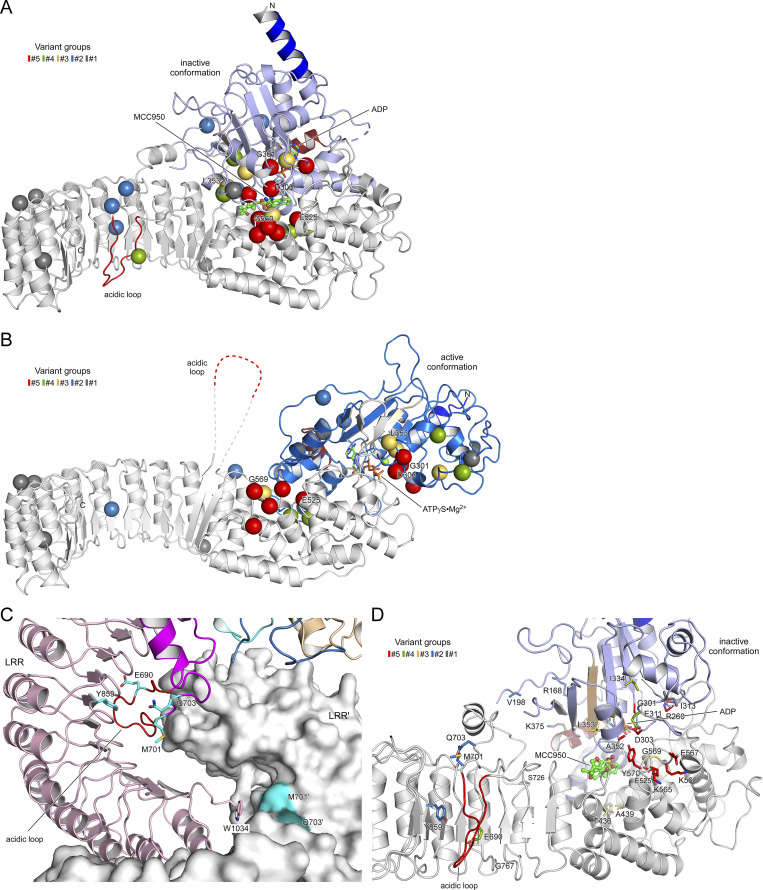
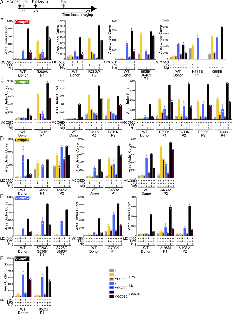
NLRP3-associated autoinflammatory disease is a heterogenous group of monogenic conditions caused by NLRP3 gain-of-function mutations. The poor functional characterization of most NLRP3 variants hinders diagnosis despite efficient anti-IL-1 treatments. Additionally, while NLRP3 is controlled by priming and activation signals, gain-of-functions have only been investigated in response to priming. Here, we characterize 34 NLRP3 variants in vitro, evaluating their activity upon induction, priming, and/or activation signals, and their sensitivity to four inhibitors. We highlight the functional diversity of the gain-of-function mutants and describe four groups based on the signals governing their activation, correlating partly with the symptom severity. We identify a new group of NLRP3 mutants responding to the activation signal without priming, associated with frequent misdiagnoses. Our results identify key NLRP3 residues controlling inflammasome activity and sensitivity to inhibitors, and antagonistic mechanisms with broader efficacy for therapeutic strategies. They provide new insights into NLRP3 activation, an explanatory mechanism for NLRP3-AID heterogeneity, and original tools for NLRP3-AID diagnosis and drug development.


Conflict of interest statement

Disclosures: A. Belot reported personal fees from Boehringer Ingelheim, Novartis, AbbVie, Kabi, and GlaxoSmithKline outside the submitted work. O. Lambotte reported personal fees from MSD, BMS, Boehringer, AbbVIe, and Gilead outside the submitted work. B.F. Py reported a patent to European patent application N°23306463.3 filed on September 4, 2023, entitled “Methods for the diagnosis, treatment and characterization of NLRP3-associated autoinflammatory diseases” pending. No other disclosures were reported.

Figures

Figure 1.
Figure 1.
NLRP3 expression, priming, and/or activation triggers pyroptosis in reconstituted U937 depending on the NLRP3 variants. (A–G) NLRP3-deficient U937 cells reconstituted with doxycycline-inducible NLRP3 variants were treated with doxycycline (1 μg/ml, 3 h), LPS (40 ng/ml, 2 h), and nigericin (15 μg/ml) before cell death was monitored by PI incorporation over time quantified by time-lapse high content microscopy. (B) Example of microscopy images for WT and K568N (60 min, objective 10×, scale bar 100 μm). (C) K568N (group#5). (D) E311K (group#4). (E) A439V (group#3). (F) R168Q (group#2). (G) T952M (group#1). Means of duplicates and 1 SD (left panel) and means of AUC of duplicates and 1 SD (right panel) for cells expressing NLRP3 variants (full square) and WT (open circle) are represented. One experiment done in duplicates representative of two to eight independent experiments is shown (as indicated for each variant on the right side). Results obtained with one variant typical of each functional group are represented. Results obtained with all tested variants are presented in Fig. S2, B–F. Statistical analysis including all independent experiments are represented in Fig. 2.
Figure S1.
Figure S1.
Reconstitution of NLRP3-deficient U937 with doxycycline-inducible NLRP3 variants. NLRP3-deficient U937 reconstituted with doxycycline-inducible WT NLRP3 (pInd-WT), indicated NLRP3 variants, and the empty vector (pInd) were treated with doxycycline (indicated doses, 4 h). U937 treated with LPS (40 ng/ml, 3 h) was used as a control. NLRP3 protein levels were analyzed by WB. Source data are available for this figure: SourceData FS1.
Figure 2.
Figure 2.
Relative cell death of U937 expressing NLRP3 variants as compared with U937 expressing WT NLRP3 in response to NLRP3 expression, priming, and/or activation. Regression coefficient (RC) heatmap from the glmm for each variant as compared with the WT. Positive RC denotes increased cell death in the considered variant as compared with the WT, and conversely. For significant values (P < 0.05), RC corresponding to increases (magenta) or decreases (cyan) in cell death are color-coded. In gray, not significant values (P > 0.05) correspond to conditions (variant * treatment * time point) in which the percentage of dead cells among U937 expressing NLRP3 variant is not statistically different from those among U937 expressing WT NLRP3. Based on the results, variants are classified into five functional groups: constitutive active variants (group#5, red), variants active upon either priming or activation signal (group#4, green), variants active upon priming signal (group#3, yellow), variants active upon activation signal (group#2, blue), and mutants active upon priming and activation signals (no gain-of-function, group#1, black). Results of two to eight independent experiments done in duplicates.
Figure S2.
Figure S2.
NLRP3 expression, priming, and/or activation triggers pyroptosis in reconstituted U937 depending on the NLRP3 variants. (A–F) NLRP3-deficient U937 cells reconstituted with doxycycline-inducible NLRP3 variants were treated with doxycycline (1 μg/ml, 3 h), LPS (40 ng/ml, 2 h), and nigericin (15 μg/ml) before cell death was monitored by PI incorporation over time quantified by time-lapse high content microscopy. (A) Unsupervised clustering of the variants according to RC from the glmm applied on AUC for each variant as compared with the WT. Positive RC denotes increased cell death in the considered variant as compared with the WT, and conversely. RC corresponding to increases (magenta) or decreases (cyan) in cell death are color-coded. Based on the results, variants are classified into five functional groups: constitutive active variants (group#5, red), variants active upon either priming or activation signal (group#4, green), variants active upon priming signal (group#3, yellow), variants active upon activation signal (group#2, blue), and mutants active upon priming and activation signals (no gain-of-function, group#1, black). Results of two to eight independent experiments done in duplicates. (B) Group#5. (C) Group#4. (D) Group#3. (E) Group#2. (F) Group#1. Means of duplicates and 1 SD are represented. One experiment done in duplicates representative of two to eight independent experiments is shown. Statistical analysis including all independent experiments are represented in Fig. 3. (G) NLRP3-deficient U937 cells reconstituted with doxycycline-inducible group#5 NLRP3 variants were treated with doxycycline (1 μg/ml) before cell death was monitored by PI incorporation over time quantified by time-lapse high content microscopy. One experiment done in duplicates representative of two independent experiments is shown. Statistical analysis including all independent experiments is represented in Fig. 2. (H–J) U937 cells transduced with vectors encoding indicated doxycycline-inducible NLRP3 variants or empty vector (empty) and not transduced controls (−) were treated with doxycycline (1 μg/ml, 4 h) or LPS (40 ng/ml, 3 h) and NLRP3 protein levels were analyzed by WB (H). Cells were treated with doxycycline (1 μg/ml, 3 h), LPS (40 ng/ml, 2 h), and/or nigericin (15 μg/ml) before monitoring PI incorporation (I). Means of duplicates and 1 SD are represented. One experiment done in duplicates representative of two to three independent experiments is shown. LDH release was assessed 2 h after nigericin treatment (J). Means of two independent experiments done in duplicates and 1 SD are represented. Source data are available for this figure: SourceData FS2.
Figure 3.
Figure 3.
NLRP3 expression, priming, and/or activation triggers IL-18 and IL-1β secretion in reconstituted U937 depending on the NLRP3 variants. (A) U937 cells were differentiated in PMA (50 or 100 ng/ml, 4 or 16 h) and treated with LPS (50 ng/ml, 4 or 16 h). Protein levels of pro-IL-18, pro-IL-1β, and NLRP3 were assessed by WB. Actin is used as loading control. (B–H) NLRP3-deficient U937 cells reconstituted with doxycycline-inducible NLRP3 variants were differentiated in PMA (50 ng/ml) and treated with doxycycline (1 μg/ml), LPS (40 ng/ml), and nigericin (15 μg/ml) as indicated. NLRP3, pro-IL-1β, pro-IL-18, and actin levels (as control) were assessed by WB in lysates of NLRP3-deficient U937 cells reconstituted with doxycycline-inducible WT NLRP3 (C). IL-18, IL-1β, and TNF levels (as control) were assessed in cell supernatant by ELISA in K568N (group#5) (D), E311K (group#4) (E), A439V (group#3) (F), R168Q (group#2) (G), and T952M (group#1) (H). Means of duplicates and 1 SD are represented. One experiment done in duplicates representative of 3–18 independent experiments is shown. Results obtained with one variant typical of each functional group are represented. Results obtained with all tested variants and statistical analysis including all independent experiments are represented in Fig. S3. Source data are available for this figure: SourceData F3.
Figure S3.
Figure S3.
NLRP3 expression, priming, and/or activation triggers IL-18 and IL-1β secretion in reconstituted U937 depending on the NLRP3 variants. (A–E) NLRP3-deficient U937 cells reconstituted with doxycycline-inducible NLRP3 variants were differentiated in PMA (50 ng/ml) and treated with doxycycline (1 μg/ml), LPS (40 ng/ml), and nigericin (15 μg/ml) as indicated. IL-18, IL-1β, and TNF (as control) levels were assessed in cell supernatant by ELISA. Group#5 (A), group#4 (B), group#3 (C), group#2 (D), group#1 (E). Means of duplicates and 1 SD are represented. One experiment done in duplicates representative of 3–18 independent experiments is shown.
Figure S4.
Figure S4.
Statistical analysis of IL-18 and IL-1β secretion in reconstituted U937 upon NLRP3 expression, priming, and/or activation. RC heatmap from the lmm modeling for each variant as compared with the WT. Positive RC denotes increased cytokine concentrations by the variant as compared to NLRP3 WT, and conversely. RC corresponding to statistically significant increases (magenta) or decreases (cyan) in cytokine secretions are color-coded (P < 0.05, and RC > +1 or RC < −1, respectively). Not significant variations are color-coded in gray (P > 0.05). Results of 3–18 independent experiments done in duplicates (as indicated for each variant on the right side).
Figure S5.
Figure S5.
Sensitivity of NLRP3 variants to NLRP3 inhibitors (groups#5 and 4). (A) Dose-dependent inhibition of pyroptosis by NLRP3 inhibitors (Inh). NLRP3-deficient U937 cells reconstituted with doxycycline-inducible WT NLRP3 and treated with doxycycline (1 μg/ml, 3 h) were treated with indicated doses (μM) of MCC950, CY-09, G5, and CRT0066101 or vehicle (2h15), LPS (40 ng/ml, 2 h), and nigericin (15 μg/ml) before cell death was monitored by PI incorporation over time quantified by time-lapse high content microscopy. Means of AUC of duplicates and 1 SD are represented. Two-way ANOVA multiple comparisons of each variant with WT control with corresponding treatment, ***, P < 0.001; ****, P < 0.0001. (B and C) NLRP3-deficient U937 cells reconstituted with doxycycline-inducible NLRP3 variants were treated with doxycycline (1 μg/ml, 3 h), LPS (40 ng/ml, 2 h), and/or nigericin (15 μg/ml) in the presence of MCC950 (1 μM), CY-09 (50 μM), G5 (1 μM), and CRT006101 (0.5 μM) NLRP3 inhibitors or DMSO vehicle (added 20 min before the last treatment), and cell death was monitored by PI incorporation over time quantified by time-lapse high content microscopy for 2 h. Group#5 (B), group#4 (C). Means of AUC of duplicates and 1 SD are represented. One experiment done in duplicates representative of two independent experiments is shown. Statistical analysis including all independent experiments are represented in Fig. 4 H.
Figure 4.
Figure 4.
Cell death of U937 expressing NLRP3 variants in response to NLRP3 expression, priming, and/or activation in the presence of NLRP3 inhibitors. (A–G) NLRP3-deficient U937 cells reconstituted with doxycycline-inducible NLRP3 variants were treated with doxycycline (1 μg/ml, 3 h), LPS (40 ng/ml, 2 h), and nigericin (15 μg/ml) in the presence of MCC950 (1 μM), CY-09 (50 μM), G5 (1 μM), and CRT006101 (0.5 μM) NLRP3 inhibitors (Inh), or DMSO vehicle (added 20 min before the last treatment) and cell death was monitored by PI incorporation over time quantified by time-lapse high content microscopy for 2 h (A). (B) K568N (group#5). (C) E311K (group#4). (D) A439V (group#3). (E) R168Q (group#2). (F) T952M (group#1). (G) WT as control. Means of AUC of duplicates and 1 SD are represented. One experiment done in duplicates representative of two independent experiments is shown. Results obtained with one variant typical of each functional group are represented (B–F). Results obtained with all tested variants are presented in Fig. S4, B–F. (H) Statistical analysis of the inhibition of each variant in each relevant condition by MCC950, CY-09, G5, and CRT0066101 compounds. Inhibition heatmap from the linear mixed model of PI incorporation AUC for each NLRP3 variant with a specific treatment for each inhibitor, as compared with vehicle only. Inhibition is expressed as RC corresponding to Log10(1−%inhibition/100). The negative coefficient denoted inhibition of cell death, and the null coefficient corresponded to no inhibition. For significant values (P < 0.05), RC between −2 (99% inhibition) and 0.1 (20% inhibition) are color-coded with decreasing blue intensities. In gray, not significant values (P > 0.05) correspond to conditions (variant * treatment) in which the inhibitors do not decrease PI incorporation. In black, the variant * treatment combinations do not trigger cell death and therefore inhibitors were not tested. Results of two independent experiments done in duplicates.
Figure S6.
Figure S6.
Sensitivity of NLRP3 variants to NLRP3 inhibitors (groups#3–1). (A–C) NLRP3-deficient U937 cells reconstituted with doxycycline-inducible NLRP3 variants were treated with doxycycline (1 μg/ml, 3 h), LPS (40 ng/ml, 2 h), and/or nigericin (15 μg/ml) in the presence of MCC950 (1 μM), CY-09 (50 μM), G5 (1 μM), and CRT006101 (0.5 μM) NLRP3 inhibitors (Inh) or DMSO vehicle (added 20 min before the last treatment), and cell death was monitored by PI incorporation over time quantified by time-lapse high content microscopy for 2 h. Group#3 (A), group#2 (B), and group#1 (C). Means of AUC of duplicates and 1 SD are represented. One experiment done in duplicates representative of two independent experiments is shown. Statistical analysis including all independent experiments are represented in Fig. 4 H.
Figure S7.
Figure S7.
Details of the disease-causing mutant sites in NLRP3 and MCC950 inhibition. (A) Localization of gain-of-function mutations in the MCC950-inhibited state of human NLRP3 (7PZC), color-coded according to variant groups. The five mutant positions that are not responsive to MCC950 treatment, G301D, E303H, L353P, E525K, and G569R, are labeled. (B) Localization of gain-of-function mutations in the adenosine triphosphate-bound active state of human NLRP3 (8EJ4). (C) The three gain-of-function mutations in the acidic loop of NLRP3, E690, M701, and Q703, are in the dimer interface of the interlaced LRRs, while Y859 in the concave surface of the LRR interacts with the acidic loop. (D) MCC950 is in the center of the NACHT and LRR domains in proximity to many disease-causing mutations.
Figure 5.
Figure 5.
Patient monocytes respond differently to NLRP3 priming and/or activation, and NLRP3 inhibitor MCC950 depending on NLRP3 variants. (A) Monocytes of 22 autoinflammatory patients and healthy donors (WT) were treated with LPS (40 ng/ml, 3 h) and nigericin (5 μg/ml) in the presence of MCC950 (1 μM, added 15 min before LPS), and cell death was monitored by PI incorporation over time quantified by time-lapse high content microscopy for 2 h. (B) R260W, E525K+D646Y, and K565E (group#5). (C) E311K and E690K (group#4). (D) T348M and A439V (group#3). (E) S726G+S896P, Q703K, and V198M (group#2). (F) T952M (group#1). Means of AUC of two to six replicates and 1 SD are represented. One experiment done in two to six replicates is shown. Data represented in the same graph correspond to one same experiment on relatives. Due to limited amount of blood from patients E525K/D646Y P1, K565E P1 and P2, and T348M P1, some conditions could not be tested.
Figure 6.
Figure 6.
Localization of disease mutant sites in the structure of human NLRP3. (A) Bar diagram showing the domain architecture of NLRP3 with the mutant sites of the five variant groups indicated. (B) Display of the inactive spherical NLRP3-ADP decamer (7PZC) (Hochheiser et al., 2022) and the active radial NLRP3-ATP decamer bound to NEK7 (in gray) (8EJ4) (Xiao et al., 2023). (C) The conformational transition from the inactive to the active state in NLRP3 involves an 85° rotation of the FISNA-NBD-HD1 subdomains in the NACHT relative to the remainder. The missense mutations of the five variant groups are highlighted as spheres in the inactive conformation. (D) Close-up of residues E690, M701, and Q703 in the loop section of the transition LRR and Y859 in the concave side of the LRR. (E) Localization of the constitutively active group#5 mutant sites in the NACHT domain of NLRP3. (F) Residues T436 and A439 are at the pivot point of the FISNA-NBD-HD1 rotation. The active state (8EJ4) is shown in gray relative to the colored inactive state (7PZC). (G) Variant group#2 residues R168 and V198 in the FISNA domain and S726 in the trLRR are located in variable regions of NLRP3.

References

    1. Andreeva, L., David L., Rawson S., Shen C., Pasricha T., Pelegrin P., and Wu H.. 2021. NLRP3 cages revealed by full-length mouse NLRP3 structure control pathway activation. Cell. 184:P6299-6312.E22. 10.1016/j.cell.2021.11.011 - DOI - PMC - PubMed
    1. Bauernfeind, F.G., Horvath G., Stutz A., Alnemri E.S., MacDonald K., Speert D., Fernandes-Alnemri T., Wu J., Monks B.G., Fitzgerald K.A., et al. . 2009. Cutting edge: NF-kappaB activating pattern recognition and cytokine receptors license NLRP3 inflammasome activation by regulating NLRP3 expression. J. Immunol. 183:787–791. 10.4049/jimmunol.0901363 - DOI - PMC - PubMed
    1. Booshehri, L.M., and Hoffman H.M.. 2019. CAPS and NLRP3. J. Clin. Immunol. 39:277–286. 10.1007/s10875-019-00638-z - DOI - PMC - PubMed
    1. Brinkschulte, R., Fußhöller D.M., Hoss F., Rodríguez-Alcázar J.F., Lauterbach M.A., Kolbe C.-C., Rauen M., Ince S., Herrmann C., Latz E., and Geyer M.. 2022. ATP-binding and hydrolysis of human NLRP3. Commun. Biol. 5:1176. 10.1038/s42003-022-04120-2 - DOI - PMC - PubMed
    1. Coll, R.C., Robertson A.A.B., Chae J.J., Higgins S.C., Muñoz-Planillo R., Inserra M.C., Vetter I., Dungan L.S., Monks B.G., Stutz A., et al. . 2015. A small-molecule inhibitor of the NLRP3 inflammasome for the treatment of inflammatory diseases. Nat. Med. 21:248–255. 10.1038/nm.3806 - DOI - PMC - PubMed

MeSH terms

Substances