. 2024 Mar 26;134(9):e173537.
doi: 10.1172/JCI173537.

Satellite glial GPR37L1 and its ligand maresin 1 regulate potassium channel signaling and pain homeostasis

Affiliations

Satellite glial GPR37L1 and its ligand maresin 1 regulate potassium channel signaling and pain homeostasis

Sangsu Bang et al. J Clin Invest. .

Abstract

G protein-coupled receptor 37-like 1 (GPR37L1) is an orphan GPCR with largely unknown functions. Here, we report that Gpr37l1/GRP37L1 ranks among the most highly expressed GPCR transcripts in mouse and human dorsal root ganglia (DRGs) and is selectively expressed in satellite glial cells (SGCs). Peripheral neuropathy induced by streptozotoxin (STZ) and paclitaxel (PTX) led to reduced GPR37L1 expression on the plasma membrane in mouse and human DRGs. Transgenic mice with Gpr37l1 deficiency exhibited impaired resolution of neuropathic pain symptoms following PTX- and STZ-induced pain, whereas overexpression of Gpr37l1 in mouse DRGs reversed pain. GPR37L1 is coexpressed with potassium channels, including KCNJ10 (Kir4.1) in mouse SGCs and both KCNJ3 (Kir3.1) and KCNJ10 in human SGCs. GPR37L1 regulates the surface expression and function of the potassium channels. Notably, the proresolving lipid mediator maresin 1 (MaR1) serves as a ligand of GPR37L1 and enhances KCNJ10- or KCNJ3-mediated potassium influx in SGCs through GPR37L1. Chemotherapy suppressed KCNJ10 expression and function in SGCs, which MaR1 rescued through GPR37L1. Finally, genetic analysis revealed that the GPR37L1-E296K variant increased chronic pain risk by destabilizing the protein and impairing the protein's function. Thus, GPR37L1 in SGCs offers a therapeutic target for the protection of neuropathy and chronic pain.

Keywords: G protein–coupled receptors; Homeostasis; Neurological disorders; Neuroscience.


Conflict of interest statement

Conflict of interest: The authors have declared that no conflict of interest exists.

Figures

Figure 1
Figure 1. Gpr37l1 and GPR37L1 transcripts are highly expressed in mouse and human DRGs.
(A and B) Highly expressed GPCR transcripts in mouse DRGs. RNA-Seq shows mRNA expression levels (FPKM) of the top 10 GPCR transcripts (A, n = 3) and the top 10 orphan GPCR transcripts (B, n = 3). Gpr37l1 expression is highlighted in red bars. Note that the Gpr37 expression is much lower than Gpr37l1. (C and D) Highly expressed GPCR transcripts in human DRGs. Normalized microarray shows mRNA expression levels (intensity) of the top 10 GPCR transcripts (C, n = 214) and the top 10 orphan GPCR transcripts (D, n = 214). GRP37L1 expression is highlighted in red bars. GPR37 expression is included for comparison. Data are represented as means ± SEM.
Figure 2
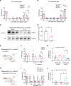
Figure 2. Mouse and human SGCs express Gpr37l1/GPR37L1 mRNA and GPR37L1 protein.
(A) Double staining of RNAscope ISH (for Gpr37l1) and IHC (for Tuj1) show nonoverlapping expression of Gpr37l1 mRNA (red) and Tuj1 (white) in mouse DRGs. Right, enlarged image from the box in the left panel. Yellow arrows indicate Gpr37l1+ cells surrounding DRG neurons. Scale bars: 25 μm. (B) Double IHC staining shows colocalization of GPR37L1 (red) with FABP7 (white). Scale bar: 25 μm. (C) Western blot showing GPR37L1 expression in DRGs of Gpr37l1+/+, Gpr37l1+/–, and Gpr37l1–/– mice. GAPDH was included as a loading control from the same gel. (D) Double staining of ISH (GPR37L1, red) and IHC (GS green) shows colocalization of GPR37L1 mRNA and GS in human SGCs. (E) Double staining of IHC for GPR37L1 (red) and FABP7 (green) shows heavy colocalization of GPR37L1 and FABP7 in human SGCs. Note that GPR37L1 is enriched on the inner side of SGCs in close contact with neurons. Asterisks indicate neurons, and arrows indicate SGCs surrounding the neurons. Scale bars: 25 μm. (F) Top, Western blots showing PM and intracellular cytosol (IC) fractions of GPR37L1 and GAPDH loading control (from the same gel) in human DRGs of neuropathic pain patients and controls (Con) (n = 3). Bottom, the ratio of PM/IC GPR37L1 expression in human DRGs of control and neuropathic pain patients. Data are represented as means ± SEM and analyzed by t test. **P < 0.05, unpaired Student’s t test. n = 3.
Figure 3
Figure 3. GPR37L1 is dysregulated in pain and protects against neuropathic pain.
(A and B) Neuropathic pain (mechanical allodynia) induced by STZ (75 mg/kg) and PTX (6 mg/kg) in WT and Gpr37l1 mutant mice. (A) Time course of STZ (75mg/kg)-induced mechanical allodynia in Gpr37l1+/+ mice (n = 5), Gpr37l1+/– mice (n = 7), and Gpr37l1–/– mice (n = 6). (B) Time course of PTX (6 mg/kg)-induced mechanical allodynia in Gpr37l1+/+ mice (n = 7), Gpr37l1+/– mice (n = 10), and Gpr37l1–/– mice (n = 8). (C and D) GPR37L1 expression in PM fraction of DRG tissues of control mice and mice with STZ treatment (n = 3). Transferrin (TfR) was used as a loading control from a parallel gel. (D) Quantification of GPR37L1. (EG) Unilateral i.g. microinjection of Gpr37l1-targeting siRNA reduces Gpr37l1 expression and induces persistent mechanical allodynia in naive animals. (E) Schematic of i.g. microinjection of siRNA or scRNA in the L4 and L5 DRGs, followed by von Frey testing and tissue collection for quantitative reverse-transcription PCR (RT-PCR) analysis. (F) Mechanical allodynia is induced by siRNA (n = 10), not scRNA (n = 10). (G) RT-PCR analyses of Gpr37l1 in DRGs (n = 10 mice). (HJ) Unilateral i.g. microinjection of Fabp7-Gpr37l1 (AAV-L1) or Fabp7-mock AAV virus (AAV-Con) rescued Gpr37l1 expression and reduced persistent mechanical allodynia in CIPN mice (6 mg/kg PTX). (H) Schematic of i.g. microinjection of AAV virus in the L4 and L5 DRGs, given 1 week after PTX, followed by von Frey testing and tissue collection for RT-qPCR analysis. (I) PTX-mediated mechanical allodynia is reduced by AAV-L1 application (n = 8) but not AAV-Con (n = 8). (J) RT-PCR analyses showing expression of Gpr37l1 (n = 4 mice). Data are represented as mean ± SEM and statistically analyzed by 2-way ANOVA with Tukey’s post hoc test (A, B, and I) or Bonferroni’s post hoc test (F), 1-way ANOVA with Tukey’s post hoc test (G and J), and 2-tailed t test (D). *P < 0.05; **P < 0.01; ****P < 0.0001.
Figure 4
Figure 4. MaR1 binds GPR37L1.
(A and B) Lipid overlay assay shows GPR37L1 binding to MaR1. (A) Schematic for detecting GPR37L1-binding lipid mediators. hGPR37L1 is FLAG tagged and expressed in HEK293 cells after hGPR37L1 cDNA transfection. (B) Specific binding of hGPR37L1 to MaR1. The plate was coated with 10 different lipid mediators (1 µg/ml, nos. 2–11) as well as vehicle control (0.1% ethanol, no. 1) and no coating control (NC, no. 12) and then incubated with cell lysates of hGPR37L1- or mock-transfected cells. Yellow asterisk indicates a positive response. (C) Upper panel: representative blot of MaR1-coated PVDF membrane. Lower panel: quantification of dot intensity in DRG lysates from WT and Gpr37l1–/– mice. n = 3 repeats. (D and E) Overall structure of hGPR37L1 (green) in complex with MaR1 (magenta). (E) 100 ns RMSD graph was obtained with a 1000 ns simulation of the GPR37L1-MaR1 complex (red) or GPR37L1(blue). Data are expressed as means ± SEM and were analyzed by unpaired t test (C) *P < 0.05.
Figure 5
Figure 5. Injection i.t. or i.g. of MaR1 reduces neuropathic pain via GPR37L1 expressed on SGCs.
(A and B) MaR1 injected i.t. (100 ng) reduces mechanical allodynia in Gpr37l1+/+ and Gpr37l1+/– mice induced by 75 mg/kg of STZ and 6 mg/kg of PTX. (A) Paw withdrawal thresholds were assessed in Gpr37l1+/+ mice (left, n = 7), Gpr37l1+/– mice (middle, n = 10), and Gpr37l1–/– mice (n = 8) after i.t. MaR1 injection in the STZ model. (B) Paw withdrawal thresholds in Gpr37l1+/+ mice (left, n = 5), Gpr37l1+/– mice (middle, n = 7), and Gpr37l1–/– mice (n = 6) after i.t. MaR1 injection in the PTX model. Behavior was assessed 1 hour after MaR1 injection on post-PTX and post-STZ day 3. (C) Upper panel, schematic of i.g. injection. Lower panel, knockdown of Gpr37l1 expression in the L4 DRGs after siRNA treatment compared with scRNA treatment (n = 8). (D) Injection of MaR1 (i.g., 10 ng, 2 µl) reduces PTX-induced mechanical allodynia in control animals treated with scRNA, but not in animals treated with Gpr37l1 siRNA (n = 10). (E) MaR1 inhibits PTX-induced IL-1β release in SGC-neuron cocultures via GPR37L1. IL-1β release in cocultures from DRG of WT mice (n = 6) and Gpr37l1–/– mice (n = 6) was analyzed by ELISA. The cultures were stimulated with 1 μM PTX for 24 hours in the absence or presence of MaR1 (100 nM). Data are expressed as means ± SEM and were statistically analyzed by paired t test (A and B), 2-way ANOVA with Bonferroni’s post hoc test (D and E), Tukey’s post hoc test (D and E), or unpaired t test (C). *P < 0.05, **P < 0.01, ***P < 0.001, ****P < 0.0001.
Figure 6
Figure 6. MaR1 increases surface expression of GPR37L1 and KCNJ10/Kir4.1 and regulates K+ currents in SGCs via GPR37L1.
(AC) Western blot analysis of surface GPR37L1 in whole-mount DRG. (A) Schematic of whole-mount DRG treatment with PTX or MaR1, followed by PM preparation and Western blot. (B) Western blot showing effects of PTX (1 μM) and MaR1 (100 ng/ml, 2 hours) on PM and total fraction of GPR37L1. (C) Quantification of GRP37L1 expression (n = 6 preps). (DJ) Patch-clamp recordings of K+ currents in SGCs in whole-mount DRG. (D) Micrograph showing patch-clamp recording in SGC (red arrow). Scale bar: 10 μm. (E) Representative trace for total K+ currents in SGCs. (F) Average current-voltage (I/V) curves in SGCs treated with vehicle (n = 5 cells) and PTX (1 μM, n = 18 cells). (G) Quantification of amplitude of K+ currents (Ik-160). (H) Representative traces for total K+ currents in SGCs of Gpr37l1+/+ or Gpr37l1+/– DRGs treated with PTX (1 μM, 1 hour) or PTX plus MaR1 (100 ng/ml, 1 hour) or in Gpr37l1+/– DRGs. (I) Average I/V curves in WT SGCs after treatment with PTX (n = 18 cells) or PTX plus MaR1 (n = 15 cells). (J) Amplitude of Κ+ currents (Ik-160) for H and I. Gpr37l1+/– DRG preps: n = 17 for PTX; n = 14 for PTX plus MaR1. (K) Triple IHC staining colocalization of GPR37L1 (green), GS (white), and KCNJ10 (red) in mouse DRG SGCs. Scale bar: 25 μm. (L) Protein pull-down and Co-IP show GPR37L1 and KCNJ10 interaction in mouse DRGs from parallel gels. (M) Quantification of pull-down results in L for GPR37L1 and KCNJ10. n = 5 mice. (N) Dose-response curve of Ti+ influx based on AUC using Richard’s 5-parameter methods (10 minutes, n = 6). Data are expressed as mean ± SEM and were analyzed by 2-way ANOVA with Bonferroni’s post hoc test (F, I, and J), 1-way ANOVA with Tukey’s post hoc test (C and M), or 2-tailed t test (G). *P < 0.05; **P < 0.01; ***P < 0.001.
Figure 7
Figure 7. GPR37L1 regulates KCNJ3/GIRK1 signaling in human SGCs.
(A and B) Single-cell RNA-Seq meta-analysis of KCNJ transcript expression in mouse (A) and human (B) TGs from a published database (36). (A) Mouse SGCs predominantly express Gpr37l1 and Kcnj10, which are clustered together. (B) Human SGCs express GPR37L1, KCNJ3, and KCNJ10, which are clustered together. Note greater expression of KCNJ3 than KCNJ10. (C) Transcriptomic meta-analysis of human DRGs, from a published database (29), reveals downregulations of GPR37L1 and KCNJ3, but not KCNJ10, in patients with painful DPN. (D) Triple RNAscope ISH showing the expression of KCNJ10 (red), KCNJ3 mRNA (white), and TUBB3 (green) in nondiseased human DRGs. Note colocalization of KCNJ10 and KCNJ3 in SGCs surrounding the TUBB3+ neurons. Scale bar: 50 μm. (E) Western blots showing PM fractions of KCNJ3 levels in human DRGs of neuropathic pain patients and controls from parallel gels. (F) Quantification of the Western blot results in E (n = 4). (G and H) MaR1 increases K+ levels in human SGCs. (G) Images of Ti+ influx in human DRG cocultures for SGCs and neurons treated with vehicle and MaR1. SGCs are indicated with white arrows. *: neuron, 20x magnification. Scale bar: 50 μm. (H) Quantification of fluorescence intensity for G at 10 minutes after 500 μM Ti+ simulation in human SGCs. n = 5 cultures from 2 donors. (I) Time course of Ti+ influx in HEK293 cells expressing GPR37L1 with vehicle (n = 16) or MaR1 (100 nM, 30 minutes n = 16) and GPR37L1/KCNJ3 in the presence of vehicle (n = 13) and MaR1 (100 nM, 30 minutes, n = 12). (J) Quantification of Ti+ influx in I after 10 minutes of 500 μM Ti+ stimulation (n = 6 culture). Data are expressed as means ± SEM and were analyzed by 2-tailed t test (C, F, and H) or 1-way ANOVA with Tukey’s post hoc test (J). *P < 0.05; **P < 0.01; ***P < 0.001.
Figure 8
Figure 8. GPR37L1 rare variant effects in chronic pain.
(A) The 8 chronic pain sites found in the UK Biobank project. Abdo, abdomen. (B) Volcano plot showing rare variants’ significance as a function of effect size. Each dot represents a variant. The vertical bar indicates the null effect, while the dotted horizontal bar indicates the threshold for an FDR of 20%. Two significant variants were identified: rs148475636 (blue) and rs767987863 (pink). (C) Forest plot for variant rs148475636. (D) Forest plot for variant rs767987863. The forest plots show variants’ effects at the 8 chronic pain sites. Segments track a 95% CI for point estimates of odds ratios. Estimates are not provided for allele counts of less than 5. Meta estimate from a meta-analysis of all available pain sites. Segments are colored (blue/pink) when P < 0.05.

Update of

References

    1. Huang LY, et al. Communication between neuronal somata and satellite glial cells in sensory ganglia. Glia. 2013;61(10):1571–1581. doi: 10.1002/glia.22541. - DOI - PMC - PubMed
    1. Hanani M, Spray DC. Emerging importance of satellite glia in nervous system function and dysfunction. Nat Rev Neurosci. 2020;21(9):485–498. doi: 10.1038/s41583-020-0333-z. - DOI - PMC - PubMed
    1. Vit JP, et al. Silencing the Kir4.1 potassium channel subunit in satellite glial cells of the rat trigeminal ganglion results in pain-like behavior in the absence of nerve injury. J Neurosci. 2008;28(16):4161–4171. doi: 10.1523/JNEUROSCI.5053-07.2008. - DOI - PMC - PubMed
    1. Warwick RA, Hanani M. The contribution of satellite glial cells to chemotherapy-induced neuropathic pain. Eur J Pain. 2013;17(4):571–580. doi: 10.1002/j.1532-2149.2012.00219.x. - DOI - PubMed
    1. Jasmin L, et al. Can satellite glial cells be therapeutic targets for pain control? Neuron Glia Biol. 2010;6(1):63–71. doi: 10.1017/S1740925X10000098. - DOI - PMC - PubMed

Publication types

Substances