. 2024 Mar 26;134(11):e178351.
doi: 10.1172/JCI178351.

IgG hexamers initiate complement-dependent acute lung injury

Affiliations

IgG hexamers initiate complement-dependent acute lung injury

Simon J Cleary et al. J Clin Invest. .

Abstract

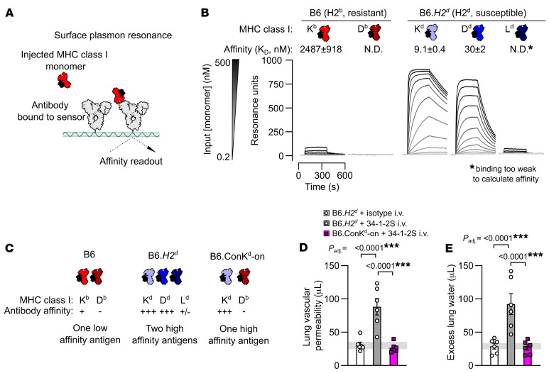
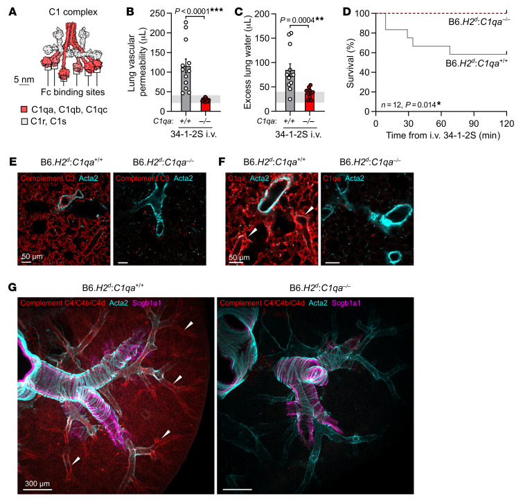
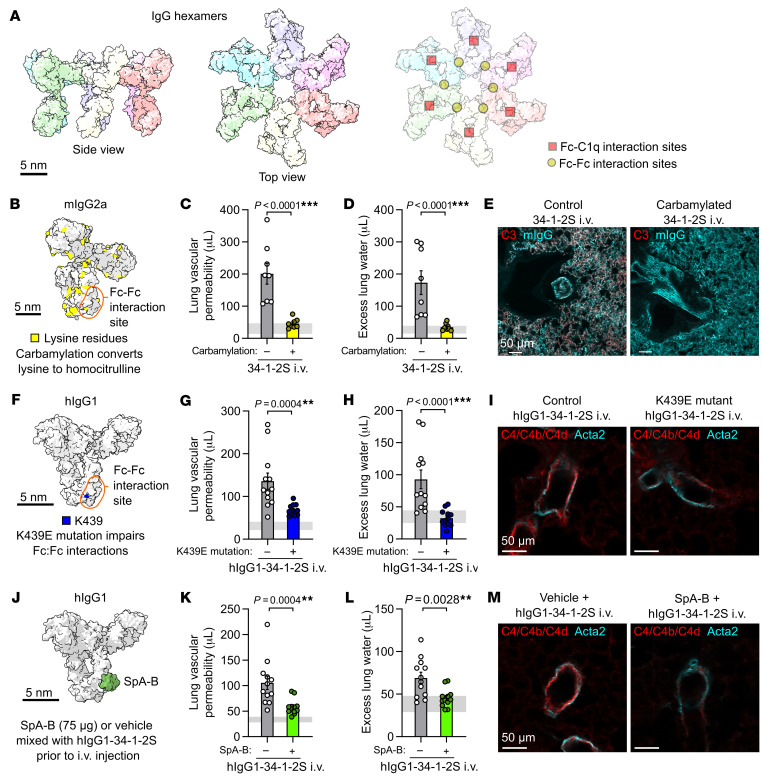
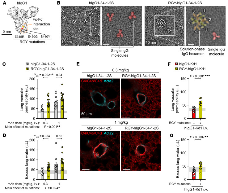
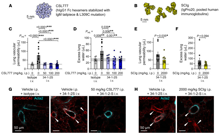
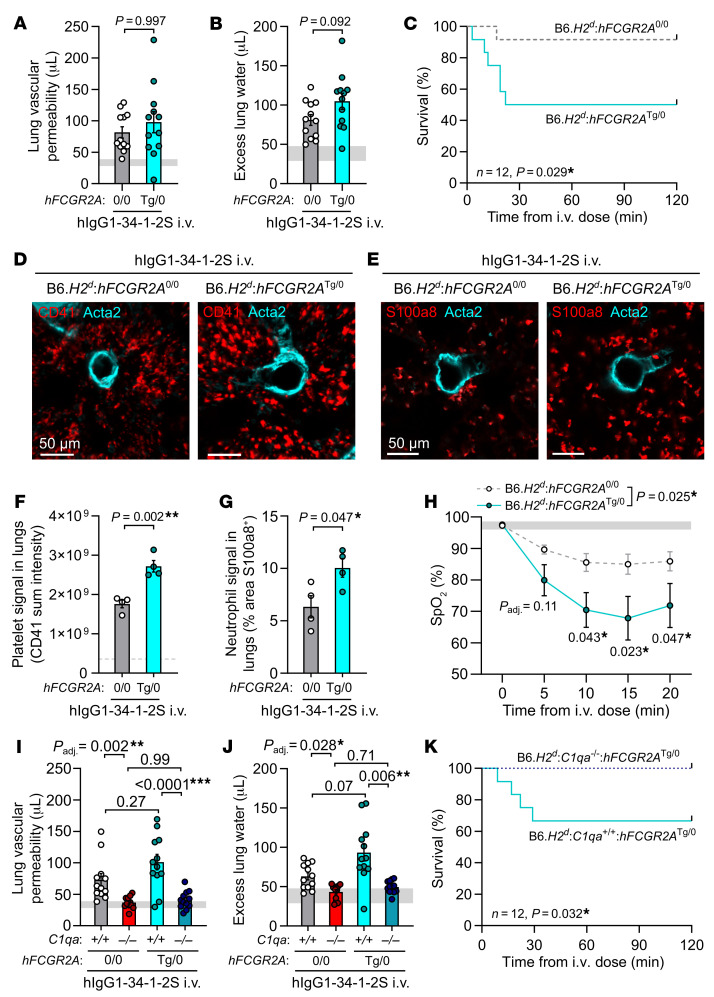
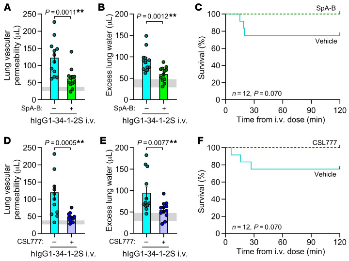
Antibodies can initiate lung injury in a variety of disease states such as autoimmunity, in reactions to transfusions, or after organ transplantation, but the key factors determining in vivo pathogenicity of injury-inducing antibodies are unclear. Harmful antibodies often activate the complement cascade. A model for how IgG antibodies trigger complement activation involves interactions between IgG Fc domains driving the assembly of IgG hexamer structures that activate C1 complexes. The importance of IgG hexamers in initiating injury responses was not clear, so we tested their relevance in a mouse model of alloantibody- and complement-mediated acute lung injury. We used 3 approaches to block alloantibody hexamerization (antibody carbamylation, the K439E Fc mutation, or treatment with domain B from staphylococcal protein A), all of which reduced acute lung injury. Conversely, Fc mutations promoting spontaneous hexamerization made a harmful alloantibody into a more potent inducer of acute lung injury and rendered an innocuous alloantibody pathogenic. Treatment with a recombinant Fc hexamer "decoy" therapeutic protected mice from lung injury, including in a model with transgenic human FCGR2A expression that exacerbated pathology. These results indicate an in vivo role of IgG hexamerization in initiating acute lung injury and the potential for therapeutics that inhibit or mimic hexamerization to treat antibody-mediated diseases.

Keywords: Antigen; Immunoglobulins; Innate immunity; Pulmonology; Transplantation.


Conflict of interest statement

Conflict of interest: RS is an employee of CSL Behring (Bern, Switzerland).

Figures

Figure 1
Figure 1. The 34-1-2S alloantibody binds to multiple MHC class I antigens to trigger acute lung injury.
(A) Schematic showing the approach for measuring affinity of the MHC class I alloantibody 34-1-2S for MHC class I monomers using SPR. (B). SPR sensorgrams showing detection of the binding of 34-1-2S to the Kb MHC class I antigen from H2b mice and the Kd, Dd, and Ld antigens from mice with the H2d haplotype. (C) Classical MHC class I antigens present in B6, B6.H2d and B6.Con-Kd-on mice with a summary of the results from B. (D) Lung vascular permeability and (E) excess lung water measurements from LPS-primed B6.H2d mice given i.v. doses of either 34-1-2S or isotype control versus B6.Con-Kd-on mice given i.v. 34-1-2S. Depictions of IgG and MHC class I in AC are based on the Protein Data Bank (PDB) entries 1HZH and 1RK1. Data in B, D and E indicate the mean ± SEM. P values adjusted for multiple comparisons (Padj.) in D and E were determined by ordinary 1-way ANOVA with Dunnett’s test for differences relative to the B6.H2d plus 34-1-2S i.v. group, with data being log10-transformed prior to analysis. ***P < 0.0001.
Figure 2
Figure 2. Classical complement activation on the pulmonary endothelium initiates 34-1-2S–induced acute lung injury.
(A) Molecular model of the C1 complex based on the small-angle scattering database entry SASDB38 (12). (B) Lung vascular permeability and (C) excess lung water measurements from LPS-primed B6.H2d C1qa–/– mice and B6.H2d C1qa+/+ littermates given i.v. 34-1-2S at 1 mg/kg. Horizontal gray lines are SDs of values from “no-injury” controls (B6.H2d mice given LPS i.p. plus mIgG2a isotype control i.v.). (D) Survival of LPS-primed B6.H2d C1qa–/– mice and B6.H2d C1qa+/+ littermates given i.v. 34-1-2S at 4.5 mg/kg. (EG) Immunofluorescence staining for (E) complement C3, (F) C1qa, and (G) C4/C4b/C4d (red) as well as for α–smooth muscle actin (Acta2, cyan) and Scgb1a1 (club cell secretory protein, magenta) in lung sections from LPS-primed B6.H2d C1qa–/– mice and B6.H2d C1qa+/+ mice fixed 5 minutes after i.v. injection of 1 mg/kg 34-1-2S. Images in G are maximum-intensity projections sampling 240 μm from a cleared thick section. White arrowheads point to arterioles positive for complement components. Scale bars: 50 um (E and F); 300 μm (G). Data in B and C show the mean ± SEM. **P < 0.01 and ***P < 0.0001, by unpaired, 2-tailed t test on log10-transformed data (B and C) or log-rank test (D). n = 12/group; n = 3 samples/group for immunofluorescence staining.
Figure 3
Figure 3. Inhibiting IgG hexamer assembly reduces 34-1-2S-induced acute lung injury responses.
(A) Molecular models of IgG hexamers based on the PDB entry 1HZH, showing Fc-Fc and Fc-C1q interaction sites. Scale bar: 5 nm. (B) Molecular model showing lysine residues in mouse IgG2a (mIgG2a), per PDB entry 1IGT. (C) Lung vascular permeability, (D) excess lung water measurements, and (E) lung complement C3 and mIgG immunostains from LPS-primed BALB/c mice after i.v. injection of carbamylated or control 34-1-2S. (F) Molecular model showing the location of Fc domain lysine 439 (K439) in hIgG1, per PDB entry 1HZH. (G) Lung vascular permeability, (H) excess lung water measurements, and (I) lung complement C4/C4b/C4d and Acta2 immunostains from LPS-primed B6.H2d mice after i.v. injection with hIgG1-34-1-2S or hIgG1-34-1-2S with the K439E mutation.(J) Molecular model showing the binding site of SpA-B to the Fc domain of hIgG1, per the PDB entries 1HZH and 5U4Y. (K) Lung vascular permeability, (L) excess lung water measurements, and (M) lung complement C4/C4b/C4d and Acta2 immunostains from LPS-primed B6.H2d mice after i.v. injection with hIgG1-34-1-2S and 75 μg SpA-B or vehicle control. Samples for the injury measurements were collected 2 hours after antibody injections, and lungs were fixed for immunostaining 5 minutes after antibody injections. Graphs show the mean ± SEM, with horizontal gray lines showing 95% CIs of measurements from no-injury control mice given LPS and nonreactive isotype antibodies. **P < 0.01 and ***P < 0.0001, by unpaired, 2-tailed t tests on log10-transformed data, with n = 8/group (C and D) and n = 12/group (G, H, K, and L), or are representative of 3 samples/group (E, I, and M). Scale bars: 50 μm (E, I, and M).
Figure 4
Figure 4. Fc mutations promoting IgG hexamer assembly increase the in vivo pathogenicity of alloantibodies.
(A) Molecular model showing amino acids mutated in RGY-hIgG1 antibodies, per PDB entry 1HZH. (B) Negative stain electron micrographs showing single hIgG1-34-1-2S molecules and spontaneous solution-phase hexamers formed by RGY-hIgG1-34-1-2S (colored overlay highlights structures in expanded images). Numbers 1–6 label individual IgG molecules forming a hexamer. Scale bars: 10 nm (including enlarged insets). (C) Lung vascular permeability and (D) excess lung water measurements from LPS-primed B6.H2d mice injected with control or RGY-mutated hIgG1-34-1-2S mAbs at i.v. doses of either 0.3 or 1 mg/kg. (E) Immunofluorescence staining showing pulmonary arterioles stained for complement C4/C4b/C4d (red) and Acta2 (cyan) in lung sections from LPS-primed B6.H2d mice given the indicated treatments, representative of 3 samples per group fixed 5 minutes after antibody injections. Scale bars: 50 μm. (F) Lung vascular permeability and (G) excess lung water measurements from LPS-primed B6.H2d mice injected with control or RGY-mutated hIgG1-Kd1 (a novel mAb targeting only the H-2Kd MHC class I antigen) at 1 mg/kg i.v. Graphs show the mean ± SEM, with the horizontal line representing 95% CIs from no-injury controls (LPS-primed B6.H2d mice given hIgG1 isotype control i.v.). *P < 0.05, **P < 0.01 and ***P < 0.0001; log10-transformed data were analyzed using an ordinary 2-way ANOVA with Šídák’s multiple-comparison test for the effect of the Fc mutation within the dose level (C and D) or unpaired, 2-tailed t test (F and G). n = 12/group.
Figure 5
Figure 5. Treatment with recombinant Fc hexamer decoys prevents alloantibody-mediated acute lung injury.
(A) Molecular representation of CSL777, an investigational recombinant Fc hexamer decoy treatment that inhibits classical complement activation, based on PDB entry 7X13 (52). (B) Molecular representation of SCIg, a pooled hIg therapeutic with antiinflammatory properties at high doses, based on PDB entry 1HZH. Scale bar: 5 nm. (C) Lung vascular permeability and (D) excess lung water measurements from LPS-primed BALB/c mice given i.p. vehicle or CSL777 at the indicated doses 1 hour before i.v. injection of 34-1-2S or mIgG2a isotype control. (E) Lung vascular permeability and (F) excess lung water measurements from LPS-primed BALB/c mice given i.p. vehicle or 2,000 mg/kg SCIg 1 hour before i.v. injection of 34-1-2S or mIgG2a isotype control. (G and H) Immunofluorescence showing pulmonary arterioles stained for complement C4/C4b/C4d (red) and Acta2 (cyan) in lung sections from LPS-primed BALB/c mice given the indicated treatments, representative of 3 samples per group fixed 5 minutes after antibody injections. White arrowheads point to arterioles with endothelial positivity for C4/C4b/C4d. Scale bars: 50 μm. Graphs show the mean ± SEM, with the horizontal line representing 95% CIs of data from no-injury controls (from vehicle plus isotype control group). *P < 0.05, **P < 0.01 and ***P < 0.0001; log10-transformed data were analyzed using either (C and D) an ordinary 1-way ANOVA (C and D), Dunnett’s multiple-comparison test for differences relative to the vehicle plus 34-1-2S group (C and D), or 2-tailed, unpaired t test (E and F). n = 10/group.
Figure 6
Figure 6. Acute lung injury is complement dependent in a model incorporating human FCGR2A–mediated pathology.
(A) Lung vascular permeability, (B) excess lung water, and (C) survival readouts from LPS-primed B6.H2d:hFCGR2aTg/0 mice and littermate controls lacking hFCGR2A expression that were given i.v. hIgG1-34-1-2S at 1 mg/kg. (D) Immunofluorescence images of platelet sequestration (CD41, red, with Acta2 in cyan) and (E) neutrophils (S100a8, red, with Acta2 in cyan) in lungs from LPS-primed B6.H2d:hFCGR2aTg/0 mice and littermates without hFCGR2A expression, fixed at 20 minutes after hIgG1-34-1-2S injections and (F and G) quantification. Scale bars: 50 μm. (H) SpO2 measurements from LPS-primed B6.H2d:hFCGR2aTg/0 mice and littermate controls without hFCGR2A expression before and after hIgG1-34-1-2S injections. (I) Lung vascular permeability, (J) excess lung water, and (K) survival readouts for LPS-primed B6.H2d C1qa+/+ and B6.H2d C1qa–/– mice, as well as for hFCGR2A-expressing littermates of each genotype, that were given i.v. hIgG1-34-1-2S at 1 mg/kg. Data in A, B, F, G, H, I, and J show the mean ± SEM, with horizontal gray lines showing values from the no-injury controls (baseline readings or values from B6.H2d mice given LPS i.p. plus hIgG1 isotype control i.v.), and were log10 transformed prior to analysis. *P < 0.05, **P < 0.01 and ***P < 0.0001, by unpaired, 2-tailed t tests (A, B, F, and G); 2-way ANOVA with Šídák’s multiple-comparison test (I and J); log-rank test (C and K); or 2-way, repeated-measures mixed-model approach with tests for main effect of the genotype and for post-baseline effects of the genotype within time levels with Holm’s adjustment for multiple comparisons (H). n = 4/group (DG); n = 10/group (H); n = 12/group (other graphs).
Figure 7
Figure 7. Approaches to inhibit or mimic IgG hexamerization reduce antibody-mediated acute lung injury in mice expressing human FCGR2A.
(A) Lung vascular permeability, (B) excess lung water, and (C) survival readouts for LPS-primed B6.H2d:hFCGR2aTg/0 mice given i.v. hIgG1-34-1-2S at 1 mg/kg with either the IgG hexamerization inhibitor SpA-B or vehicle. (D) Lung vascular permeability, (E) excess lung water, and (F) survival readouts for LPS-primed B6.H2d:hFCGR2aTg/0 mice given either vehicle or 50 mg/kg CSL777 i.p. before i.v. injection of hIgG1-34-1-2S at 1 mg/kg. Data in A, B, D, and E show the mean ± SEM, with horizontal gray lines showing SDs of the values from the no-injury controls (B6.H2d mice given LPS i.p. plus hIgG1 isotype control i.v.) and were log10 transformed prior to analysis. **P < 0.01, by unpaired, 2-tailed t test (A, B, D, and E) or log-rank test (C and F) (all, n = 12/group).

Update of

References

    1. Vlaar APJ, Juffermans NP. Transfusion-related acute lung injury: a clinical review. Lancet. 2013;382(9896):984–994. doi: 10.1016/S0140-6736(12)62197-7. - DOI - PubMed
    1. Panch SR, et al. Hemolytic transfusion reactions. N Engl J Med. 2019;381(2):150–162. doi: 10.1056/NEJMra1802338. - DOI - PubMed
    1. Loupy A, Lefaucheur C. Antibody-mediated rejection of solid-organ allografts. N Engl J Med. 2018;379(12):1150–1160. doi: 10.1056/NEJMra1802677. - DOI - PubMed
    1. Delaney M, Matthews DC. Hemolytic disease of the fetus and newborn: managing the mother, fetus, and newborn. Hematology Am Soc Hematol Educ Program. 2015;2015(1):146–151. doi: 10.1182/asheducation-2015.1.146. - DOI - PubMed
    1. Strait RT, et al. MHC class I-specific antibody binding to nonhematopoietic cells drives complement activation to induce transfusion-related acute lung injury in mice. J Exp Med. 2011;208(12):2525–2544. doi: 10.1084/jem.20110159. - DOI - PMC - PubMed