. 2024 May;13(5):1383-1398.
doi: 10.1007/s40123-024-00925-y. Epub 2024 Mar 26.

Enhancing Diabetic Macular Edema Treatment Outcomes: Exploring the ESASO Classification and Structural OCT Biomarkers

Collaborators, Affiliations

Enhancing Diabetic Macular Edema Treatment Outcomes: Exploring the ESASO Classification and Structural OCT Biomarkers

Giacomo Panozzo et al. Ophthalmol Ther. 2024 May.

Abstract

Introduction: This study assessed the European School of Advanced Studies in Ophthalmology (ESASO) classification's prognostic value for diabetic macular edema (DME) in predicting intravitreal therapy outcomes.

Methods: In this retrospective, multicenter study, patients aged > 50 years with type 1 or 2 diabetes and DME received intravitreal antivascular endothelial growth factor (anti-VEGF) agents (ranibizumab, bevacizumab, and aflibercept) or steroids (dexamethasone). The primary outcome was visual acuity (VA) change post-treatment, termed as functional response, measured 4-6 weeks post-third anti-VEGF or 12-16 weeks post-steroid injection, stratified by initial DME stage.

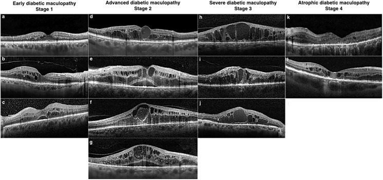
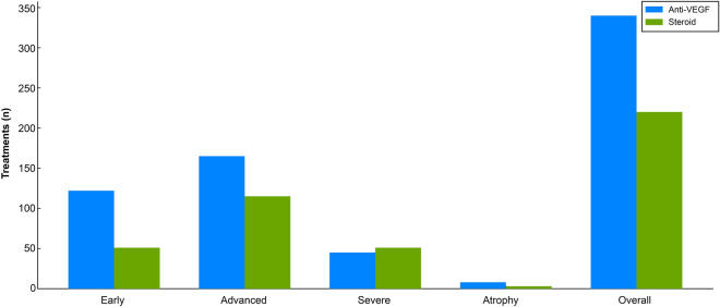
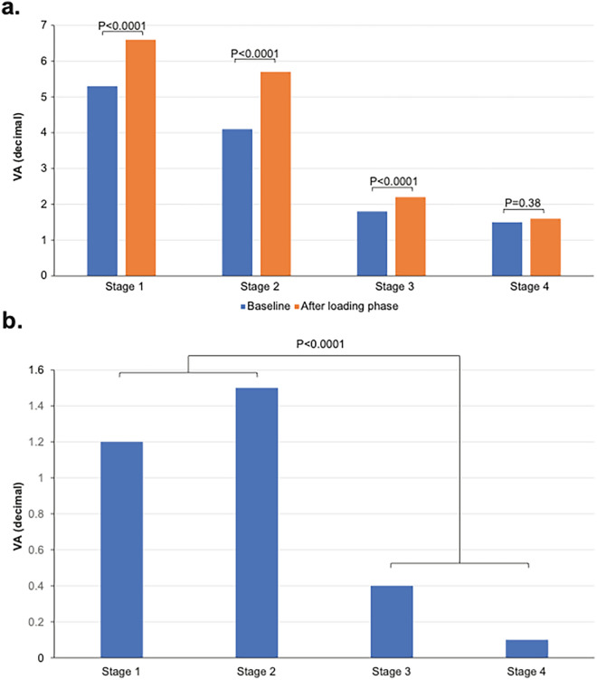
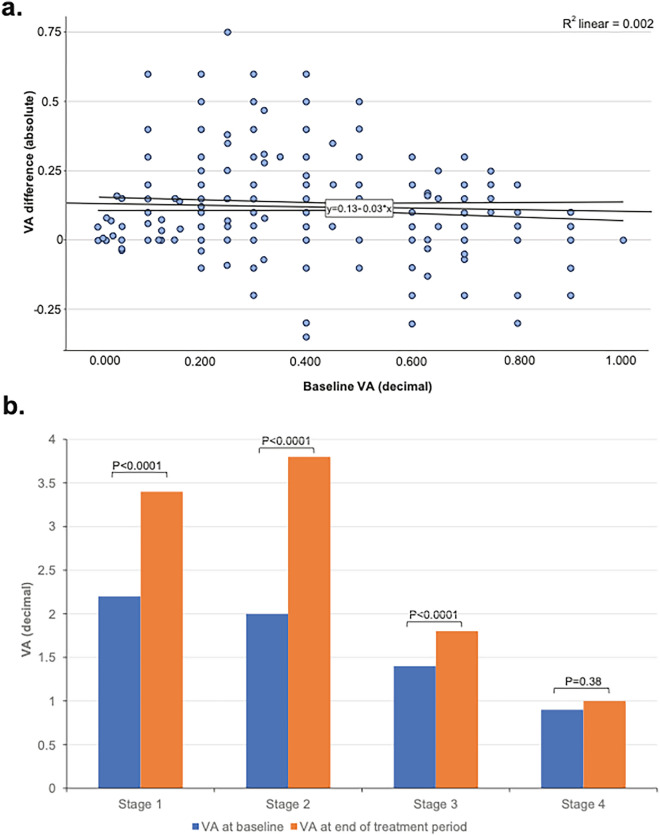
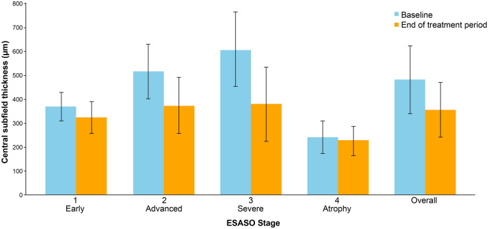
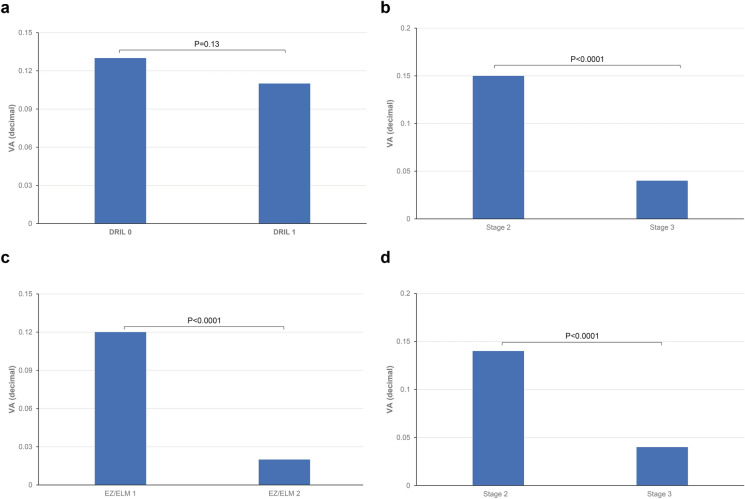
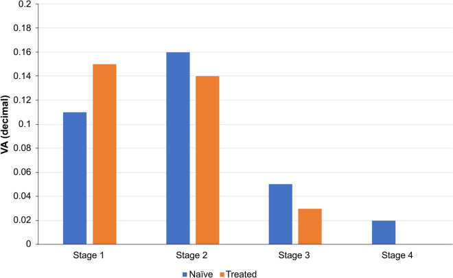
Results: Of the 560 eyes studied (62% male, mean age 66.7 years), 31% were classified as stage 1 (early), 50% stage 2 (advanced), 17% stage 3 (severe), and 2% stage 4 (atrophic). Visual acuity (VA; decimal) improved by 0.12-0.15 decimals in stages 1-2 but only 0.03 decimal in stage 3 (all p < 0.0001) and 0.01 in stage 4 (p = 0.38). Even in eyes with low baseline VA ≤ 0.3, improvements were significant only in stages 1 and 2 (0.12 and 0.17 decimals, respectively). Central subfield thickness (CST) improvement was greatest in stage 3 (-229 µm, 37.6%, p < 0.0001), but uncorrelated with VA gains, unlike stages 1 and 2 (respectively: -142 µm, 27.4%; - 5 µm, 12%; both p < 0.0001). Stage 4 showed no significant CST change. Baseline disorganization of retinal inner layers and focal damage of the ellipsoid zone/external limiting membrane did not influence VA improvement in stages 1 and 2. Treatment patterns varied, with 61% receiving anti-VEGF and 39% dexamethasone, influenced by DME stage, with no significant differences between therapeutic agents.

Conclusion: The ESASO classification, which views the retina as a neurovascular unit and integrates multiple biomarkers, surpasses single biomarkers in predicting visual outcomes. Significant functional improvement occurred only in stages 1 and 2, suggesting reversible damage, whereas stages 3 and 4 likely reflect irreversible damage.

Keywords: Anti-VEGF; Diabetic macular edema; ESASO classification; OCT Biomarkers; Optical coherence tomography; Retinal Neurovascular Unit; Steroid; Treatment outcomes in DME.


Conflict of interest statement

Neither Giacomo Panozzo nor Maria Vittoria Cicinelli, Giulia Dalla Mura, Diana Giannarelli, Maria Vadalà, Vincenza Bonfiglio, Giovanni Bellisario, nor Francesco Bandello received financial support for the research,authorship, and/or publication of this article.

Figures

Fig. 1
Fig. 1
Illustrations of ESASO DME classification stages. Early diabetic maculopathy (stage 1) includes: a Small cystoid spaces near the fovea, with all retinal layers intact and normal central subfoveal thickness and macular volume. TCED-HFV: T = 1; C = 1; E = 0; D = 0; H = 0; F = 0; V = 0. b Multiple perifoveal cystoid spaces with mild macular thickening and incomplete vitreous detachment; over 30 hyperreflective foci. TCED-HFV: T = 1; C = 2; E = 0; D = 0; H = 1; F = 0; V = 1. c Mild macular edema with intact retinal profile, cystoid spaces, and initial epiretinal membrane; over 30 hyperreflective foci. TCED-HFV: T = 1; C = 2; E = 0; D = 0; H = 1; F = 1; V = 4. Advanced diabetic maculopathy (stage 2) includes: d cystoid spaces with retinal thickening and central macrocyst; damaged subfoveal ellipsoid zone. TCED-HFV: T = 2; C = 3; E = 1; D = 0; H = 1; F = 0; V = 0. e Intermediate cystoid spaces with disrupted external limiting membrane and visible internal retinal layers; no adhesion or traction. TCED-HFV: T = 2; C = 2; E = 1; D = 0; H = 0; F = 1; V = 0. f Large pseudocyst in the fovea with parafoveal cystoid spaces; damaged subfoveal external limiting membrane and partial vitreous detachment. TCED-HFV: T = 2; C = 3; E = 1; D = 1; H = 0; F = 0; V = 1. g Large cystoid spaces with shallow subfoveal detachment; non-gradable ellipsoid zone and discontinuous external limiting membrane; visible inner retinal segmentation and slightly elevated vitreous. TCED-HFV: T = 2; C = 3; E = 1; D = 0; H = 1; F = 1; V = 1. Severe diabetic maculopathy (stage 3) includes: h multiple central macrocysts with inner retinal layer disorganization; absent subfoveal external limiting membrane and ellipsoid zone. TCED-HFV: T = 2; C = 3; E = 2; D = 1; H = 0; F = 0; V = 1. i Central macrocyst with large surrounding cystoid spaces, severe DRIL, and incomplete vitreous detachment; absent subfoveal external limiting membrane and ellipsoid zone. TCED-HFV: T = 2; C = 3; E = 2; D = 1; H = 0; F = 0; V = 1. j Central macrocyst and multiple large cysts with a few hyperreflective foci; damaged but visible retinal inner layers and normal vitreoretinal relationship. Atrophic diabetic maculopathy (stage 4) includes: k Central retinal thinning with DRIL, atrophic retinal pigment epithelium, and below-normal thickness and volume; some parafoveal microcysts and peripheral cystoid spaces. TCED-HFV: T = 0; C = 1; E = 2; D = 1; H = 1; F = 0; V = 0. l Central thinning with DRIL, irregular and focally atrophic retinal pigment epithelium, and below-normal thickness and volume, but with some moderate intraretinal cysts. TCED-HFV: T = 0; C = 1; E = 2; D = 1; H = 0; F = 0; V = 0. Adapted from reference [4]. DME diabetic macular edema, DRIL disorganization of the inner retinal layers, ESASO European School of Advanced Studies in Ophthalmology, TCED-HDF Thickening–cysts–ellipsoid–DRIL–hyperreflective foci–fluid (subretinal)–vitreoretinal relationship
Fig. 2
Fig. 2
Treatment patterns in diabetic macular edema. VEGF vascular endothelial growth factor
Fig. 3
Fig. 3
Mean VA improvement after the loading phase, any therapy (a), and absolute mean VA improvement after the loading phase, any therapy (b). VA visual acuity
Fig. 4
Fig. 4
VA improvement in the function of VA from baseline (a), stratified by ESASO DME classification in eyes with VA < 0.3 decimal at baseline (b). DME diabetic macular edema, ESASO European School of Advanced Studies in Ophthalmology, VA visual acuity
Fig. 5
Fig. 5
Correlation between ESASO DME stage and changes in CST at the end of follow-up. CST central subfield thickness, DME diabetic macular edema, ESASO European School of Advanced Studies in Ophthalmology
Fig. 6
Fig. 6
Absolute variation in VA in DRIL at baseline and in DRIL at the end of the treatment period (a), stage 2 and in stage 3 (b), VA in EZ/ELM Grade1 (n = 224), in EZ/ELM grade 2 (c), and VA in stage 2 and stage 3. DRIL disorganization of the inner retinal layers, EZ ellipsoid zone, ELM external limiting membrane, VA visual acuity
Fig. 7
Fig. 7
Absolute difference in (VA) according to stage and treatment naivety. VA visual acuity

References

    1. International Diabetes Federation. IDF Diabetes Atlas Ninth Edition. 2019.
    1. Ciulla TA, Amador AG, Zinman B. Diabetic retinopathy and diabetic macular edema: pathophysiology, screening, and novel therapies. Diabetes Care. 2003;26(9):2653–2664. doi: 10.2337/diacare.26.9.2653. - DOI - PubMed
    1. Wong TY, Cheung CM, Larsen M, Sharma S, Simo R. Diabetic retinopathy. Nat Rev Dis Primers. 2016;2:16012. doi: 10.1038/nrdp.2016.12. - DOI - PubMed
    1. Panozzo G, Cicinelli MV, Augustin AJ, et al. An optical coherence tomography-based grading of diabetic maculopathy proposed by an international expert panel: the European School for Advanced Studies in Ophthalmology classification. Eur J Ophthalmol. 2020;30(1):8–18. doi: 10.1177/1120672119880394. - DOI - PubMed
    1. Panozzo G, Franzolin E, Giannarelli D, et al. Validation of ESASO classification of diabetic maculopathy. Eur J Ophthalmol. 2023 doi: 10.1177/11206721231186649. - DOI - PubMed

LinkOut - more resources