. 2024 Apr 4;128(13):3090-3101.
doi: 10.1021/acs.jpcb.3c07363. Epub 2024 Mar 26.

Sequence Dependence in Nucleosome Dynamics

Affiliations

Sequence Dependence in Nucleosome Dynamics

Prabir Khatua et al. J Phys Chem B. .

Abstract

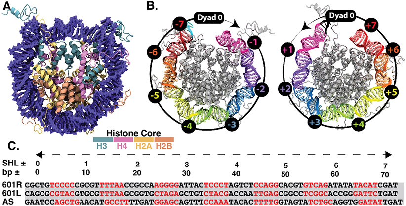
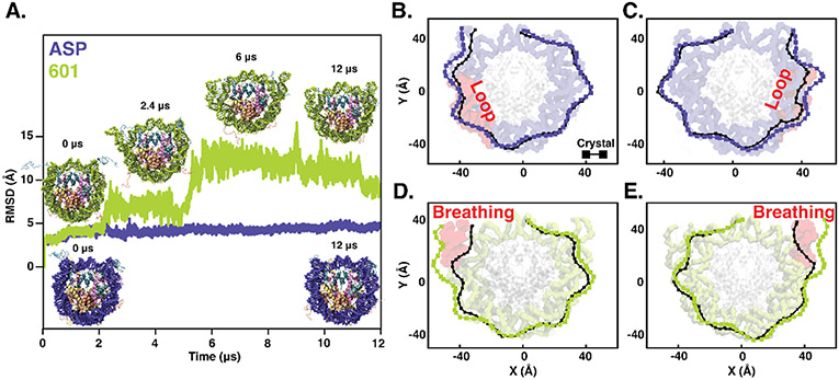
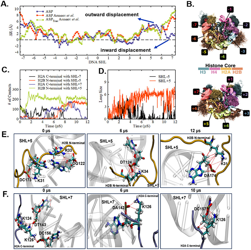
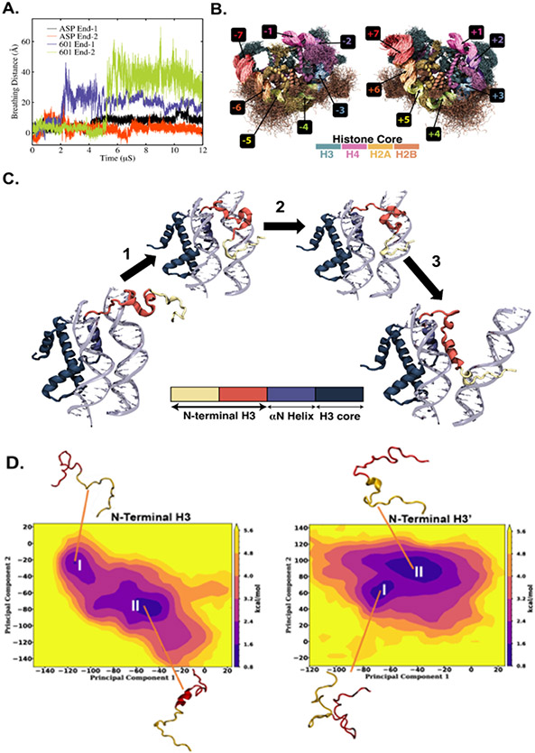
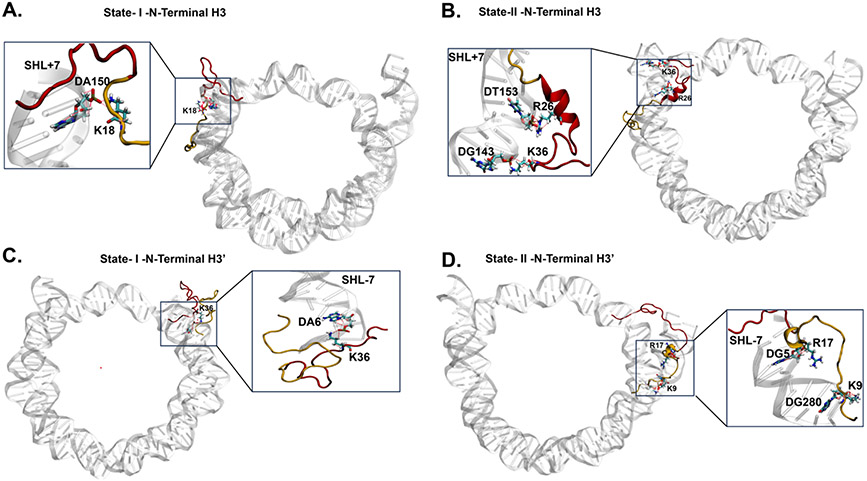
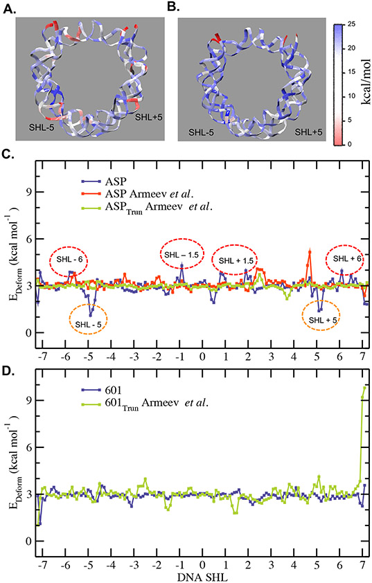
The basic packaging unit of eukaryotic chromatin is the nucleosome that contains 145-147 base pair duplex DNA wrapped around an octameric histone protein. While the DNA sequence plays a crucial role in controlling the positioning of the nucleosome, the molecular details behind the interplay between DNA sequence and nucleosome dynamics remain relatively unexplored. This study analyzes this interplay in detail by performing all-atom molecular dynamics simulations of nucleosomes, comparing the human α-satellite palindromic (ASP) and the strong positioning "Widom-601" DNA sequence at time scales of 12 μs. The simulations are performed at salt concentrations 10-20 times higher than physiological salt concentrations to screen the electrostatic interactions and promote unwrapping. These microsecond-long simulations give insight into the molecular-level sequence-dependent events that dictate the pathway of DNA unwrapping. We find that the "ASP" sequence forms a loop around SHL ± 5 for three sets of simulations. Coincident with loop formation is a cooperative increase in contacts with the neighboring N-terminal H2B tail and C-terminal H2A tail and the release of neighboring counterions. We find that the Widom-601 sequence exhibits a strong breathing motion of the nucleic acid ends. Coincident with the breathing motion is the collapse of the full N-terminal H3 tail and formation of an α-helix that interacts with the H3 histone core. We postulate that the dynamics of these histone tails and their modification with post-translational modifications (PTMs) may play a key role in governing this dynamics.


Figures

Fig. 1.
Fig. 1.
(A) Crystal structure of the nucleosome core particle. Histone subunits H2A, H2B, H3, and H4, are represented in four distinct colors with DNA in blue. (B) Left and right DNA halves of the nucleosome core particle. Various DNA turns are highlighted in distinct colors, while the histone octamer is represented in gray color. The rotational orientation of each turn corresponding to the DNA double helix is represented relative to the central base pair (super helix location zero, SHL-0, where the major groove faces the histone octamer) following the standard SHL notation, where the negative and positive values correspond to right and left halves, respectively. These SHL locations are marked on a representative circle with an arrow pointing from the histone core. (C) Comparison of DNA sequence for the crystal structure of nucleosome structure used in this study, 'Widom-601' and human α-satellite sequence (ASP). The red DNA sequence represents minor grooves, while black indicates the major grooves. Only one-half of the sequence is shown for the human α-satellite sequence as it is a palindromic sequence, whereas both right (601R) and left (601L) half sequence for the 'Widom-601' sequence are shown.
Fig. 2.
Fig. 2.
(A) Time evolution of root mean square deviation (RMSD) of DNA non-hydrogen atoms for the nucleosome with human α-satellite (ASP, shown in blue) and 'Widom-601' sequence (shown in green). The representative structures corresponding to the sharp changes associated with RMSD values and the structures at the beginning and end of 12 μs simulations are highlighted. Two-dimensional projection of DNA for (B-C) the human α-satellite sequence (ASP, shown in blue) and (D-E) for the 'Widom-601' sequence (shown in green) as obtained at the end of 12 μs simulations. For comparison, a similar projection for the respective crystal structure is shown in black. The corresponding simulated configurations are also shown in each panel B-E to highlight the regions with a loop or breathing (shown in red). Two DNA halves, left (SHL-0 to SHL-7, panels B and D) and right (SHL-0 to SHL+7, panels C and E), are shown separately.
Fig. 3.
Fig. 3.
(A) Average distance (R) of each DNA base pair center (represented in SHL notation) with respect to the center of mass of non-hydrogen atoms of DNA in the crystal structure as obtained over the last 1 μs simulations of ASP sequence presented as the difference (ΔR) between the simulated and crystal structure. Simultaneous inward and outward displacement of DNA at SHL+6 and SHL+5 suggest a “wave-like” motion. Comparison with last 1 μs of trajectories from Armeev et al. (B) Superimposed simulated configurations of ASP sequence by highlighting various DNA SHL regions and histone proteins in distinct colors. For easy visualization, the superimposed configurations are presented separately for left (SHL0 to SHL-7; top panel) and right (SHL0 to SHL+7; bottom panel) DNA halves. (C) The number of contacts between the H2A C-terminal/H2B N-terminal tail with SHL±7/SHL±5 as a function of time for the ASP sequence. (D) The loop size formed at SHL-5 and SHL+5 locations for ASP sequence as a function of time. (E) H2B N-terminal tail at 0 μs, 6 μs, and 12 μs forms a hydrogen bond between LYS/ARG with SHL+5 region of DNA. At 0 μs, there is a hydrogen bond between LYS31 and ARG33 with the phosphate backbone of DC173 and DG122. At 6 μs and 12 μs, there is a hydrogen bond between LYS34 and LYS31 with the phosphate backbone of DT124 and DA174 respectively. (F) H2A C-terminal tail at 0 μs, 6 μs, and 10 μs exhibits hydrogen bond between positively charged residues LYS/ARG with SHL+7 region of DNA. At 0 μs, it shows hydrogen bond of LYS124 and LYS126 with the phosphate backbone of DT143 and DC156. Similarly at 6 μs and 10 μs, it shows hydrogen bond of LYS126 with the phosphate backbone of DA142 and DC157.
Fig. 4.
Fig. 4.
(A) Time evolution of breathing distance for nucleosomal DNA End-1 and End-2 for both Widom-601 and ASP sequences. (B) Superimposed simulated configurations of Widom-601 sequence with highlighting various DNA SHL regions and histone proteins in distinct colors. For easy visualization, the superimposed configurations are presented for left (SHL0 to SHL-7; left panel) and right (SHL0 to SHL+7; right panel) DNA halves separately. (C) Mechanism for the role of H3 tail in governing the DNA end breathing. There are three major steps. Step 1: Condensation of extremely flexible H3 N-terminal tail (residues 1-15; shown in yellow) on the minor groove of SHL ± 6, Step 2: Condensation of H3 N-terminal residues 16 to 43 (shown in red) on SHL ± 7. This results in the movement of the DNA end region away from the histone core. Step 3: Helix-helix linkage formation between α-N core helix (blue) and N-terminal tail residues 16 to 43 (red) of H3 protein. This helix-helix linkage formation further leads to DNA outward movement, resulting in a notable breathing motion. D) PCA analysis was performed to study collective modes of the full H3 tails in the Widom-601 NCP over 12 μs. The free energy landscape constructed using the first two principal components for the H3 N-terminal Tail 1 (left) exhibits two major minima (I and II) corresponding to disordered and collapsed tail. For the H3’ N-terminal Tail 2 (right), the free energy landscape also exhibits to major minima (I and II).
Fig. 5.
Fig. 5.
Conformations of the N-Terminal H3 tail extracted from the PCA free energy landscape as shown in Fig. 4 D I and II with neighboring DNA shows interaction between the SHL+7 base pairs and positively charged residues of the N-terminal H3 tail. (A) PCA conformation I of H3 N-terminal Tail 1 exhibits hydrogen bonds LYS18 and the phosphate backbone of DA150 of the SHL+7 region. (B) PCA conformation II of the H3 N-terminal Tail 1 exhibits hydrogen bonds of ARG26 and LYS36 with DT153 and DG143 of SHL+7, respectively. (C) PCA conformation I of the H3’ N-terminal Tail 2 shows hydrogen bond formation between LYS36 and DA6 of the SHL-7 region. (D) PCA conformation II of the H3’ N-terminal Tail 2 shows hydrogen bond formations between ARG17 and LYS9 with DG5 and DG280 of the SHL-7 region.
Fig. 6.
Fig. 6.
A color map for the average force constant (kcal/mol) associated with the rise deformation for (A) the ASP and (B) the ‘Widom-601’ nucleosome sequences as obtained from diagonal elements corresponding to the force component of the average 6X6 stiffness matrix. (C) Total deformation energy score as a function of DNA base pair calculated for the selected simulated conformations (see the text) for the ASP (blue) sequence compared with Armeev et al. (red and green). The SHL locations with significantly high deformation scores (red circles) and those with low deformation scores (orange circles) for the ASP sequence are marked. (D) Total deformation energy score as a function of DNA base pair calculated for the selected simulated conformations (see the text) for the ‘Widom-601’ (blue) sequence compared with Armeev et al. (green)

References

    1. Kornberg RD; Lorch Y Twenty-five years of the nucleosome, fundamental particle of the eukaryote chromosome. Cell 1999, 98 (3), 285–294. - PubMed
    1. Segal E; Fondufe-Mittendorf Y; Chen L; Thåström A; Field Y; Moore IK; Wang J-PZ; Widom J A genomic code for nucleosome positioning. Nature 2006, 442 (7104), 772–778. - PMC - PubMed
    1. McGinty RK; Tan S Nucleosome structure and function. Chemical reviews 2015, 115 (6), 2255–2273. - PMC - PubMed
    1. Müller MM; Muir TW Histones: At the crossroads of peptide and protein chemistry. Chemical reviews 2015, 115 (6), 2296–2349. - PMC - PubMed
    1. Davey CA; Sargent DF; Luger K; Maeder AW; Richmond TJ Solvent mediated interactions in the structure of the nucleosome core particle at 1.9 å resolution. Journal of molecular biology 2002, 319 (5), 1097–1113. - PubMed

Publication types