Review
. 2024:42:103585.
doi: 10.1016/j.nicl.2024.103585. Epub 2024 Mar 5.

ENIGMA's simple seven: Recommendations to enhance the reproducibility of resting-state fMRI in traumatic brain injury

Affiliations
Review

ENIGMA's simple seven: Recommendations to enhance the reproducibility of resting-state fMRI in traumatic brain injury

Karen Caeyenberghs et al. Neuroimage Clin. 2024.

Abstract

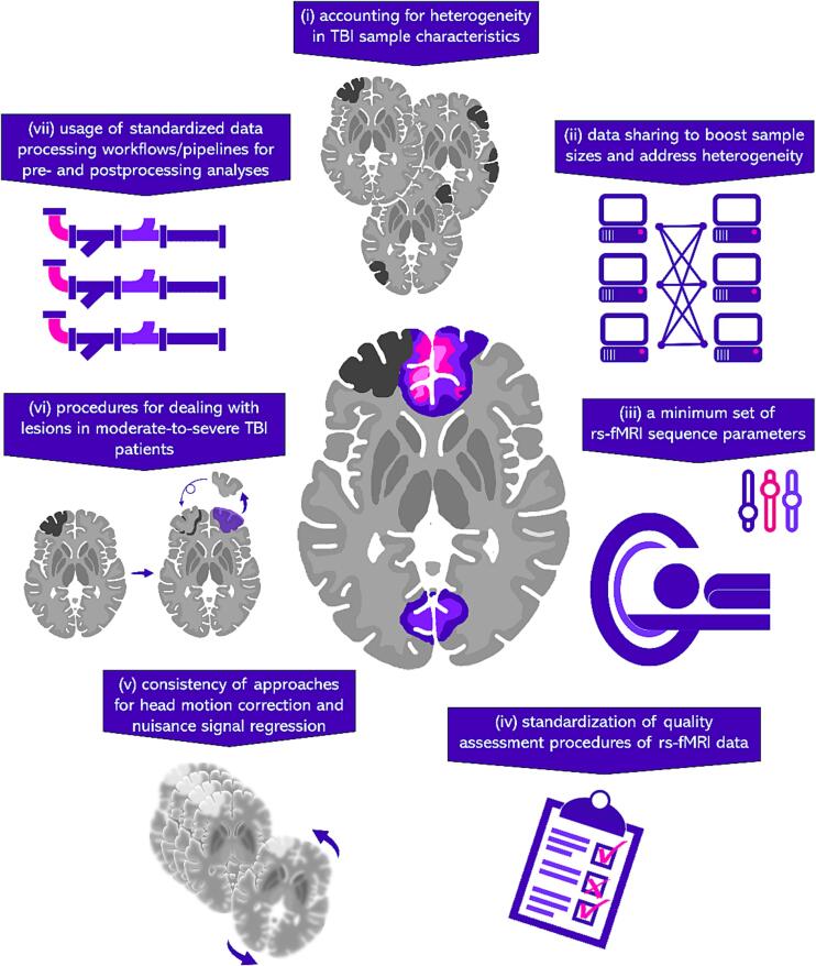
Resting state functional magnetic resonance imaging (rsfMRI) provides researchers and clinicians with a powerful tool to examine functional connectivity across large-scale brain networks, with ever-increasing applications to the study of neurological disorders, such as traumatic brain injury (TBI). While rsfMRI holds unparalleled promise in systems neurosciences, its acquisition and analytical methodology across research groups is variable, resulting in a literature that is challenging to integrate and interpret. The focus of this narrative review is to address the primary methodological issues including investigator decision points in the application of rsfMRI to study the consequences of TBI. As part of the ENIGMA Brain Injury working group, we have collaborated to identify a minimum set of recommendations that are designed to produce results that are reliable, harmonizable, and reproducible for the TBI imaging research community. Part one of this review provides the results of a literature search of current rsfMRI studies of TBI, highlighting key design considerations and data processing pipelines. Part two outlines seven data acquisition, processing, and analysis recommendations with the goal of maximizing study reliability and between-site comparability, while preserving investigator autonomy. Part three summarizes new directions and opportunities for future rsfMRI studies in TBI patients. The goal is to galvanize the TBI community to gain consensus for a set of rigorous and reproducible methods, and to increase analytical transparency and data sharing to address the reproducibility crisis in the field.

Keywords: Functional MRI; Functional connectivity; Lesions; Reproducibility; Resting state fMRI; Traumatic brain injury.


Conflict of interest statement

Declaration of competing interest The authors declare that they have no known competing financial interests or personal relationships that could have appeared to influence the work reported in this paper.

Figures

Fig. 1
Fig. 1
Schematic overview of the seven recommendations from the ENIGMA rsfMRI working group.
Fig. 2
Fig. 2
EPI Confounds output from a HALFpipe QC report for two mild TBI patients with good (top) and bad (bottom) data (from an ongoing study led by KC). Displayed are time courses (x-axis) of the magnitude (y-axis) of the global signal (GS), global signal in CSF (GSCSF) and white matter (GSWM), DVARS (D: temporal derivative of time courses; VARS: RMS variance over voxels) and the framewise displacement (FD). The time courses are followed by a carpet plot, a two-dimensional heatmap of the BOLD time series, with time on the x-axis and voxels on the y-axis. Voxels are arranged into cortical (blue) and subcortical (orange) grey matter, cerebellum (green), and white matter and cerebro-spinal fluid (red). QC involves looking for fluctuations in intensity in the carpet plot with reference to motion and signal changes in the time courses. Sudden changes in the carpet plot are likely to be caused by abrupt movement, whereas prolonged signal changes may be indicative of motion or acquisition artifacts. Sustained and substantial changes in the carpet plot of TBI03 (red arrows), particularly around the halfway mark, corresponding to changes in the time courses of all other quality metrics (with a maximum FD > 5 mm), are associated with movement (determined by visual inspection). (For interpretation of the references to colour in this figure legend, the reader is referred to the web version of this article.)
Fig. 3
Fig. 3
Ability of different preprocessing pipelines (rows) to mitigate spurious head-motion related noise, under different participant exclusion regimens (i.e., censoring-based, lenient exclusion threshold, stringent exclusion threshold), as captured by two quality control metrics (top: the correlation between head motion parameters and functional connectivity, QC-FC; bottom: the degree to which the QC-FC correlation depends on how distant two ROIs are, QC-FC Distance dependence). As discussed in the text, stringent rejection of datasets (i.e., exclude if: <4 min of the data; mFD > 0.25 mm; FD > 0.2 mm in more than 20% of volumes; or any volume has FD > 5 mm; cf., Satterthwaite et al., 2013) minimizes the spurious effects of motion and makes the choice of pipeline secondary, albeit at the cost of potentially large data loss. (Figure adapted from Weiler et al., 2023, OHBM).
Fig. 4
Fig. 4
Models of edema (red), low-density tissue (yellow), and cerebral spinal fluid (blue) in a male subject with moderate-severe TBI (49 years old at time of scan, 15 years since injury). Lesion tracing indicates large areas of edema involving anterior and inferior frontal lobes, right lateral temporal and parietotemporal regions extending to the posterior frontal lobe. Low tissue density is observed in the anteromedial aspect of the right thalamus, and the anterior body and genu of the corpus callosum. MRI volumes (FLAIR, T1-weighted, and averaged rsfMRIs) are provided below the model. Canonical volumetric views (axial, sagittal, and coronal) are displayed in radiological convention for all images. Five trained lesion raters traced edema on FLAIR volumes, and low-density tissue was traced on T1-weighted MRI. (For interpretation of the references to colour in this figure legend, the reader is referred to the web version of this article.)

References

    1. Adhikari B.M., Jahanshad N., Shukla D., Turner J., Grotegerd D., Dannlowski U., Kochunov P. A resting state fMRI analysis pipeline for pooling inference across diverse cohorts: an ENIGMA rs-fMRI protocol. Brain Imaging Behav. 2019;13:1453–1467. doi: 10.1007/s11682-018-9941-x. - DOI - PMC - PubMed
    1. Aerts H., Fias W., Caeyenberghs K., Marinazzo D. Brain networks under attack: robustness properties and the impact of lesions. Brain J. Neurol. 2016;139(Pt 12):3063–3083. doi: 10.1093/brain/aww194. - DOI - PubMed
    1. Agcaoglu O., Wilson T.W., Wang Y.P., Stephen J., Calhoun V.D. Resting state connectivity differences in eyes open versus eyes closed conditions. Hum. Brain Mapp. 2019;40(8):2488–2498. doi: 10.1002/hbm.24539. - DOI - PMC - PubMed
    1. Almansour M., Ghanem N., Bassiouny S. IEEE EMBS International Conference on Biomedical and Health Informatics (BHI) 2021 Athens, Greece. Vol. 1. 2021. High-resolution MRI brain inpainting; p. 6. - DOI
    1. Amgalan, A., Maher, A., Ghosh, S., Chui, H., Bogdan, P., & Irimia, A. (2022). Brain age estimation reveals older adults' accelerated senescence after traumatic brain injury. Geroscience. 2022, 44(5), 2509-2525. doi: 10.1007/s11357-022-00597-1. - PMC - PubMed

Further reading

    1. Abbas K., Shenk T.E., Poole V.N., Breedlove E.L., Leverenz L.J., Nauman E.A., Talavage T.M., Robinson,, M., E. Alteration of default mode network in high school football athletes due to repetitive subconcussive mild traumatic brain injury: a resting-state functional magnetic resonance imaging study. Brain Connect. 2015;5(2):91–101. doi: 10.1089/brain.2014.0279. - DOI - PubMed
    1. Zhou Y. Small world properties changes in mild traumatic brain injury. Journal of Magnetic Resonance Imaging: JMRI. 2017;46(2):518–527. doi: 10.1002/jmri.25548. - DOI - PMC - PubMed

Publication types

MeSH terms