. 2024 Mar 27;9(1):69.
doi: 10.1038/s41392-024-01766-8.

Rapid development of double-hit mRNA antibody cocktail against orthopoxviruses

Affiliations

Rapid development of double-hit mRNA antibody cocktail against orthopoxviruses

Hang Chi et al. Signal Transduct Target Ther. .

Abstract

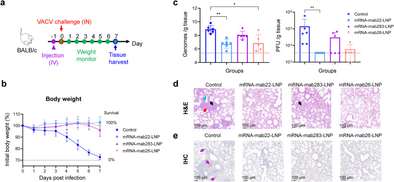
The Orthopoxvirus genus, especially variola virus (VARV), monkeypox virus (MPXV), remains a significant public health threat worldwide. The development of therapeutic antibodies against orthopoxviruses is largely hampered by the high cost of antibody engineering and manufacturing processes. mRNA-encoded antibodies have emerged as a powerful and universal platform for rapid antibody production. Herein, by using the established lipid nanoparticle (LNP)-encapsulated mRNA platform, we constructed four mRNA combinations that encode monoclonal antibodies with broad neutralization activities against orthopoxviruses. In vivo characterization demonstrated that a single intravenous injection of each LNP-encapsulated mRNA antibody in mice resulted in the rapid production of neutralizing antibodies. More importantly, mRNA antibody treatments showed significant protection from weight loss and mortality in the vaccinia virus (VACV) lethal challenge mouse model, and a unique mRNA antibody cocktail, Mix2a, exhibited superior in vivo protection by targeting both intracellular mature virus (IMV)-form and extracellular enveloped virus (EEV)-form viruses. In summary, our results demonstrate the proof-of-concept production of orthopoxvirus antibodies via the LNP-mRNA platform, highlighting the great potential of tailored mRNA antibody combinations as a universal strategy to combat orthopoxvirus as well as other emerging viruses.


Conflict of interest statement

C.F.Q., Y.Q.D., H.C., S.Q.Z., R.Y.C., and X.X.S. have filed patents related to the finding reported in this manuscript.

Figures

Fig. 1
Fig. 1
Construction and characterization of anti-orthopoxvirus monoclonal antibody-encoding mRNAs. a Schematic of mRNAs encoding the heavy and light chains of the anti-orthopoxvirus monoclonal antibody (mAb). IgK Sp, human IgK light chain signal peptide. mAb22, mAb283, mAb301 and mAb26 target VACV A33, B5, MPXV M1 and VACV A27 proteins, respectively. b Validation of mAb heavy and light chain expression in the corresponding mRNA-transfected cell supernatants by Western blot. BHK-21 cells were transfected with HC mRNA and LC mRNA. The supernatants were harvested at 24 h post transfection. c Quantification of mAb levels in mRNA transfection supernatants using human IgG ELISA. BHK-21 cells and 293TN cells were respectively transfected with HC mRNA and LC mRNA. Transfection supernatants were harvested at 24 h post transfection. d The evaluation of binding activity respectively to MPXV proteins A35, B6, M1, and A29 of the BHK-21 cell transfection supernatants using indirect ELISA. The absorbance at 450 nm /630 nm was measured. Data are shown as the mean ± SD. Data were obtained using GraphPad Prism version 9.0 (GraphPad software)
Fig. 2
Fig. 2
In vivo expression of LNP-encapsulated mRNA encoding protective antibodies against Orthopoxvirus. a Schematic of the experimental design. Six- to eight-week-old specific pathogen-free female BALB/c mice (n = 6 per group) were intravenously (i.v.) injected with a single dose of 1 mg/kg of mRNA-mab22-LNP, mRNA-mab283-LNP, mRNA-mab26-LNP, mRNA-mab301-LNP or placebo. Orbital blood from each group of mice were collected at 24 h post injection. Sera were harvested and heat inactivated before further test. b Quantification of mAb levels in sera were performed using human IgG ELISA. Statistical significance was analyzed by ordinary one-way ANOVA (***P < 0.001 and ****P < 0.0001). c EEV neutralization activity of mRNA-mab22-LNP and mRNA-mab283-LNP were measured using EEV forms of VACV by PRNT assay in BS-C-1 cells. d IMV neutralization activity of mRNA-mab26-LNP and mRNA-mab301-LNP were measured using IMV forms of VACV by PRNT assay in BS-C-1 cells. Data are shown as the mean ± SD. Statistical significance was analyzed by Kruskal‒Wallis one-way ANOVA (*p < 0.05, **p < 0.01 and ***p < 0.001). Data were obtained using GraphPad Prism version 9.0 (GraphPad software)
Fig. 3
Fig. 3
Protection efficiency of the candidate mRNA-encoded antibody component in a VACV challenge mouse model. a Schematic of the experimental design. Six- to eight-week-old specific pathogen-free female BALB/c mice (n = 5-6 per group) were intravenously (i.v.) administered a single dose of mRNA-mab22-LNP, mRNA-mab283-LNP, mRNA-mab26-LNP or placebo. All mice were intranasally challenged with 7.5 × 10⁴ PFU VACV (WR strain) at 24 h post administration and monitored daily for body weight. Tissues were harvested at 7 d.p.i. Part of lung tissues were homogenized using a tissue homogenizer. The other part of lung tissues from mice were fixed with 4% formaldehyde and embedded in paraffin. Sections of the paraffin-embedded tissues were cut and placed on glass histology slides. b Body weight and survival were monitored daily for 7 d.p.i. c Viral genome copies and VACV titers in the lung at 7 d.p.i. were measured by qPCR and standard plaque assay in BS-C-1 cells, respectively. Viral genome copies were expressed as genomes/g tissue. Virus titers were expressed as PFU/g of tissue. Data are shown as the mean ± SD. Statistical significance was analyzed by Kruskal‒Wallis one-way ANOVA (*p < 0.05 and **p < 0.01). Data were obtained using GraphPad Prism version 9.0 (GraphPad software). d H&E staining of the lung sections from mice infected with VACV (WR strain) at 7 d.p.i. Diffuse degeneration and necrosis of the epithelial lining (black arrow), accompanied by hemorrhage, edema (red arrow), and fibrin exudation into surrounding alveoli (blue arrow). e Immunohistochemistry (IHC) analysis was performed with a human anti-VACV D8L monoclonal antibody. Purple arrows indicate VACV infection foci. The slides were scanned with a Pannoramic MIDI histoscanner (3DHISTECH), and images were analyzed using Pannoramic Viewer software. Scale bars (100 μm) are indicated for each picture. Brown-colored staining indicates positive results
Fig. 4
Fig. 4
Injection of BALB/c mice with mRNA antibody cocktail contributes to neutralizing activity against Orthopoxvirus. a Schematic of the experimental design for the characterization of the mRNA antibody cocktail in two batches of animals. Six- to eight-week-old specific pathogen-free female BALB/c mice (n = 6 per group) were intravenously (i.v.) administered 2 mg/kg Mix2a (1 mg/kg mRNA-mab22-LNP plus 1 mg/kg mRNA-mab26-LNP), Mix2b (1 mg/kg mRNA-mab22-LNP plus 1 mg/kg mRNA-mab301-LNP) or placebo. In batch I, orbital blood from each group of mice were collected on Days 1, 3, and 7 post injection. Sera were harvested and heat inactivated before further test. In batch II, mice were intranasally challenged with 7.5 × 10⁴ PFU VACV (WR strain) at 24 h post administration and monitored daily for body weight. Tissues were harvested at 7 d.p.i. Part of lung tissues were homogenized using a tissue homogenizer. The other part of lung tissues from mice were fixed with 4% formaldehyde and embedded in paraffin. Sections of the paraffin-embedded tissues were cut and placed on glass histology slides. b The anti-VACV IgG antibody titers of sera were evaluated by indirect ELISA at the indicated times. c Neutralizing activity of sera at 24 h post administration was assessed using VACV IMV- and EEV-neutralization assays in BS-C-1 cells. d Body weight and survival were monitored daily for 7 days. e Viral genome copies in the lung were measured by qPCR and are expressed as genomes/g tissue. VACV titers in the lung were assessed using a standard plaque assay in BS-C-1 cells. Viral genome copies were expressed as genomes/g tissue. Virus titers were expressed as PFU/g of tissue. Data are shown as the mean ± SD. Statistical significance was analyzed by Kruskal‒Wallis one-way ANOVA (*p < 0.05, **p < 0.01, ***p < 0.001 and ****p < 0.0001). Data were obtained using GraphPad Prism version 9.0 (GraphPad software). f H&E staining of the lung sections from mice infected with VACV (WR strain) at 7 d.p.i. Diffuse degeneration and necrosis of the epithelial lining (black arrow), accompanied by hemorrhage, edema (red arrow), and fibrin exudation into surrounding alveoli (blue arrow). IHC analysis was performed with a human anti-VACV D8L monoclonal antibody. Purple arrows indicate VACV infection foci. The slides were scanned with a Pannoramic MIDI histoscanner (3DHISTECH), and images were analyzed using Pannoramic Viewer software. Scale bars (100 μm) are indicated for each picture. Brown-colored staining indicates positive results

References

    1. Tiecco G, et al. Monkeypox, a Literature Review: What Is New and Where Does This concerning Virus Come From? Viruses. 2022;14:1894. doi: 10.3390/v14091894. - DOI - PMC - PubMed
    1. Mühlemann B, et al. Diverse variola virus (smallpox) strains were widespread in northern Europe in the Viking Age. Science. 2020;369:eaaw8977. doi: 10.1126/science.aaw8977. - DOI - PubMed
    1. Vorou RM, Papavassiliou VG, Pierroutsakos IN. Cowpox virus infection: an emerging health threat. Curr. Opin. Infect. Dis. 2008;21:153–156. doi: 10.1097/QCO.0b013e3282f44c74. - DOI - PubMed
    1. Trindade GS, Emerson GL, Carroll DS, Kroon EG, Damon IK. Brazilian vaccinia viruses and their origins. Emerg. Infect. Dis. 2007;13:965–972. doi: 10.3201/eid1307.061404. - DOI - PMC - PubMed
    1. Townsend MB, et al. Humoral immunity to smallpox vaccines and monkeypox virus challenge: proteomic assessment and clinical correlations. J. Virol. 2013;87:900–911. doi: 10.1128/JVI.02089-12. - DOI - PMC - PubMed

Publication types

Substances