

Haemodynamics of stent-mounted neural interfaces in tapered and deformed blood vessels
- PMID: 38532013
- PMCID: PMC10965969
- DOI: 10.1038/s41598-024-57460-w
Haemodynamics of stent-mounted neural interfaces in tapered and deformed blood vessels
Abstract
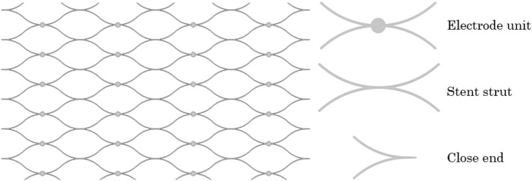
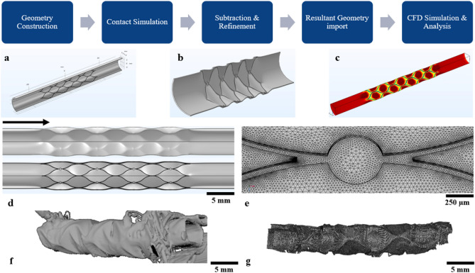
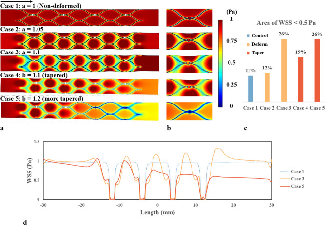
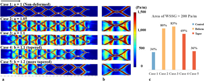
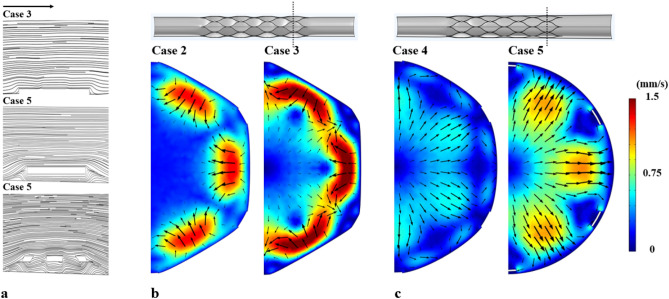
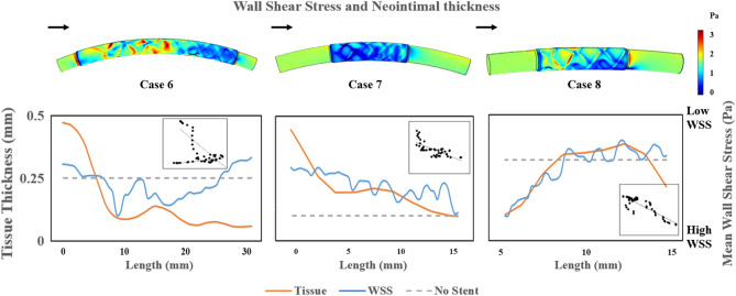
The endovascular neural interface provides an appealing minimally invasive alternative to invasive brain electrodes for recording and stimulation. However, stents placed in blood vessels have long been known to affect blood flow (haemodynamics) and lead to neointimal growth within the blood vessel. Both the stent elements (struts and electrodes) and blood vessel wall geometries can affect the mechanical environment on the blood vessel wall, which could lead to unfavourable vascular remodelling after stent placement. With increasing applications of stents and stent-like neural interfaces in venous blood vessels in the brain, it is necessary to understand how stents affect blood flow and tissue growth in veins. We explored the haemodynamics of a stent-mounted neural interface in a blood vessel model. Results indicated that blood vessel deformation and tapering caused a substantial change to the lumen geometry and the haemodynamics. The neointimal proliferation was evaluated in sheep implanted with an endovascular neural interface. Analysis showed a negative correlation with the mean Wall Shear Stress pattern. The results presented here indicate that the optimal stent oversizing ratio must be considered to minimise the haemodynamic impact of stenting.
© 2024. The Author(s).
Conflict of interest statement
NLO is a director and holds stock options in Synchron Inc. DBG, NLO and SEJ have patents related to the Stentrode technology. Other authors do not have any competing interests.
Figures
Similar articles
-
Computational fluid dynamics analysis of balloon-expandable coronary stents: influence of stent and vessel deformation.Med Eng Phys. 2014 Aug;36(8):1047-56. doi: 10.1016/j.medengphy.2014.05.011. Epub 2014 Jun 20. Med Eng Phys. 2014. PMID: 24953569
-
Segmental vessel wall shear stress and neointimal formation after sirolimus-eluting stent implantation: physiological insights in a porcine coronary model.Cardiovasc Revasc Med. 2005 Apr-Jun;6(2):58-64. doi: 10.1016/j.carrev.2005.05.004. Cardiovasc Revasc Med. 2005. PMID: 16263360
-
Computational Fluid Dynamics of Stent-Mounted Neural Interfaces in an Idealized Cerebral Venous Sinus.Annu Int Conf IEEE Eng Med Biol Soc. 2023 Jul;2023:1-4. doi: 10.1109/EMBC40787.2023.10341099. Annu Int Conf IEEE Eng Med Biol Soc. 2023. PMID: 38082814
-
Local Hemodynamic Forces After Stenting: Implications on Restenosis and Thrombosis.Arterioscler Thromb Vasc Biol. 2017 Dec;37(12):2231-2242. doi: 10.1161/ATVBAHA.117.309728. Epub 2017 Nov 9. Arterioscler Thromb Vasc Biol. 2017. PMID: 29122816 Review.
-
[The role of haemodynamic factors in the development of in-stent restenosis].Kardiol Pol. 2012;70(11):1194-8. Kardiol Pol. 2012. PMID: 23180536 Review. Polish.
Cited by
-
The association between stent design and patient exercise intensity: structural coupling effects and hemodynamic analysis.Front Bioeng Biotechnol. 2024 Dec 18;12:1514929. doi: 10.3389/fbioe.2024.1514929. eCollection 2024. Front Bioeng Biotechnol. 2024. PMID: 39744594 Free PMC article.
References
MeSH terms
Grants and funding
LinkOut - more resources
Full Text Sources
