. 2024 Jul;51(8):2216-2228.
doi: 10.1007/s00259-024-06686-x. Epub 2024 Mar 27.

Macrophage mannose receptor CD206 targeting of fluoride-18 labeled mannosylated dextran: A validation study in mice

Affiliations

Macrophage mannose receptor CD206 targeting of fluoride-18 labeled mannosylated dextran: A validation study in mice

Putri Andriana et al. Eur J Nucl Med Mol Imaging. 2024 Jul.

Abstract

Purpose: Aluminum fluoride-18-labeled 1,4,7-triazacyclononane-1,4,7-triacetic acid-conjugated mannosylated dextran derivative (Al[18F]F-NOTA-D10CM) is a new tracer for PET imaging. We report here on in vitro and in vivo validation of the tracer's ability to target the macrophage mannose receptor CD206.

Methods: First, the uptake of intravenously (i.v.) administered Al[18F]F-NOTA-D10CM was compared between wild-type (WT) and CD206-/- knockout (KO) mice. C57BL/6N mice were injected with complete Freund's adjuvant (CFA) in the left hind leg and the uptake of Al[18F]F-NOTA-D10CM after i.v. or intradermal (i.d.) injection was studied at 5 and 14 days after CFA induction of inflammation. Healthy C57BL/6N mice were studied as controls. Mice underwent PET/CT on consecutive days with [18F]FDG, i.v. Al[18F]F-NOTA-D10CM, and i.d. Al[18F]F-NOTA-D10CM. After the last imaging, Al[18F]F-NOTA-D10CM was i.v. injected for an ex vivo biodistribution study and autoradiography of inflamed tissues. Blood plasma samples were analyzed using high-performance liquid chromatography. To evaluate the specificity of Al[18F]F-NOTA-D10CM binding, an in vitro competitive displacement study was performed on inflamed tissue sections using autoradiography. CD206 expression was assessed by immunohistochemical staining.

Results: Compared with WT mice, the uptake of Al[18F]F-NOTA-D10CM was significantly lower in several CD206-/- KO mice tissues, including liver (SUV 8.21 ± 2.51 vs. 1.06 ± 0.16, P < 0.001) and bone marrow (SUV 1.63 ± 0.37 vs. 0.22 ± 0.05, P < 0.0001). The uptake of i.v. injected Al[18F]F-NOTA-D10CM was significantly higher in inflamed ankle joint (SUV 0.48 ± 0.13 vs. 0.18 ± 0.05, P < 0.0001) and inflamed foot pad skin (SUV 0.41 ± 0.10 vs. 0.04 ± 0.01, P < 0.0001) than in the corresponding tissues in healthy mice. The i.d.-injected Al[18F]F-NOTA-D10CM revealed differences between CFA-induced lymph node activation and lymph nodes in healthy mice. Ex vivo γ-counting, autoradiography, and immunohistochemistry supported the results, and a decrease of ~ 80% in the binding of Al[18F]F-NOTA-D10CM in the displacement study with excess NOTA-D10CM confirmed that tracer binding was specific. At 60 min after i.v. injection, an average 96.70% of plasma radioactivity was derived from intact Al[18F]F-NOTA-D10CM, indicating good in vivo stability. The uptake of Al[18F]F-NOTA-D10CM into inflamed tissues was positively associated with the area percentage of CD206-positive staining.

Conclusion: The uptake of mannosylated dextran derivative Al[18F]F-NOTA-D10CM correlated with CD206 expression and the tracer appears promising for inflammation imaging.

Keywords: Fluorine-18; Inflammation; Macrophage mannose receptor CD206; Mannosylated dextran; PET/CT.


Conflict of interest statement

AS received fees for consultancy or lecturing from Abbott, AstraZeneca, Janssen, Novartis, and Pfizer outside the current study. The remaining authors have no conflicts of interest to disclose.

Figures

Fig. 1
Fig. 1
Study design
Fig. 2
Fig. 2
A Representative coronal maximum intensity projection images of Al[18F]F-NOTA-D10CM PET/CT of wild-type (WT) and CD206−/− knockout (KO) mice. The difference in radioactivity concentration after intravenous injection is most prominent in the liver. Quantification of Al[18F]F-NOTA-D10CM uptake in tissues of interest by (B) in vivo PET and (C) ex vivo γ-counting at 2 h post-injection. Bars represent mean values and error bars are standard deviations. *P < 0.05, **P < 0.01, ***P < 0.001. D CD206 immunohistochemical staining shows abundant positivity in the bone marrow, liver, and pancreas of WT mice, whereas no specific staining is seen in CD206−/− KO mice
Fig. 3
Fig. 3
A Representative maximum intensity projection PET/CT images of Al[18F]F-NOTA-D10CM intravenously administered into the hind legs of a healthy mouse and a mouse on day 5 after complete Freund’s adjuvant (CFA) inflammation induction, and coronal views of inflamed lymph nodes (LN). In the photographs, the inflamed hind leg appears clearly swollen. The blue arrow denotes the ankle joint, the yellow arrow denotes the foot pad, and the red and white arrows denote LNs. B Quantification of Al[18F]F-NOTA-D10CM uptake by in vivo PET 50 − 60 min post-injection as standardized uptake value (SUV). *P < 0.05, **P < 0.01, ***P < 0.001. (C) Time-activity curves for Al[18F]F-NOTA-D10CM uptake. Bars and curves represent means, and error bars are standard deviation
Fig. 4
Fig. 4
A Representative Al[18F]F-NOTA-D10CM autoradiographs after intravenous injection, hematoxylin–eosin (H&E) staining, and CD206 immunohistochemical staining of inflamed foot pad skin (upper panel) and inflamed popliteal lymph node (lower panel) at 5 days and 14 days after complete Freund’s adjuvant-induced inflammation. The high focal Al[18F]F-NOTA-D10CM uptake co-localizes with inflammatory foci seen in H&E and CD206-positive staining (in brown). PSL, photostimulated luminescence. B Target-to-background ratio derived from Al[18F]F-NOTA-D10CM autoradiographs as CD206high area/CD206low area. (C) Correlation between Al[18F]F-NOTA-D10CM uptake and CD206 area-%
Fig. 5
Fig. 5
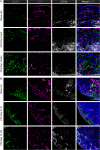
Representative immunofluorescence staining of healthy and inflamed foot pad skin (A) shows an increase in CD206+ macrophages in the dermis at days 5 and 14 after induction with complete Freund’s adjuvant (CFA). CD31 labels blood and lymphatic vessels in the dermis (positive staining of epidermis is nonspecific), and CD11b labels myeloid cells. B In the draining popliteal lymph node (LN), CD206 is expressed in the CD31+ medullary sinus in both healthy and inflamed lymph nodes. Scale bars are 100 µm
Fig. 6
Fig. 6
A In vivo PET/CT after intradermal injection of Al[18F]F-NOTA-D10CM (white arrows) reveals a change in the lymphatic drainage pathway after induction of inflammation by complete Freund’s adjuvant (CFA). B In healthy mice (n = 4), the tracer was distributed from the popliteal lymph node to the iliac and renal lymph nodes, whereas the route to the inguinal lymph node was low. Five days after CFA induction, the tracer was distributed from the popliteal lymph node to the inguinal lymph node, with the route to the iliac lymph node being increased. Fourteen days after CFA induction, the tracer’s route to the inguinal lymph node was significantly increased (P = 0.002). We also noted uptake of the tracer in inflamed axillary lymph node. Bars represent standardized uptake values (SUV) at 2 h after intradermal Al[.18F]F-NOTA-D10CM injection and error bars are standard deviations. *P < 0.05, **P < 0.01
Fig. 7
Fig. 7
A Representative digital autoradiographs from the in vitro competitive displacement assay on adjacent sections of an inflamed popliteal lymph node confirmed by H&E and CD206 immunohistochemical staining. Positive CD206 staining is shown as a brown color. B Co-incubation with excess of unlabeled NOTA-D10CM significantly reduced the binding of Al[18F]F-NOTA-D10CM by 79.48% ± 2.63. *P < 0.05

Similar articles

Cited by

References

    1. Martinez-Pomares L. The mannose receptor. J Leukoc Biol. 2012;92:1177–1186. doi: 10.1189/jlb.0512231. - DOI - PubMed
    1. Sturge J, Todd SK, Kogianni G, McCarthy A, Isacke CM. Mannose receptor regulation of macrophage cell migration. J Leukoc Biol. 2007;82:585–593. doi: 10.1189/jlb.0107053. - DOI - PubMed
    1. Marttila-Ichihara F, Turja R, Miiluniemi M, et al. Macrophage mannose receptor on lymphatics controls cell trafficking. Blood. 2008;112:64–72. doi: 10.1182/blood-2007-10-118984. - DOI - PubMed
    1. Lee SJ, Evers S, Roeder D, et al. Mannose receptor-mediated regulation of serum glycoprotein homeostasis. Science. 2002;295:1898–1901. doi: 10.1126/science.1069540. - DOI - PubMed
    1. Leong SP, Kim J, Ross M, et al. A phase 2 study of 99mTc-tilmanocept in the detection of sentinel lymph nodes in melanoma and breast cancer. Ann Surg Oncol. 2011;18:961–969. doi: 10.1245/s10434-010-1524-z. - DOI - PMC - PubMed

Publication types