. 2024 Mar 26;14(1):7199.
doi: 10.1038/s41598-024-57941-y.

The C-type lectin DCIR contributes to the immune response and pathogenesis of colorectal cancer

Affiliations

The C-type lectin DCIR contributes to the immune response and pathogenesis of colorectal cancer

Giulia Trimaglio et al. Sci Rep. .

Abstract

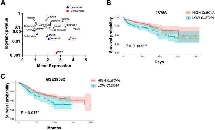
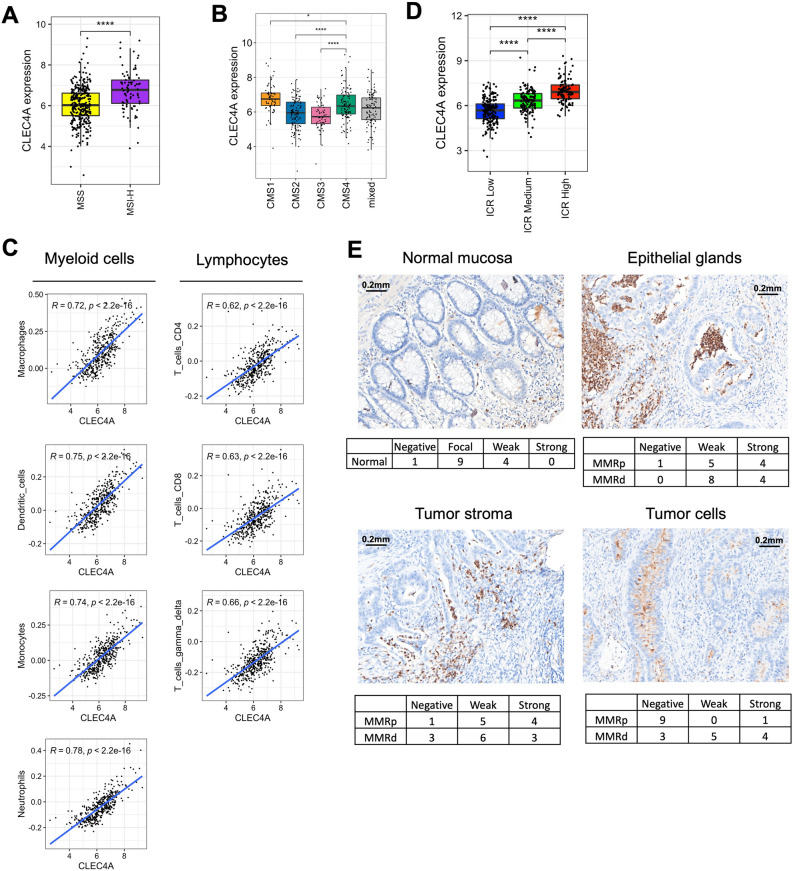
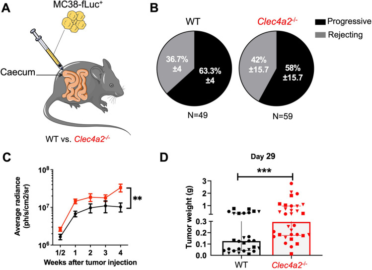
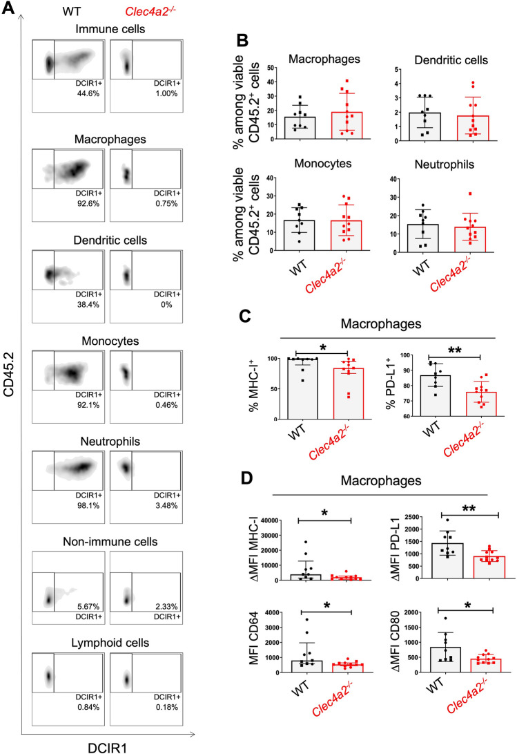
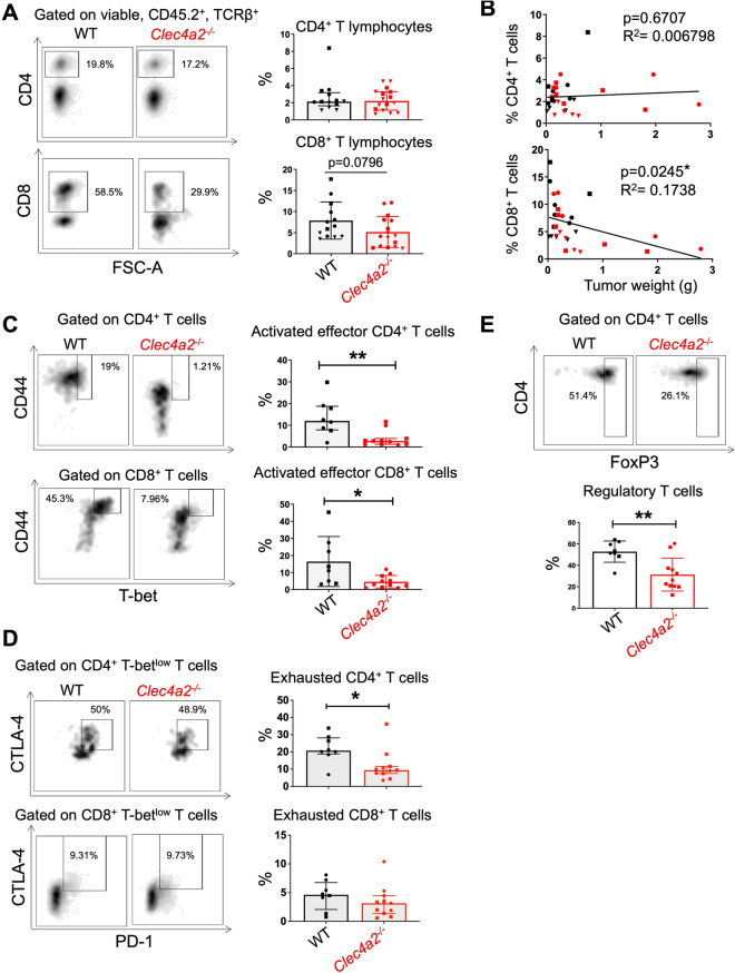
Development and progression of malignancies are accompanied and influenced by alterations in the surrounding immune microenvironment. Understanding the cellular and molecular interactions between immune cells and cancer cells has not only provided important fundamental insights into the disease, but has also led to the development of new immunotherapies. The C-type lectin Dendritic Cell ImmunoReceptor (DCIR) is primarily expressed by myeloid cells and is an important regulator of immune homeostasis, as demonstrated in various autoimmune, infectious and inflammatory contexts. Yet, the impact of DCIR on cancer development remains largely unknown. Analysis of available transcriptomic data of colorectal cancer (CRC) patients revealed that high DCIR gene expression is associated with improved patients' survival, immunologically "hot" tumors and high immunologic constant of rejection, thus arguing for a protective and immunoregulatory role of DCIR in CRC. In line with these correlative data, we found that deficiency of DCIR1, the murine homologue of human DCIR, leads to the development of significantly larger tumors in an orthotopic murine model of CRC. This phenotype is accompanied by an altered phenotype of tumor-associated macrophages (TAMs) and a reduction in the percentage of activated effector CD4+ and CD8+ T cells in CRC tumors of DCIR1-deficient mice. Overall, our results show that DCIR promotes antitumor immunity in CRC, making it an attractive target for the future development of immunotherapies to fight the second deadliest cancer in the world.

Keywords: C-type lectin; Colorectal cancer; Dendritic cell immunoreceptor; Immune response; Tumor microenvironment.


Conflict of interest statement

The authors declare no competing interests.

Figures

Figure 1
Figure 1
CLEC4A expression correlates with an improved CRC patient’s survival. (A) CLEC4A expression in 17 different types of cancer and its association with cancer patient’s survival. Y-axis represents the mean of CLEC4A Fragments Per Kilobase Million (FPKM) in the 694 cancer patients. X-axis (in log scale) represents the log-rank p-value indicating the predictive power of CLEC4A expression for cancer patient’s survival. CLEC4A expression correlates with an improved (Favorable) or with a worse (Unfavorable) patients survival probability. (B,C) Overall survival curves from CRC patients exhibiting high and low CLEC4A expression in their tumors. Transcriptomic data were obtained from the (B) TCGA-COREAD dataset (colon and rectal cohorts combined) (high CLEC4A, n = 516, low CLEC4A, n = 178) and from the (C) GSE39582 dataset (high CLEC4A, n = 486, low CLEC4A, n = 93). A log-rank test was used for the statistical analysis. *P < 0.05, **P < 0.01.
Figure 2
Figure 2
CLEC4A expression is associated with immunologically “hot” CRC tumors. (A,B) CLEC4A expression in primary tumor samples of the TCGA-COAD dataset by (A) microsatellite instability status (Microsatellite Stable (MSS), n = 331 and high microsatellite instability (MSI-H), n = 82) and by (B) consensus molecular subtypes of colorectal cancer (CMS1, n = 64, CMS2, n = 121, CMS3, n = 55, CMS4, n = 103, mixed, n = 96). An unpaired Student’s t-test was used for the statistical analysis. (C) Pearson correlation between CLEC4A expression in primary tumor samples of the TCGA-COAD dataset (n = 439) and estimated relative abundance of immune cells subsets using deconvolution by ConsensusTME. (D) CLEC4A expression in TCGA-COAD dataset by Immunologic Constant of Rejection (ICR) clusters (ICR High, n = 107, ICR Medium, n = 146, ICR Low, n = 186). (E) Representative immunohistochemical images of DCIR expression detected in the normal mucosa, epithelial glands, tumor stroma and tumor cells of a CRC patients’ cohort (n = 22). Values indicate the numbers of cores presenting negative, focal, weak or strong positive staining for DCIR in MMR-proficient (MMRp) and MMR-deficient (MMRd) CRC. *P < 0.05, ****P < 0.0001.
Figure 3
Figure 3
DCIR1 deficient mice developed larger CRC tumors than WT mice. (A) Schematic illustrating the orthotopic CRC mouse model used in this study. (B) Pie charts showing the mean ± SD of the percentage of progressive and rejecting mice in WT and DCIR1-deficient (Clec4a2−/−) mice at day 29 after IC injection of MC38-fLuc+ tumor cells (3 independent experiments pooled). The total number of mice (N) per genotype is indicated below. (C) Longitudinal bioluminescence emission (in ph/s/cm2/sr; photon per second per square centimeter per steradian) from imaging of WT and Clec4a2−/− progressive mice following IC injection of MC38-fLuc+ cells (3 independent experiments pooled, n = 31 WT and n = 34 Clec4a2−/− mice). Mean ± SEM in log10 scale is represented. (D) Quantification of tumor weight in grams (g) from WT and Clec4a2−/− progressive mice on day 29 following IC injection of MC38-fLuc+ cells (3 independent experiments pooled, n = 28 WT and n = 32 Clec4a2−/− mice). Each symbol corresponds to a single mouse and the different symbols (i.e., dots, squares, triangles) are used to indicate independent experiments. **P < 0.01, ***P < 0.001.
Figure 4
Figure 4
DCIR1 deficiency modulates innate immune response during CRC development in mouse. FACS analysis of tumor-infiltrating myeloid cells in WT and DCIR1 deficient (Clec4a2−/−) mice at day 29 after IC injection of MC38-fLuc+ cells. (A) Representative FACS plots of DCIR1 expression at the cell surface of tumor-infiltrating myeloid cell subsets, non-immune cells (CD45.2) and lymphoid cells (CD45.2+ CD11b CD11c) from WT and Clec4a2−/− mice. (B) Percentage of tumor-infiltrating myeloid cell subsets. (C) Percentage of MHC-I+ or PD-L1+ tumor-associated macrophages. (D) Median fluorescence intensity (MFI) or differential median fluorescence intensity (ΔMFI) of MHC-I, PD-L1, CD64 and CD80 at the cell surface of tumor-associated macrophages. Panels B to D were generated from two independent experiments (n = 9 WT and n = 11 Clec4a2−/− mice). Different symbols (i.e., dots and squares) are used to indicate independent experiments. *P < 0.05, **P < 0.01.
Figure 5
Figure 5
DCIR1 deficiency alters the adaptive immune response during CRC development in mouse. FACS analysis of tumor-infiltrating T lymphocytes in WT and DCIR1 deficient (Clec4a2−/−) mice at day 29 after IC injection of MC38-fLuc+ cells. (A) Representative FACS plots (left panels) and percentage (right panels) of tumor infiltrating CD4+ and CD8+ T cells (n = 13 WT and n = 16 Clec4a2−/− mice). (B) Correlation between tumor weight and the percentage of tumor-infiltrating CD4+ or CD8+ T cells. (C,D) Representative FACS plots and percentage of tumor infiltrating (C) activated effector (D) exhausted CD4+ and CD8+ T lymphocytes (n = 8 WT and n = 11 Clec4a2−/− mice) and (E) regulatory T cells. *P < 0.05, **P < 0.01.

References

    1. Hinshaw DC, Shevde LA. The tumor microenvironment innately modulates cancer progression. Cancer Res. 2019;79:4557–4566. doi: 10.1158/0008-5472.CAN-18-3962. - DOI - PMC - PubMed
    1. Demaria O, et al. Harnessing innate immunity in cancer therapy. Nature. 2019;574:45–56. doi: 10.1038/s41586-019-1593-5. - DOI - PubMed
    1. Li D-K, Wang W. Characteristics and clinical trial results of agonistic anti-CD40 antibodies in the treatment of malignancies. Oncol. Lett. 2020;20:176. - PMC - PubMed
    1. Anfray C, Ummarino A, Andón FT, Allavena P. Current strategies to target tumor-associated-macrophages to improve anti-tumor immune responses. Cells. 2019;9:46. doi: 10.3390/cells9010046. - DOI - PMC - PubMed
    1. Raymond, B. B. A., Neyrolles, O. & Rombouts, Y. C-type Lectins in Immunity to Lung Pathogens. Curr. Top Microbiol. 1–44. 10.1007/82_2020_197 (2020). - PubMed