. 2024 May;11(5):1135-1147.
doi: 10.1002/acn3.52029. Epub 2024 Mar 26.

Optimized microburst VNS elicits fMRI responses beyond thalamic-specific response from standard VNS

Collaborators, Affiliations

Optimized microburst VNS elicits fMRI responses beyond thalamic-specific response from standard VNS

Jerzy P Szaflarski et al. Ann Clin Transl Neurol. 2024 May.

Abstract

Objective: In parallel to standard vagus nerve stimulation (VNS), microburst stimulation delivery has been developed. We evaluated the fMRI-related signal changes associated with standard and optimized microburst stimulation in a proof-of-concept study (NCT03446664).

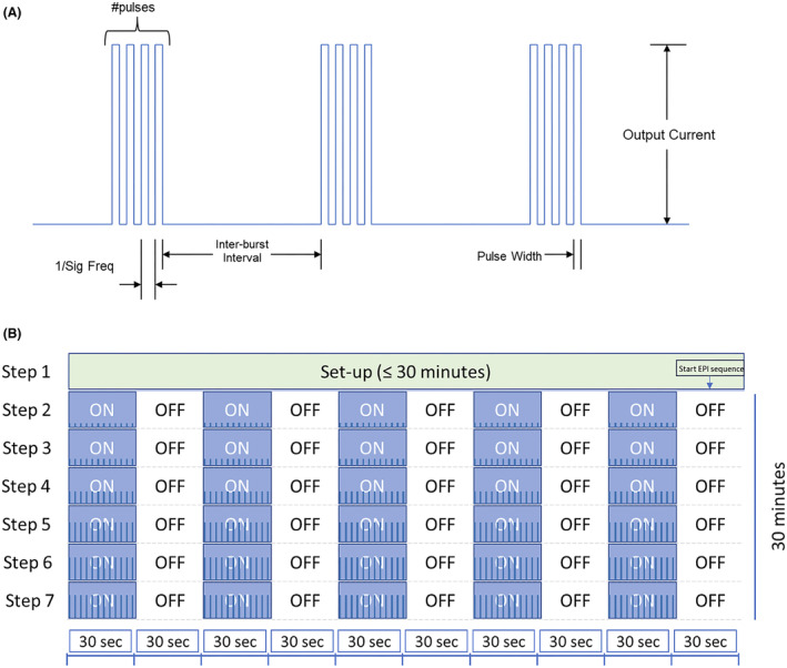
Methods: Twenty-nine drug-resistant epilepsy patients were prospectively implanted with VNS. Three 3T fMRI scans were collected 2 weeks postimplantation. The maximum tolerated VNS intensity was determined prior to each scan starting at 0.125 mA with 0.125 mA increments. FMRI scans were block-design with alternating 30 sec stimulation [ON] and 30 sec no stimulation [OFF]: Scan 1 utilized standard VNS and Scan 3 optimized microburst parameters to determine target settings. Semi-automated on-site fMRI data processing utilized ON-OFF block modeling to determine VNS-related fMRI activation per stimulation setting. Anatomical thalamic mask was used to derive highest mean thalamic t-value for determination of microburst stimulation parameters. Paired t-tests corrected at P < 0.05 examined differences in fMRI responses to each stimulation type.

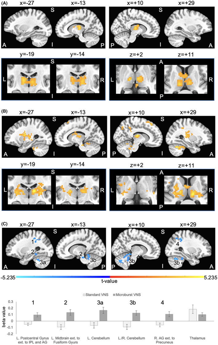
Results: Standard and microburst stimulation intensities at Scans 1 and 3 were similar (P = 0.16). Thalamic fMRI responses were obtained in 28 participants (19 with focal; 9 with generalized seizures). Group activation maps showed standard VNS elicited thalamic activation while optimized microburst VNS showed widespread activation patterns including thalamus. Comparison of stimulation types revealed significantly greater cerebellar, midbrain, and parietal fMRI signal changes in microburst compared to standard VNS. These differences were not associated with seizure responses.

Interpretation: While standard and optimized microburst VNS elicited thalamic activation, microburst also engaged other brain regions. Relationship between these fMRI activation patterns and clinical response warrants further investigation.

Clinical trial registration: The study was registered with clinicaltrials.gov (NCT03446664).


Figures

Figure 1
Figure 1
(A) Schematic of microburst stimulation train with specific parameter characteristics including number of pulses per burst, frequency of pulses, pulse width, interburst interval (time between bursts), and output current. (B) Schematic of typical scan during an MRI visit. For each of the three MRI scans, Step 1 (VNS is OFF) lasted up to 30 min to allow for participant placement in MRI scanner and acquisition of localizer and T1‐weighted 3D anatomical scans. The start of the parameter sweep at Step 2 was manually synced to the start of the 30 min fMRI scan. Starting at Step 2, each fMRI scan was a blocked design where stimulation was provided for 30 sec (ON), followed by no stimulation for 30 sec (OFF). Each stimulation step was assessed for 5 min (i.e., 5 ON and 5 OFF blocks). The protocol allowed up to six stimulation steps (Steps 2–7) during the 30‐min fMRI. Blue bars within the ON steps depict increasing intensity of stimulation parameters as typical for scan 2 (microburst optimization step).
Figure 2
Figure 2
Group data for vagus nerve stimulation (VNS) during fMRI (n = 28 subjects). The maximum tolerated stimulation output was determined out‐of‐scanner and used as the maximum setting for fMRI assessment. Group composite maps are based on peak thalamic activation for the (A) standard VNS parameters and (B) optimized novel microburst VNS parameters. Orange/yellow clusters illustrate VNS‐related fMRI signal increases. Standard and microburst VNS elicit overlapping fMRI activation in the left lateral posterior nucleus and right medial dorsal nucleus of the thalamus as shown in the second row of panels in A and B. Coordinates for peak activation and extent of each cluster are provided in Table 4. Top rows show 4 sagittal slices at x = −27 and x = −13 in the left hemisphere and x = +10 and x = 29 in the right hemisphere; bottom rows focus on thalamic overlap and show two coronal slice at y = −19 and y = −14, and two axial slices at z = +2 and z = +11. (C) Significant differences in fMRI activations between the two stimulation types. Blue clusters indicate higher VNS‐related activation for optimized microburst VNS compared to standard VNS. Row of 4 sagittal slices correspond with coordinates in A and B. A binary mask of clusters showing significant differences was created, and beta values within this mask were extracted from each subject's general linear modeling of the fMRI BOLD response of peak thalamic activation for each stimulation type. The numbers in the sagittal slices correspond to the cluster numbers in the graph. Beta values within the thalamus mask used to determine peak activation were also extracted and graphed. Group fMRI activation maps are overlaid onto a standard average brain in Talairach coordinate space. Activations are significant at corrected P  < 0.05 (voxelwise P  < 0.01 (t‐value is at least 2.7991 per voxel), cluster extent threshold of 46 voxels (1242 mm3) in which sides of voxels must touch). Note that some clusters are large and within those clusters distinct anatomical brain regions coalesce. L = left, R = right, A = anterior, P = posterior, S = superior, I = inferior, IPL = inferior parietal lobule, AG = angular gyrus.

References

    1. Bolden L, Pati S, Szaflarski J. Neurostimulation, neuromodulation, and the treatment of epilepsies. J Epileptol. 2015;23:45‐59.
    1. Verner R, Szaflarski JP, Allendorfer JB, Vonck K, Giannicola G, Microburst Study Group . Modulation of the thalamus by microburst vagus nerve stimulation: a feasibility study protocol. Front Neurol. 2023;14:1169161. - PMC - PubMed
    1. Touma L, Dansereau B, Chan AY, et al. Neurostimulation in people with drug‐resistant epilepsy: systematic review and meta‐analysis from the ILAE Surgical Therapies Commission. Epilepsia. 2022;63:1314‐1329. - PubMed
    1. Denison T, Morrell MJ. Neuromodulation in 2035: the neurology future forecasting series. Neurology. 2022;98:65‐72. - PMC - PubMed
    1. Ziemann U, Hallett M, Cohen LG. Mechanisms of deafferentation‐induced plasticity in human motor cortex. J Neurosci. 1998;18:7000‐7007. - PMC - PubMed

Associated data

Grants and funding