. 2024 Mar 27;31(1):33.
doi: 10.1186/s12929-024-01023-8.

Association of TRAIL receptor with phosphatase SHP-1 enables repressing T cell receptor signaling and T cell activation through inactivating Lck

Affiliations

Association of TRAIL receptor with phosphatase SHP-1 enables repressing T cell receptor signaling and T cell activation through inactivating Lck

I-Tsu Chyuan et al. J Biomed Sci. .

Abstract

Background: T cell receptor (TCR) signaling and T cell activation are tightly regulated by gatekeepers to maintain immune tolerance and avoid autoimmunity. The TRAIL receptor (TRAIL-R) is a TNF-family death receptor that transduces apoptotic signals to induce cell death. Recent studies have indicated that TRAIL-R regulates T cell-mediated immune responses by directly inhibiting T cell activation without inducing apoptosis; however, the distinct signaling pathway that regulates T cell activation remains unclear. In this study, we screened for intracellular TRAIL-R-binding proteins within T cells to explore the novel signaling pathway transduced by TRAIL-R that directly inhibits T cell activation.

Methods: Whole-transcriptome RNA sequencing was used to identify gene expression signatures associated with TRAIL-R signaling during T cell activation. High-throughput screening with mass spectrometry was used to identify the novel TRAIL-R binding proteins within T cells. Co-immunoprecipitation, lipid raft isolation, and confocal microscopic analyses were conducted to verify the association between TRAIL-R and the identified binding proteins within T cells.

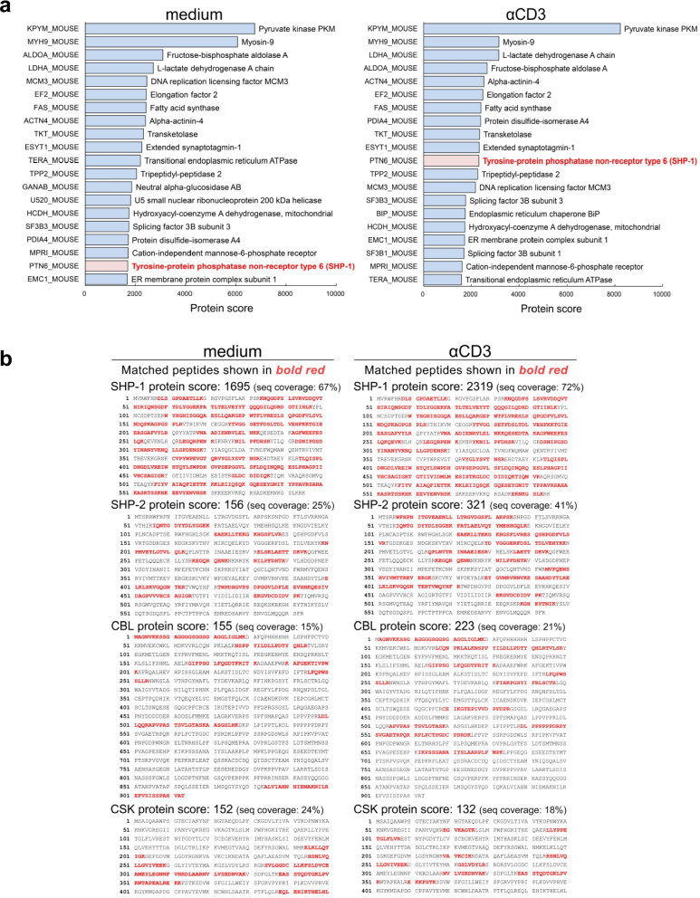
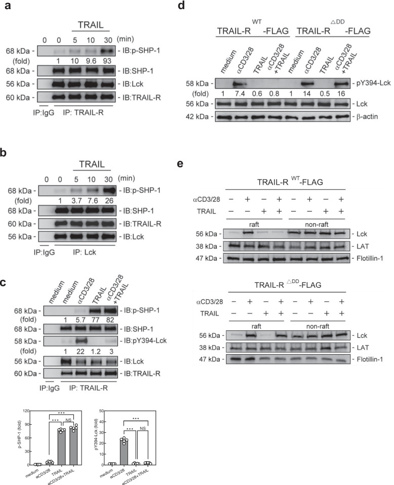
Results: TRAIL engagement downregulated gene signatures in TCR signaling pathways and profoundly suppressed phosphorylation of TCR proximal tyrosine kinases without inducing cell death. The tyrosine phosphatase SHP-1 was identified as the major TRAIL-R binding protein within T cells, using high throughput mass spectrometry-based proteomics analysis. Furthermore, Lck was co-immunoprecipitated with the TRAIL-R/SHP-1 complex in the activated T cells. TRAIL engagement profoundly inhibited phosphorylation of Lck (Y394) and suppressed the recruitment of Lck into lipid rafts in the activated T cells, leading to the interruption of proximal TCR signaling and subsequent T cell activation.

Conclusions: TRAIL-R associates with phosphatase SHP-1 and transduces a unique and distinct immune gatekeeper signal to repress TCR signaling and T cell activation via inactivating Lck. Thus, our results define TRAIL-R as a new class of immune checkpoint receptors for restraining T cell activation, and TRAIL-R/SHP-1 axis can serve as a potential therapeutic target for immune-mediated diseases.

Keywords: Phosphatase; SHP-1; T cell receptor signaling; TRAIL receptor.


Conflict of interest statement

The authors declare that they have no competing interests.

Figures

Fig. 1
Fig. 1
TRAIL-R inhibits proximal TCR signaling and T cell activation. a Flow cytometry analyses of T cell activation markersCD69 and CD25 (24 h), (b) carboxyfluorescein diacetate succinimidyl diester dilution assays (96 h), and (c) intracellular staining of IL-2 (24 h) on Trail-r+/+ or Trail-r–/– murine splenic CD4+ T cells stimulated with anti-CD3 (3 µg/ml) and anti-CD28 (2 µg/mL) Abs in the presence/absence of TRAIL (0–1 µg/mL). Data in a–c represent analyses from three independent experiments; statistical significance determined by Mann–Whitney U test; NS, no significance; * p < 0.05; *** p < 0.001. d Trail-r+/+ murine splenic CD4+ T cells were stimulated with CD3 (3 µg/mL) and CD28 (2 µg/ml) Abs in the presence/absence of TRAIL (10 µg/mL) for 24 h. Total RNA was extracted and sequenced on the Illumina platform. Principal component analysis represented correlations among groups. Network analysis of biological process Gene Ontology term enrichment among significant genes in the indicated comparisons. Node color is proportional to p-adjusted enrichment value; node size proportional to number of genes in each Gene Ontology term. Heatmap showing two differentially expressed genes in TCR signaling pathway. Color bars indicate scores of log2-fold change for each comparison. e ELISAs of lysates from Trail-r+/+ or Trail-r–/– murine splenic CD4+ T cells stimulated with CD3 (3 µg/mL) and CD28 (2 µg/ml) Abs in the presence/absence of TRAIL (10 µg/mL) for 24 h. Data represent analyses from three independent experiments; statistical significance determined by Mann–Whitney U test; NS, no significance; * p < 0.05; *** p < 0.001. f Immunoblotting of phosphorylation of TCR tyrosine kinases in Trail-r+/+ or Trail-r−/− murine splenic CD4+ T cells at indicated time points. Quantification of phosphorylated protein levels are shown at the bottom of the panel. Data represent analyses from at least three independent experiments; statistical significance determined by two-tailed Student’s t test; NS, no significance; *** p < 0.001. g Trail-r+/+ or Trail-r−/− CD4+ T cells were stimulated with anti-CD3 (3 µg/mL) and anti-CD28 (2 µg/mL) Abs in presence/absence of TRAIL (10 µg/mL) for 30 min, then lysed and fractionated as raft and non-raft layers; Lck, ZAP70, LAT, and PLCγ1 were immunoblotted from pooled raft or non-raft fractions. h Representative confocal images of Trail-r+/+ murine splenic CD4+ T cells following staining with anti-GM-1 (red) and anti-Lck (green), or anti-ZAP70 (green) Abs, as indicated. Colocalization of GM-1 with Lck or ZAP70 is indicated by yellow areas. Scale bar: 4 µm
Fig. 2
Fig. 2
TRAIL-R inhibits proximal TCR signaling without activation of apoptosis signaling. a Immunoblotting of caspase-8, caspase-3, and proximal TCR signaling molecules (30 min), and (b) Flow cytometry of Annexin V+ apoptotic cells (24 h) from murine splenic CD4.+ T cells (WT) or Jurkat cells stimulated with anti-CD3 (3 µg/mL) and anti-CD28 (2 µg/mL) Abs with or without TRAIL (10 µg/mL) in the presence or absence of pan-caspase inhibitor, Z-VAD-FMK (10 µg/mL). Data in (b) represent analyses from three independent experiments. Statistical significance was determined using the Mann–Whitney U test; NS, no significance; *** p < 0.001
Fig. 3
Fig. 3
Phosphatase SHP-1 is the major intracellular TRAIL-R binding protein in TRAIL-R IP-MASS proteomics analysis. EL4 cells were transfected with plasmid encoding TRAIL-R, followed by stimulation with anti-CD3 (5 µg/mL) Ab for 30 min. Cell lysates were immunoprecipitated with an anti-TRAIL-R Ab, digested with trypsin, and subjected to LC–MS/MS. Proteins were identified, and scores were mapped using a Mascot MS/MS Ion Search. Protein scores were calculated as the sum of the highest ion scores for each peptide. a Ranking of putative TRAIL-R-interacting proteins in the order of protein scores. b Protein scores for SHP-1 and other major regulatory proteins involved in proximal TCR signaling
Fig. 4
Fig. 4
SHP-1 is associated with TRAIL-R in T cells. a, b Immunoprecipitation analyses of tyrosine phosphatases SHP-1, SHP-2, Cbl, and Csk with TRAIL-R in murine splenic CD4+ T cells in the presence of TRAIL (10 µg/mL) at indicated time point. Quantification of phosphorylated protein levels are shown at the bottom of the panel. Data represent analyses from at least three independent experiments; statistical significance determined by two-tailed Student’s t test; NS, no significance; * p < 0.05; *** p < 0.001. c, d Co-immunoprecipitation of TRAIL-R with SHP-1 or SHP-2 from murine splenic CD4+ T cells stimulated with anti-CD3 (3 µg/mL) and anti-CD28 (2 µg/mL) Abs in the presence or absence of TRAIL (10 µg/mL) for 30 min. Quantification of phosphorylated protein levels are shown at the bottom of the panel. Data represent analyses from at least three independent experiments; statistical significance determined by two-tailed Student’s t test; NS, no significance; *** p < 0.001. e Immunoprecipitation analyses of TRAIL-R with SHP-1 or SHP-2 in TRAIL-RWT-FLAG or TRAIL-R△DD-FLAG T cells stimulated with anti-CD3 (3 µg/mL) and anti-CD28 (2 µg/mL) Abs in the presence or absence of TRAIL (10 µg/mL) for 30 min
Fig. 5
Fig. 5
TRAIL-R/SHP-1 inhibits proximal TCR signaling and T cell activation via dephosphorylation of Lck. a, b Immunoprecipitation with anti-TRAIL-R (a) or anti-Lck (b) Ab and then immunoblotting with p-SHP-1 and SHP-1 in murine splenic CD4+ T cells treated with TRAIL (10 µg/mL) at indicated time points. Quantification of phosphorylated protein levels are shown at the bottom of the panel. c Immunoprecipitation with anti-TRAIL-R Ab and then immunoblotting with p-Lck (Y394) and Lck in murine splenic CD4+ T cells stimulated with anti-CD3 (3 µg/mL) and anti-CD28 (2 µg/mL) Abs in the presence or absence of TRAIL (10 µg/mL) for 30 min. Quantification of phosphorylated protein levels are shown at the bottom of the panel. Data represent analyses from at least three independent experiments; statistical significance determined by two-tailed Student’s t test; NS, no significance; *** p < 0.001. d, e Immunoblotting of Lck in lysates (d) and pooled raft or non-raft fractions (e) of TRAIL-RWT-FLAG or TRAIL-R△DD-FLAG T cells stimulated with anti-CD3 (3 µg/mL) and anti-CD28 (2 µg/mL) Abs in the presence or absence of TRAIL (10 µg/mL) for 30 min. Quantification of phosphorylated protein levels are shown at the bottom of the panel
Fig. 6
Fig. 6
SHP-1 knockdown abolished TRAIL-R-mediated inhibition on Lck phosphorylation, lipid raft recruitment and T cell activation. a Immunoblotting of phosphorylation of Lck and other proximal TCR signaling molecules (30 min), (b) immunoblotting of Lck and other proximal TCR signaling molecules in pooled raft fractions (30 min), (c) representative confocal images of co-localization of GM-1 with Lck (30 min), (d) flow cytometry of T cell activation markers CD69 and CD25 (24 h) (Scale bar, 4 µm), and (e) intracellular staining of IL-2 from murine splenic CD4.+ T cells transfected with scramble or SHP-1 siRNA, followed by stimulation with anti-CD3 (3 µg/mL) and anti-CD28 (2 µg/mL) Abs in the presence/absence of TRAIL (0–1 µg/mL) for 24 h. Quantification of phosphorylated protein levels are shown at the bottom of the panel. Data d–e represent analyses from three independent experiments and statistics determined using Mann–Whitney U test. NS, not significant; *** p < 0.001. f Model of TRAIL-R/SHP-1 repressing TCR signaling and T cell activation through dephosphorylation of Lck at Y394. Upon TCR ligation, Lck binds to, and phosphorylates TCR and Zap70 to form an immunological synapse in the lipid raft, which transduces proximal TCR signaling downstream, resulting in T cell activation (Left). TRAIL engagement reduces Lck (Y394) phosphorylation along with the increased phosphorylated SHP-1 in the TRAIL-R/SHP-1/Lck complex, leading to the dissociation of the co-receptor-Lck complex from the lipid raft, which in turn limits downstream TCR signaling to restrain T cell activation (Right)

References

    1. Pan G, O'Rourke K, Chinnaiyan AM, Gentz R, Ebner R, Ni J, et al. The receptor for the cytotoxic ligand TRAIL. Science. 1997;276:111–113. doi: 10.1126/science.276.5309.111. - DOI - PubMed
    1. Walczak H, Degli-Esposti MA, Johnson RS, Smolak PJ, Waugh JY, Boiani N, et al. TRAIL-R2: a novel apoptosis-mediating receptor for TRAIL. EMBO J. 1997;16:5386–5397. doi: 10.1093/emboj/16.17.5386. - DOI - PMC - PubMed
    1. Wiley SR, Schooley K, Smolak PJ, Din WS, Huang CP, Nicholl JK, et al. Identification and characterization of a new member of the TNF family that induces apoptosis. Immunity. 1995;3:673–682. doi: 10.1016/1074-7613(95)90057-8. - DOI - PubMed
    1. Pitti RM, Marsters SA, Ruppert S, Donahue CJ, Moore A, Ashkenazi A. Induction of apoptosis by Apo-2 ligand, a new member of the tumor necrosis factor cytokine family. J Biol Chem. 1996;271:12687–12690. doi: 10.1074/jbc.271.22.12687. - DOI - PubMed
    1. Sprick MR, Weigand MA, Rieser E, Rauch CT, Juo P, Blenis J, et al. FADD/MORT1 and caspase-8 are recruited to TRAIL receptors 1 and 2 and are essential for apoptosis mediated by TRAIL receptor 2. Immunity. 2000;12:599–609. doi: 10.1016/S1074-7613(00)80211-3. - DOI - PubMed

MeSH terms

Substances