. 2024 Mar 26;21(1):28.
doi: 10.1186/s12987-024-00527-7.

Role of aquaporin-4 polarization in extracellular solute clearance

Affiliations

Role of aquaporin-4 polarization in extracellular solute clearance

Laura Bojarskaite et al. Fluids Barriers CNS. .

Abstract

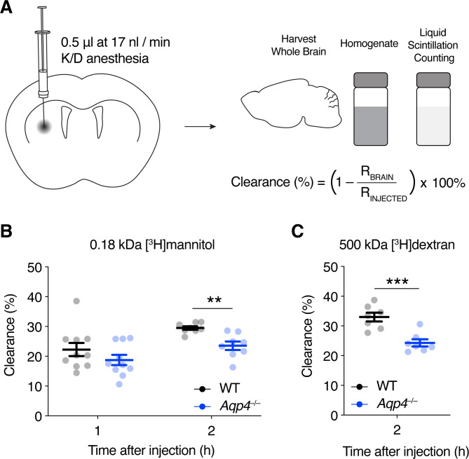
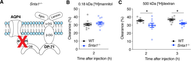
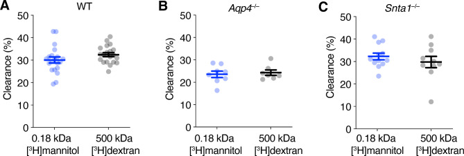
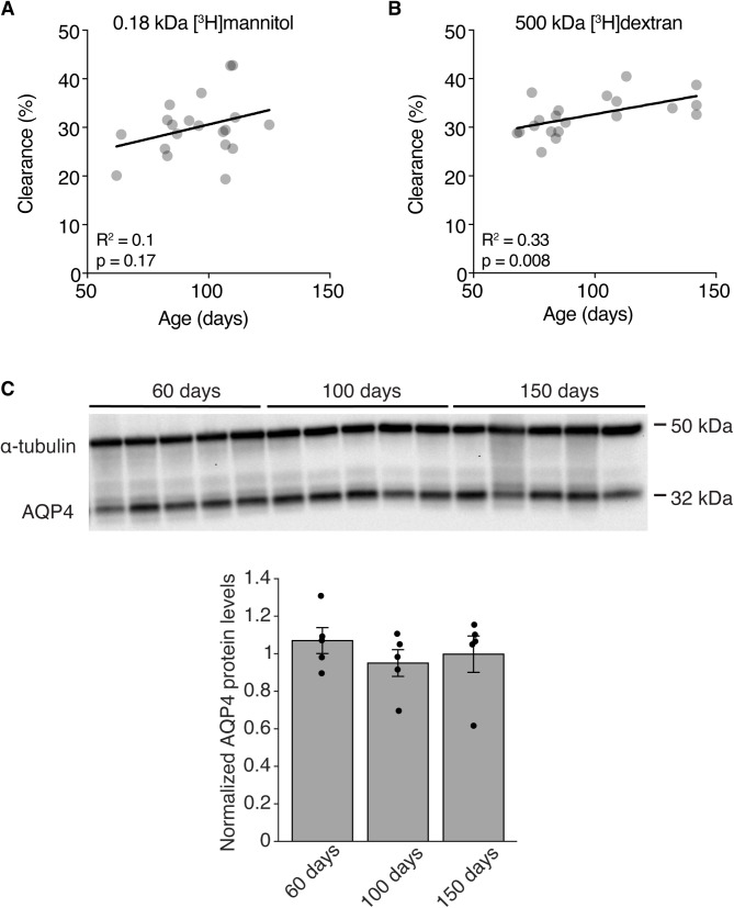
Waste from the brain has been shown to be cleared via the perivascular spaces through the so-called glymphatic system. According to this model the cerebrospinal fluid (CSF) enters the brain in perivascular spaces of arteries, crosses the astrocyte endfoot layer, flows through the parenchyma collecting waste that is subsequently drained along veins. Glymphatic clearance is dependent on astrocytic aquaporin-4 (AQP4) water channels that are highly enriched in the endfeet. Even though the polarized expression of AQP4 in endfeet is thought to be of crucial importance for glymphatic CSF influx, its role in extracellular solute clearance has only been evaluated using non-quantitative fluorescence measurements. Here we have quantitatively evaluated clearance of intrastriatally infused small and large radioactively labeled solutes in mice lacking AQP4 (Aqp4-/-) or lacking the endfoot pool of AQP4 (Snta1-/-). We confirm that Aqp4-/- mice show reduced clearance of both small and large extracellular solutes. Moreover, we find that the Snta1-/- mice have reduced clearance only for the 500 kDa [3H]dextran, but not 0.18 kDa [3H]mannitol suggesting that polarization of AQP4 to the endfeet is primarily important for clearance of large, but not small molecules. Lastly, we observed that clearance of 500 kDa [3H]dextran increased with age in adult mice. Based on our quantitative measurements, we confirm that presence of AQP4 is important for clearance of extracellular solutes, while the perivascular AQP4 localization seems to have a greater impact on clearance of large versus small molecules.

Solute clearance is reduced in mice lacking AQP4 Polarization of AQP4 to the endfeet may have a greater impact on clearance of large versus small molecules Clearance of large but not small solutes is correlated with age within adult age.

Keywords: AQP4; Astrocyte; Dystrophin; Glia; Glymphatic; Syntrophin; Waste clearance.


Conflict of interest statement

The authors declare no competing interests.

Figures

Fig. 1
Fig. 1
Clearance of extracellular solutes is reduced in Aqp4–/– mice. (A) Schematic diagram illustrating the quantification of extracellular solute clearance from the brain parenchyma. Radioactive tracers (0.18 kDa [3H]mannitol and 500 kDa [3H]dextran) infused into the striatum of anesthetized mice. Whole brains were harvested, homogenized and radioactivity counted by liquid scintillation counting. The percentage clearance was calculated based on total injected radioactivity (RINJECTED) and the remaining radioactivity in the brain (RBRAIN). (B) Clearance of intrastriatal 0.18 kDa [3H]mannitol in age-matched Aqp4–/– and WT mice 1 and 2 h after infusion. n = 10 at 1 h; n = 8 at 2 h per genotype. Average ± SD age: for 1 h – Aqp4–/– 105 ± 12 days, WT 83 ± 12 days; for 2 h – for Aqp4–/– 99 ± 14, for WT 91 ± 15. (C) Clearance of intrastriatal 500 kDa [3H]dextran in age-matched Aqp4–/– and WT mice 2 h after infusion. n = 7 per genotype. Average ± SD age: Aqp4–/– 105 ± 27 days, WT 104 ± 27 days. Data represented as mean ± s.e.m, ** = p < 0.01, *** = p < 0.001, mixed effects linear regression model (see Materials and Methods)
Fig. 2
Fig. 2
The role of perivascular AQP4 in the clearance of extracellular solutes. (A) Snta1–/– mice have severely attenuated level of AQP4 in endfeet due to the lack of α-syntrophin, which is an integral part of the anchoring of AQP4. (B) Clearance of intrastriatal [3H]mannitol in age-matched Snta1–/– and WT mice 2 h after infusion. Average ± SD age: Snta1–/– 95 ± 13 days, WT 93 ± 14 days. n = 12 mice per genotype. (C) Clearance of intrastriatal 500 kDa [3H]dextran in age-matched Snta1–/– and WT mice 2 and 3 h after infusion. n = 8 for WT and n = 10 for Snta1–/– at 2 h, n = 9 mice per genotype at 3 h. Average ± SD age: for 2 h – Snta1–/– 120 ± 12 days, WT 124 ± 17 days; for 3 h – for Snta1–/– 113 ± 22, for WT 122 ± 21. Data represented as mean ± s.e.m, * = p < 0.05, ** = p < 0.01, mixed effects linear regression model (see Materials and Methods)
Fig. 3
Fig. 3
Clearance of extracellular solutes is size independent. Clearance of intrastriatal 0.18 kDa [3H]mannitol and 500 kDa [3H]dextran 2 h after infusion in (A) WT mice, average ± SD age: [3H]mannitol 95 ± 13 days, [3H]dextran 93 ± 14 days. (B) Aqp4–/– mice that lack AQP4, average ± SD age: [3H]mannitol 95 ± 13 days, [3H]dextran 93 ± 14 days. and (C) Snta1–/– mice which have have severely attenuated level of AQP4 in endfeet due to the lack of α-syntrophin, average ± SD age: [3H]mannitol 95 ± 13 days, [3H]dextran 93 ± 14 days. Data represented as mean ± s.e.m. WT: n = 20 mice per group; Aqp4–/– mice: n = 8 for [3H]mannitol, n = 7 for [3H]dextran; Snta1–/– mice: n = 12 for [3H]mannitol, n = 10 for [3H]dextran; unpaired two tailed t-test
Fig. 4
Fig. 4
The effect of age on extracellular solute clearance. (A–B) Correlation between age and clearance of 0.18 kDa [3H]mannitol (n = 20) or 500 kDa [3H]dextran (n = 20) in WT mice (All WT controls from Figs. 1 and 2 pooled). (C) Representative immunoblots and quantification expressed in densitometric values of total protein lysates from striatum of 60, 100 and 150 day old WT mice. n = 5 for each group. (A–B) analyzed by linear regression

References

    1. Rasmussen MK, Mestre H, Nedergaard M. Fluid transport in the brain. Physiol Rev. 2022;102:1025–151. doi: 10.1152/physrev.00031.2020. - DOI - PMC - PubMed
    1. Iliff JJ, Wang M, Liao Y, Plogg BA, Peng W, Gundersen GA, et al. A paravascular pathway facilitates CSF flow through the brain parenchyma and the clearance of interstitial solutes, including amyloid β. Sci Transl Med. 2012;4:147ra111. doi: 10.1126/scitranslmed.3003748. - DOI - PMC - PubMed
    1. Cserr HF, Cooper DN, Suri PK, Patlak CS. Efflux of radiolabeled polyethylene glycols and albumin from rat brain. Am J Physiol. 1981;240:F319–28. - PubMed
    1. Cserr HF, Cooper DN, Milhorat TH. Flow of cerebral interstitial fluid as indicated by the removal of extracellular markers from rat caudate nucleus. Exp Eye Res. 1977;25:461–73. doi: 10.1016/S0014-4835(77)80041-9. - DOI - PubMed
    1. Cserr HF, Ostrach LH. Bulk flow of interstitial fluid after intracranial injection of blue dextran 2000. Exp Neurol. 1974;45:50–60. doi: 10.1016/0014-4886(74)90099-5. - DOI - PubMed

LinkOut - more resources