

Improved visualization of median, ulnar nerves, and small branches in the wrist and palm using contrast-enhanced magnetic resonance neurography
- PMID: 38532801
- PMCID: PMC10964438
- DOI: 10.1177/17562864241239739
Improved visualization of median, ulnar nerves, and small branches in the wrist and palm using contrast-enhanced magnetic resonance neurography
Abstract
Background: Magnetic resonance imaging of peripheral nerves in the wrist and palm is challenging due to the small size, tortuous course, complex surrounding tissues, and accompanying blood vessels. The occurrence of carpal palmar lesions leads to edema, swelling, and mass effect, which may further interfere with the display and identification of nerves.
Objective: To evaluate whether contrast-enhanced magnetic resonance neurography (ceMRN) improves the visualization of the morphology and pathology of the median, ulnar nerves, and their small branches in the wrist and palm.
Design: An observational study.
Methods: In total 57 subjects, including 36 volunteers and 21 patients with carpal palmar lesions, were enrolled and underwent ceMRN and non-contrast MRN (ncMRN) examination at 3.0 Tesla. The degree of vascular suppression, nerve visualization, diagnostic confidence, and lesion conspicuity was qualitatively assessed by two radiologists. Kappa statistics were obtained for inter-reader agreement. The signal-to-noise ratio, contrast ratio (CR), and contrast-to-noise ratio (CNR) of the median nerve were measured. The subjective ratings and quantitative measurements were compared between ncMRN and ceMRN.
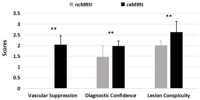
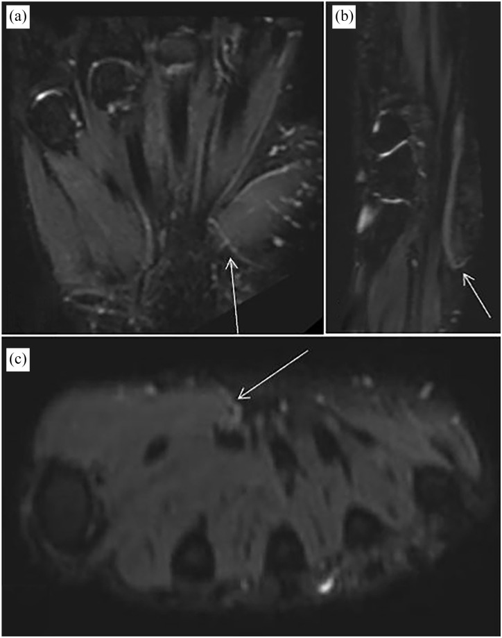
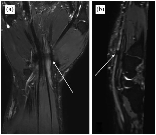
Results: The inter-reader agreement was excellent (k > 0.8) for all qualitative assessments and visualization assessment of each nerve segment. Compared with ncMRN, ceMRN significantly improved vascular suppression in volunteers and patients (both p < 0.001). The ceMRN significantly enhanced nerve visualization of each segment (all p < 0.05) and diagnostic confidence in volunteers and patients (both p < 0.05). The ceMRN improved lesion conspicuity (p = 0.003) in patients. Quantitatively, ceMRN had significantly higher CRs of nerve versus subcutaneous fat, bone marrow, and vessels and CNR of nerve versus vessel than ncMRN (all p < 0.05).
Conclusion: The ceMRN significantly improves the visualization of peripheral nerves and pathology in the wrist and palm by robustly suppressing the signals of fat, bone marrow, and especially vessels in volunteers and patients.
Keywords: common palmar digital nerve; magnetic resonance imaging; magnetic resonance neurography; median nerve; palm; proper digital nerve; thenar muscular branch; ulnar nerve; wrist.
Plain language summary
Study on the improvement of magnetic resonance imaging and lesion display of small nerves in the wrist and palm using contrast agents Why was the study done? Because the nerves and branches in the wrist and palm are numerous, small, tortuous, and surrounded by muscles, fat, bones, blood vessels and other tissues, it is difficult to show their complete shape with conventional magnetic resonance imaging. Hand lesions often lead to swelling, edema and masses, which interfere with the display of nerves. Therefore, it is difficult to directly diagnose the relationship between the lesions and nerves in clinical practice. What did the researchers do? The research team used contrast agent plus three-dimensional high-resolution magnetic resonance sequence to display the nerves of volunteers and patients with hand lesions, and used subjective and objective evaluation methods to compare the display effect of the sequence on the nerves before and after the use of contrast agent. What did the researchers find? The imaging method of contrast agent plus three-dimensional high-resolution magnetic resonance sequence can reduce the interference of fat, blood vessels, etc. on nerve display, improve the display effect of each nerve segment of the wrist and palm, increase readers’ confidence in identifying nerves, and improve the detection of lesions. What do the findings mean? This study verified the feasibility and advantages of using contrast agents for magnetic resonance imaging of nerves in the wrist and palm. It provides a new method for clinical and imaging diagnosis of hand lesions, which can simultaneously display the morphological characteristics of nerves and lesions, reducing the difficulty of clinical diagnosis and improving the efficiency of imaging diagnosis.
© The Author(s), 2024.
Conflict of interest statement
The authors declare that there is no conflict of interest.
Figures


References
-
- Chhabra A, Madhuranthakam AJ, Andreisek G. Magnetic resonance neurography: current perspectives and literature review. Eur Radiol 2018; 28: 698–707. - PubMed
-
- Filler AG, Howe FA, Hayes CE, et al. Magnetic resonance neurography. Lancet 1993; 341: 659–661. - PubMed
-
- Sneag DB, Daniels SP, Geannette C, et al. Post-contrast 3D inversion recovery magnetic resonance neurography for evaluation of branch nerves of the brachial plexus. Eur J Radiol 2020; 132: 109304. - PubMed
LinkOut - more resources
Full Text Sources
