

Uniportal video-assisted thoracoscopic surgery in the prone position for esophageal bronchogenic cyst
- PMID: 38532859
- PMCID: PMC10963949
- DOI: 10.1093/jscr/rjae186
Uniportal video-assisted thoracoscopic surgery in the prone position for esophageal bronchogenic cyst
Abstract
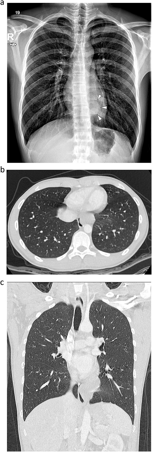
Esophageal bronchogenic cysts are very rare. A bronchogenic cyst is a congenital malformation resulting from abnormal sprouting of primitive bronchi because of a foregut bronchopulmonary malformation. An 18-year-old patient with a cystic tumor in the left posterior mediastinum was identified. The mediastinal tumor was removed by uniportal video-assisted thoracoscopic surgery. The operation was performed in the prone position through a single 4-cm incision on the lateral scapular line in the left ninth intercostal space. After tumor resection, the dissected esophageal muscle and mediastinal pleura were sutured with two continuous barbed sutures. The operation took 80 min. A biopsy confirmed the diagnosis of esophageal bronchial cyst. Diet was started on the evening of the operation. The chest tube was removed on the 1st postoperative day, and the patient was discharged without any problems on the 2nd postoperative day.
Keywords: esophageal bronchogenic cyst; prone position; uniport; video-assisted thoracic surgery.
Published by Oxford University Press and JSCR Publishing Ltd. © The Author(s) 2024.
Conflict of interest statement
None declared.
Figures


Similar articles
-
A Case of Intramural Esophageal Bronchogenic Cyst.Asian J Endosc Surg. 2025 Jan-Dec;18(1):e70055. doi: 10.1111/ases.70055. Asian J Endosc Surg. 2025. PMID: 40169167
-
[Application of artificial pneumothorax in semi-prone position to the video-assisted thoracic surgery of esophageal carcinoma].Zhonghua Zhong Liu Za Zhi. 2012 Oct;34(10):785-9. doi: 10.3760/cma.j.issn.0253-3766.2012.10.014. Zhonghua Zhong Liu Za Zhi. 2012. PMID: 23291075 Chinese.
-
Bronchogenic cyst removal via thoracoscopic surgery in the prone position: A case report and literature review.Int J Surg Case Rep. 2019;60:204-208. doi: 10.1016/j.ijscr.2019.05.064. Epub 2019 Jun 8. Int J Surg Case Rep. 2019. PMID: 31233965 Free PMC article.
-
[Esophageal bronchogenic cyst: an uncommon cause of dysphagia in adults. Case report and literature review].Cir Cir. 2018;86(2):187-190. doi: 10.24875/CIRU.M18000018. Cir Cir. 2018. PMID: 29809178 Review. Spanish.
-
[A case of mediastinal esophago-bronchogenic cyst associated with high serum level of CA 19-9].Kyobu Geka. 2001 Aug;54(9):805-8. Kyobu Geka. 2001. PMID: 11517557 Review. Japanese.
References
-
- Turkyilaz A, Eroglu A, Subasi M, Findik G. Intramural esophageal bronchogenic cyst: a review of the literature. Di Esophagus 2007;20:461–5. - PubMed
-
- Chuang KH, Huang TW, Cheng YL, et al. . Esopahgeal bronchogenic cyst: a rare entity. Z Gastroenterol 2007;45:958–60. - PubMed
-
- Jung HS, Kim DK, Lee GD, et al. . Video-assisted thoracic surgery for bronchogenic cysts: is this the surgical approach of choice? Interact Cardiovasc Thoracic Surg 2014;19:824–9. - PubMed
-
- Fievert L, D’Journo XB, Guys JM, et al. . Bronchogenic cyst: best time for surgery? Ann Thorac Surg 2012;94:1695–700. - PubMed
Publication types
LinkOut - more resources
Full Text Sources
