Case Reports
. 2024 Mar 22;19(6):2347-2350.
doi: 10.1016/j.radcr.2024.02.107. eCollection 2024 Jun.

Aluminosis pneumoconiosis presenting as hyperdense lung nodules

Affiliations
Case Reports

Aluminosis pneumoconiosis presenting as hyperdense lung nodules

Sara E Mantz et al. Radiol Case Rep. .

Abstract

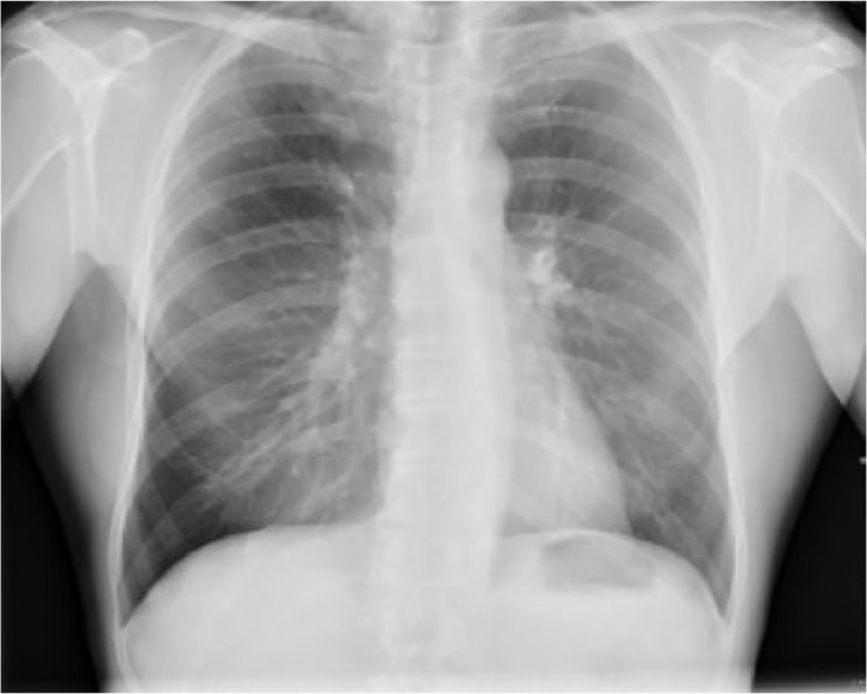
We present the case of a 66-year-old man who presented with new incidentally found hyperdense pulmonary nodules. Further workup with a PET/CT revealed that the nodules were FDG-avid and that there was associated hypermetabolic lymphadenopathy. Due to his history of aluminum toxicity from welding, aluminosis pneumoconiosis was suspected. Biopsy of one of the nodules was done which reinforced this diagnosis. Aluminosis pneumoconiosis is a rare occupational lung disease mostly associated with industrial workers with prolonged unprotected exposure to fine aluminum dust. Prognosis depends on the duration and intensity of exposure, and there is no definitive treatment other than eliminating further exposure.

Keywords: Aluminum; Hyperdense nodules; Interstitial lung disease; Occupational lung disease; Pneumoconiosis; Pulmonary nodules.


Figures

Fig 1
Fig. 1
Chest X-ray showing two small nodular densities in the right lower lung zone and one in the left lower lung zone, circled in red.
Fig 2
Fig. 2
Chest X-ray from 5 years prior.
Fig 3
Fig. 3
Chest CT with contrast, lung window, showing hyperdense nodular opacities in left lower lobe circled in red.
Fig 4
Fig. 4
Chest CT with contrast, soft tissue window, showing hyperdense nodular opacities in left lower lobe circled in red.
Fig 5
Fig. 5
Chest CT with contrast, lung window, showing hyperdense nodular opacity in right lower lobe circled in red.
Fig 6
Fig. 6
Chest CT with contrast, soft tissue window, showing hyperdense nodular opacity in right lower lobe circled in red.
Fig 7
Fig. 7
Chest CT with contrast, soft tissue window showing Hounsfield units of a blood-filled vessel, a nodule in the left lower lobe, and a lymph node. The nodule is shown to have an average reading of 67.71 Hounsfield units (HU) and the lymph node 118.85 HU, making them both hyperdense in comparison to the 50.54 HU measured in the blood.
Fig 8
Fig. 8
PET/CT showing FDG-avid nodule in right lower lobe, circled in red. Nodule SUV max 7.0; background mediastinal blood pool SUV max 2.1.
Fig 9
Fig. 9
PET/CT showing bilateral lymphadenopathy with hypermetabolic activity.

References

    1. Kraus T., Schaller K.H., Angerer J., Hilgers R.-D., Letzel S. Aluminosis – detection of an almost forgotten disease with HRCT. J Occupation Med Toxicol. 2006;1(1) doi: 10.1186/1745-6673-1-4. - DOI - PMC - PubMed
    1. Smolkova P., Nakladalova M. The etiology of occupational pulmonary aluminosis - the past and the present. Biomedic Papers. 2014;158(4):535–538. doi: 10.5507/bp.2014.017. - DOI - PubMed
    1. Niknejad M., Feger J., Hacking C., et al. Non-calcified hyperdense pulmonary nodules. Reference article, Radiopaedia.org (Accessed on 11 Oct 2023) 10.53347/rID-22013 - DOI
    1. Ceylan N., Bayraktaroglu S., Savas R., Alper H. CT findings of high-attenuation pulmonary abnormalities. Insights Imaging. 2010;1(4):287–292. doi: 10.1007/s13244-010-0039-2. - DOI - PMC - PubMed
    1. Marchiori E., Franquet T., Gasparetto T., Goncalves L., Escuissato D. Consolidation with diffuse or focal high attenuation: computed tomography findings. J Thorac Imaging. 2008;23(4):298–304. doi: 10.1097/RTI.0b013e3181788d39. - DOI - PubMed

Publication types

LinkOut - more resources