. 2024 Oct 3;147(10):3596-3610.
doi: 10.1093/brain/awae098.

Brain morphometry in former American football players: findings from the DIAGNOSE CTE research project

Affiliations

Brain morphometry in former American football players: findings from the DIAGNOSE CTE research project

Hector Arciniega et al. Brain. .

Abstract

Exposure to repetitive head impacts in contact sports is associated with neurodegenerative disorders including chronic traumatic encephalopathy (CTE), which currently can be diagnosed only at post-mortem. American football players are at higher risk of developing CTE given their exposure to repetitive head impacts. One promising approach for diagnosing CTE in vivo is to explore known neuropathological abnormalities at post-mortem in living individuals using structural MRI. MRI brain morphometry was evaluated in 170 male former American football players ages 45-74 years (n = 114 professional; n = 56 college) and 54 same-age unexposed asymptomatic male controls (n = 54, age range 45-74). Cortical thickness and volume of regions of interest were selected based on established CTE pathology findings and were assessed using FreeSurfer. Group differences and interactions with age and exposure factors were evaluated using a generalized least squares model. A separate logistic regression and independent multinomial model were performed to predict each traumatic encephalopathy syndrome (TES) diagnosis, core clinical features and provisional level of certainty for CTE pathology using brain regions of interest. Former college and professional American football players (combined) showed significant cortical thickness and/or volume reductions compared to unexposed asymptomatic controls in the hippocampus, amygdala, entorhinal cortex, parahippocampal gyrus, insula, temporal pole and superior frontal gyrus. Post hoc analyses identified group-level differences between former professional players and unexposed asymptomatic controls in the hippocampus, amygdala, entorhinal cortex, parahippocampal gyrus, insula and superior frontal gyrus. Former college players showed significant volume reductions in the hippocampus, amygdala and superior frontal gyrus compared to the unexposed asymptomatic controls. We did not observe Age × Group interactions for brain morphometric measures. Interactions between morphometry and exposure measures were limited to a single significant positive association between the age of first exposure to organized tackle football and right insular volume. We found no significant relationship between brain morphometric measures and the TES diagnosis core clinical features and provisional level of certainty for CTE pathology outcomes. These findings suggested that MRI morphometrics detect abnormalities in individuals with a history of repetitive head impact exposure that resemble the anatomic distribution of pathological findings from post-mortem CTE studies. The lack of findings associating MRI measures with exposure metrics (except for one significant relationship) or TES diagnosis and core clinical features suggested that brain morphometry must be complemented by other types of measures to characterize individuals with repetitive head impacts.

Keywords: former American football players; neuroimaging; repetitive head impact; sports-related head injury; structural MRI.


Conflict of interest statement

L.B. is the Editor-in-Chief of the Journal of Neuro-Ophthalmology. C.B. receives research support from the Ultimate Fighting Championship Top Rank promotions and Haymon Boxing. A.P.L. has an ownership interest as a co-founder of BrainSpec and consults for Agios Pharmaceuticals, Biomarin Pharmaceuticals, DesignTx and Moncon MRI. J.L.C. has provided consultation to Acadia, Actinogen, Acumen, AlphaCognition, Aprinoia, AriBio, Artery, Biogen, BioVie, Bristol-Myers Squib, Cassava, Cerecin, Diadem, EIP Pharma, Eisai, GemVax, Genentech, GAP Innovations, Janssen, Jocasta, Karuna, Lighthouse, Lilly, Lundbeck, LSP/EQT, Merck, NervGen, Novo Nordisk, Oligomerix, Optoceutics, Ono, Otsuka, PRODEO, Prothena, ReMYND, Roche, Sage Therapeutics, Signant Health, Simcere, Suven, SynapseBio, TrueBinding, Vaxxinity and Wren pharmaceutical assessment and investment companies. He owns the copyright of the Neuropsychiatric Inventory. E.M.R. is a compensated scientific advisor for Alkahest, Alzheon, Aural Analytics, Denali, Green Valley, Retromer Therapeutics and Vaxxinity and a co-founder of ALZPath. R.A.S. is a member of the Board of Directors of King-Devick Technologies Inc. (Chicago, IL, USA) and receives royalties for published neuropsychological tests from Psychological Assessment Resources Inc. (Lutz, FL, USA).

Figures

Figure 1
Figure 1
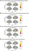
Cortical thickness group-level differences. (A) Cortical thickness group differences between former American football players (n = 170) and unexposed asymptomatic controls (n = 54). Results indicate reduced cortical thickness in the left hemisphere: entorhinal cortex and parahippocampal gyrus and right hemisphere: parahippocampal gyrus in our former American football players compared to unexposed asymptomatic controls. (B) Subgroup analysis of former professional players (n = 114) and unexposed asymptomatic controls (n = 54). Results indicate reduced cortical thickness in the left hemisphere: entorhinal cortex and parahippocampal gyrus and right hemisphere: parahippocampal gyrus in our former professional football players compared to unexposed asymptomatic controls. (C) Group differences between former college players (n = 56) and unexposed asymptomatic controls (n = 54). No group-level differences were observed. (D) Group differences between former professional players (n = 114) and former college players (n = 56). No group differences were observed. All reported P-values are corrected for multiple comparisons. n.i. = not included; n.s. = not significant.
Figure 2
Figure 2
Volumetric group-level differences. (A) Volumetric group differences between former American football players (n = 170) and unexposed asymptomatic controls (n = 58). Results indicate reduced volume in the left hemisphere: amygdala, hippocampus, entorhinal cortex, parahippocampal gyrus, insula and superior frontal gyrus; and right hemisphere: amygdala, hippocampus, temporal pole, entorhinal cortex, parahippocampal gyrus, insula and superior frontal gyrus in our former American football players compared to unexposed asymptomatic controls. (B) Group differences between former professional players (n = 114) and unexposed asymptomatic controls (n = 54). Results indicate reduced volume in the left hemisphere: amygdala, hippocampus, entorhinal cortex, parahippocampal gyrus, insula and superior frontal gyrus; and right hemisphere: amygdala, hippocampus, entorhinal cortex, insula and temporal pole in our former professional players compared to unexposed asymptomatic controls. (C) Group differences between former college players (n = 56) and unexposed asymptomatic controls (n = 54). Results indicate reduced volume in the left hemisphere: amygdala, hippocampus and superior frontal gyrus; and right hemisphere: hippocampus in our former professional players compared to unexposed asymptomatic controls. (D) Group differences between former professional players (n = 114) and former college players (n = 54). No group differences were observed. All reported P-values are corrected for multiple comparisons. n.i. = not included; n.s. = not significant.
Figure 3
Figure 3
Raw volumetric data for preselected chronic traumatic encephalopathy (CTE)-related regions in former American football players and unexposed asymptomatic controls. Violin plots showing the preselected CTE-related regions and individual raw data-points. Significant group-level differences are indicative of our volumetric main group-level result of former American football players and unexposed asymptomatic controls. Significant differences are observed in the superior frontal gyrus, temporal pole, insula, entorhinal cortex, parahippocampal gyrus, amygdala and hippocampus. Outer horizontal lines indicate the interquartile range and the middle line indicates the median. **P ≤ 0.01, *P < 0.05. P-values are corrected for multiple comparisons.

References

    1. McKee AC, Cairns NJ, Dickson DW, et al. The first NINDS/NIBIB consensus meeting to define neuropathological criteria for the diagnosis of chronic traumatic encephalopathy. Acta Neuropathol. 2016;131:75–86. - PMC - PubMed
    1. McKee AC, Stern RA, Nowinski CJ, et al. The spectrum of disease in chronic traumatic encephalopathy. Brain. 2013;136(Pt 1):43–64. - PMC - PubMed
    1. McKee AC. The neuropathology of chronic traumatic encephalopathy: The status of the literature. Semin Neurol. 2020;40:359–369. - PubMed
    1. McKee AC, Abdolmohammadi B, Stein TD. The neuropathology of chronic traumatic encephalopathy. Handb Clin Neurol. 2018;158:297–307. - PubMed
    1. McKee AC, Stein TD, Kiernan PT, Alvarez VE. The neuropathology of chronic traumatic encephalopathy. Brain Pathol. 2015;25:350–364. - PMC - PubMed