. 2024 May 2;12(5):e0353423.
doi: 10.1128/spectrum.03534-23. Epub 2024 Mar 27.

Targeting intracellular nontuberculous mycobacteria and M. tuberculosis with a bactericidal enzymatic cocktail

Affiliations

Targeting intracellular nontuberculous mycobacteria and M. tuberculosis with a bactericidal enzymatic cocktail

Helen P Bartlett et al. Microbiol Spectr. .

Abstract

To address intracellular mycobacterial infections, we developed a cocktail of four enzymes that catalytically attack three layers of the mycobacterial envelope. This cocktail is delivered to macrophages, through a targeted liposome presented here as ENTX_001. Endolytix Cocktail 1 (EC1) leverages mycobacteriophage lysin enzymes LysA and LysB, while also including α-amylase and isoamylase for degradation of the mycobacterial envelope from outside of the cell. The LysA family of proteins from mycobacteriophages has been shown to cleave the peptidoglycan layer, whereas LysB is an esterase that hydrolyzes the linkage between arabinogalactan and mycolic acids of the mycomembrane. The challenge of gaining access to the substrates of LysA and LysB provided exogenously was addressed by adding amylase enzymes that degrade the extracellular capsule shown to be present in Mycobacterium tuberculosis. This enzybiotic approach avoids antimicrobial resistance, specific receptor-mediated binding, and intracellular DNA surveillance pathways that limit many bacteriophage applications. We show this cocktail of enzymes is bactericidal in vitro against both rapid- and slow-growing nontuberculous mycobacteria (NTM) as well as M. tuberculosis strains. The EC1 cocktail shows superior killing activity when compared to previously characterized LysB alone. EC1 is also powerfully synergistic with standard-of-care antibiotics. In addition to in vitro killing of NTM, ENTX_001 demonstrates the rescue of infected macrophages from necrotic death by Mycobacteroides abscessus and Mycobacterium avium. Here, we demonstrate shredding of mycobacterial cells by EC1 into cellular debris as a mechanism of bactericide.IMPORTANCEThe world needs entirely new forms of antibiotics as resistance to chemical antibiotics is a critical problem facing society. We addressed this need by developing a targeted enzyme therapy for a broad range of species and strains within mycobacteria and highly related genera including nontuberculous mycobacteria such as Mycobacteroides abscessus, Mycobacterium avium, Mycobacterium intracellulare, as well as Mycobacterium tuberculosis. One advantage of this approach is the ability to drive our lytic enzymes through encapsulation into macrophage-targeted liposomes resulting in attack of mycobacteria in the cells that harbor them where they hide from the adaptive immune system and grow. Furthermore, this approach shreds mycobacteria independent of cell physiology as the drug targets the mycobacterial envelope while sidestepping the host range limitations observed with phage therapy and resistance to chemical antibiotics.

Keywords: AMR; LysA; LysB; NTM; arabinogalactan; capsule; enzybiotic; liposome; macrophage; mycobacteriophage; mycomembrane; peptidoglycan; phagolysosome.


Conflict of interest statement

All Endolytix employees may hold stock and/or stock options.

Figures

Fig 1
Fig 1
CFU enumeration comparing viability of LysB and EC1 enzybiotics on M. abscessus. (a) LysB from mycobacteriophage D29 is titrated in enzymatic reactions with M. abscessus for enumeration of CFU. Micromolarity of LysB and microgram per milliliter total protein are both shown on the x-axis. (b) Same as panel a for the EC1 enzymatic cocktail. (c) Time-kill kinetics (TKK) were measured via CFU for D29 LysB acting on M. abscessus over 16 h. (d) Same as panel c, instead with the EC1 of enzymes.
Fig 2
Fig 2
Measurement of the impact of EC1 on growth of M. abscessus across a 10-fold dilution series (x-axis) while titrating for pH (y-axis). M. abscessus growth is measured by absorbance with a redox-active dye that absorbs at 590 nm. All conditions are seeded with 1E5 M. abscessus cells. Killing reactions are run for 24 h and then serially diluted into media that does not contain drug. The gray vertical lines in the growth curves represent days of growth (total 121 h).
Fig 3
Fig 3
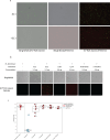
(a) Comparison of EC1-treated M. abscessus with dual overlay images of Brightfield/SYTOX Green, Brightfield/FM4-64, and SYTOX Green/FM4-64. Top row is untreated and bottom row is treated with 32 µg/mL of EC1. (b) Brightfield and fluorescence microscopy images of in vitro EC1 reactions with M. abscessus. The top panel contains brightfield images, whereas the bottom panel contains a merged image of the fluorescence of SYTOX Green that binds DNA and FM4-64 that binds to the outer leaflet of the plasma membrane. The scale bar is 10 µm. (c) Growth data after 5 days using redox-sensitive dye comparing EC1 with the individual components LysB, LysA, isoamylase, and α-amylase. All reactions contained 0.8 µg of each enzyme component.
Fig 4
Fig 4
(Top row) Gates were established by the size and features of single cells then the particles resulting from enzymatic digestion by EC1 (right column) as seen in forward- (FSC) and side-scattered (SSC) light, respectively, via flow cytometry. We detected gates that could separate M. abscessus populations of single cells, cellular debris, and aggregates after treatment with EC1 in vitro. Cells (row 2), cell debris (row 3), and aggregates (row 4) were stained with SYTOX Green DNA Stain and lipophilic FM4-64 stain. The x- and y-axes represent increasing FM4-64 and SYTOX fluorescence intensity, respectively, in logarithmic scale and range incrementally from 103 to 106.
Fig 5
Fig 5
Evaluation of EC1 effects on off-target species was performed with viability dyes SYTO9 and propidium iodide (Molecular Probes) to evaluate untreated strains from that of heat-killed and EC1-treated cells.
Fig 6
Fig 6
(a) Schematic diagram of ENTX_001 PPL. Lipids are light blue, small molecule excipients are yellow, the enzymes are PlyC in red (LysA representative), D29 LysB in black, B. licheniformis α-amylase in gray, and C. glabrata isoamylase in dark blue. (b) J774a.1 mouse macrophages or macrophages infected with M. abscessus str. 19977. IIM were treated with EC1 or ENTX_001 in a titration of protein concentrations indicated on the figure for 18 h and evaluated for necrosis by microscopy. After treatment, macrophages were stained with Hoechst (middle row, blue, total nuclei) and SYTOX Green (bottom row, green, necrotic cells) to measure necrosis. The scale bar is 50 µm.
Fig 7
Fig 7
(a) Human macrophages (THP-1 cell line) were infected with M. avium str 2285R. IIM were treated with EC1 or ENTX_001 in a titration of protein concentrations for 24 h and evaluated for necrosis by flow cytometry resulting in three classifications ranging from no macrophage protection to incomplete and strong protection. The 95% confidence intervals are indicated with dotted lines for each class. (b) The figure is identical to panel a with the drug treatment time of 48 h.

References

    1. Adjemian J, Olivier KN, Seitz AE, Holland SM, Prevots DR. 2012. Prevalence of nontuberculous mycobacterial lung disease in U.S. medicare beneficiaries. Am J Respir Crit Care Med 185:881–886. doi:10.1164/rccm.201111-2016OC - DOI - PMC - PubMed
    1. Strollo SE, Adjemian J, Adjemian MK, Prevots DR. 2015. The burden of pulmonary nontuberculous mycobacterial disease in the United States. Ann Am Thorac Soc 12:1458–1464. doi:10.1513/AnnalsATS.201503-173OC - DOI - PMC - PubMed
    1. Kalscheuer R, Palacios A, Anso I, Cifuente J, Anguita J, Jacobs WR Jr, Guerin ME, Prados-Rosales R. 2019. The Mycobacterium tuberculosis capsule: a cell structure with key implications in pathogenesis. Biochem J 476:1995–2016. doi:10.1042/BCJ20190324 - DOI - PMC - PubMed
    1. Mikušová K, Yagi T, Stern R, McNeil MR, Besra GS, Crick DC, Brennan PJ. 2000. Biosynthesis of the galactan component of the mycobacterial cell wall. J Biol Chem 275:33890–33897. doi:10.1074/jbc.M006875200 - DOI - PubMed
    1. Prasla Z, Sutliff RL, Sadikot RT. 2020. Macrophage signaling pathways in pulmonary nontuberculous mycobacteria infections. Am J Respir Cell Mol Biol 63:144–151. doi:10.1165/rcmb.2019-0241TR - DOI - PMC - PubMed

Publication types

MeSH terms

LinkOut - more resources