Review
. 2024 Mar 18;13(1):25.
doi: 10.3390/antib13010025.

vNARs as Neutralizing Intracellular Therapeutic Agents: Glioblastoma as a Target

Affiliations
Review

vNARs as Neutralizing Intracellular Therapeutic Agents: Glioblastoma as a Target

Alejandro Manzanares-Guzmán et al. Antibodies (Basel). .

Abstract

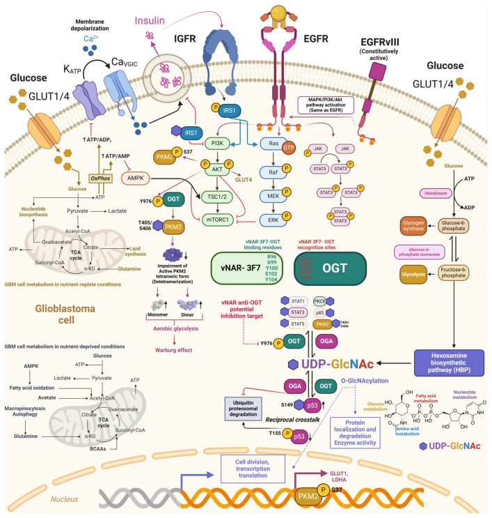
Glioblastoma is the most prevalent and fatal form of primary brain tumors. New targeted therapeutic strategies for this type of tumor are imperative given the dire prognosis for glioblastoma patients and the poor results of current multimodal therapy. Previously reported drawbacks of antibody-based therapeutics include the inability to translocate across the blood-brain barrier and reach intracellular targets due to their molecular weight. These disadvantages translate into poor target neutralization and cancer maintenance. Unlike conventional antibodies, vNARs can permeate tissues and recognize conformational or cryptic epitopes due to their stability, CDR3 amino acid sequence, and smaller molecular weight. Thus, vNARs represent a potential antibody format to use as intrabodies or soluble immunocarriers. This review comprehensively summarizes key intracellular pathways in glioblastoma cells that induce proliferation, progression, and cancer survival to determine a new potential targeted glioblastoma therapy based on previously reported vNARs. The results seek to support the next application of vNARs as single-domain antibody drug-conjugated therapies, which could overcome the disadvantages of conventional monoclonal antibodies and provide an innovative approach for glioblastoma treatment.

Keywords: cancer immunotherapy; glioblastoma; intrabodies; molecular targeted therapy; receptor tyrosine kinase; variable new antigen receptors (vNARs).


Conflict of interest statement

The authors declare no conflicts of interest.

Figures

Figure 1
Figure 1
Structural domain comparison of conventional IgG, camelid heavy chain-only Igs, and shark IgNAR. IgG antibody structure comprises two light chains (IgL) and two heavy chains (IgH). The N-terminus holds variable domains within their corresponding heavy and light chains (VH and VL). The C-terminus encompasses constant domains (CH and CL). The fragment antigen-binding region (Fab region) contains a variable domain (V-domain) and a constant domain (C-domain) corresponding to two heavy chains and constant domains (CH2 and CH3) and an antigen-binding variable single domain (VHH). IgNAR forms a homodimeric structure comprised of two layers of five constant domains (C1 to C5) sandwiched as β-sheets joined through a hidden disulfide bond. The antigen-binding variable single domain (vNAR) at the N-terminus. The 3D structures of vNAR and VHH were adapted from [48,49], Copyright © 2021 Fernández-Quintero, Seidler, Quoika and Liedl, Copyright © 2017 Gonzalez-Sapienza, Rossotti and Tabares-da Rosa.
Figure 6
Figure 6
Potential downstream regulation of vNAR-3F7 intrabody through binding to O-GlcNac transferase. O-GlcNAcylation crosstalks with other PTMs, with phosphorylation in serine/threonine residue modifications. Phosphorylation of the tyrosine residue in insulin receptor substrate 1 (IRS1) prompts its association with PI3K and promotes Akt phosphorylation and activation. IRS1 is consequently O-GlcNAcylated to insulin stimulus, hampering IRS1 interaction with PI3K and further decreasing insulin signaling. Deregulated EGF signaling and metabolic shift aerobic glycolysis (Warburg effect) through T405/S406 O-GlcNAcylation of pyruvate kinase M2 (PKM2) mediated by O-GlcNAc transferase (OGT) phosphorylation at the Y976 site further impairs the active PKM2 tetrameric form, increasing PKM2 monomeric and dimeric forms. OGT phosphorylation (Y976) also prompts the O-GlcNAcylation of downstream targets. Anti-OGT vNAR-3F7 recognizes (green) and binds to OGT (red) through specific amino acid residues [160] Copyright © 2023 Xi, Xiao, An, Liu, Liu, Hao, Wang, Song, Yu and Gu. This approach may pave the way for developing anti-OGT vNAR intrabodies that impede intracellular OGT phosphorylation (Y976) and could hamper their corresponding downstream effectors, including PKM2. Dark pink arrows depict EGFR/EGFvIII and downstream targets, whereas blue arrows depict the same for EGFR.
Figure 2
Figure 2
EGFR wild type and EGFRvIII mutated comparison. (a) The epidermal growth factor receptor (EGFR) comprises three domains: an extracellular domain, a transmembrane domain (hydrophobic), and an intracellular domain (distinctive of EGFRs from the TKI family and highly conserved). The extracellular domain comprises four smaller domains (DI-DIV). DI and DII are essential to ligand binding. EGFRvIII was obtained because the 801 bp in-frame deletion of 2–7 exons of the EGFR gene. (b) The extracellular domain interacts with numerous ligands, including epidermal growth factor (EGF) protein, EGFR-like growth factors, epiregulin (EPR), transforming growth factor-alpha (TGF-α), and betacellulin (BTC). EGFR is activated by ligand binding followed by dimerization, which provokes a conformational shift that further supports EGFR intracellular triggering by specific phosphorylated tyrosine residues (Y845, Y992, Y1068, Y1086, and Y1173) at the carboxyl-terminal domain, followed by activation of a complex program of downstream intracellular signals within the cytoplasm and nucleus, including RAS-MAPK, phosphatidylinositol-3-kinase (PI3K)/Akt and signal transducer and activator of transcription 3 (STAT3) pathways. These downstream signaling cascades prompt cell proliferation, loss of differentiation, invasion, angiogenesis, and inhibition of apoptosis. EGFRVIII maintains intact transmembrane and intracellular kinase domains, granting EGFRvIII independence of ligand binding to support further growth signaling in GBM cells and malignancy.
Figure 3
Figure 3
Therapeutic tyrosine kinase inhibitors and anti-EGFR monoclonal antibodies in glioblastoma anti-EGFR mAbs bind the EGFR extracellular domain, preventing EGFR dimerization and subsequent activation. Tyrosine kinase inhibitors (TKIs) target the intracellular TK domain, blocking subsequent receptor tyrosine kinase pathways (RTKs).
Figure 4
Figure 4
IL13Rα1/IL4Rα JAK-STAT6 pathway, IL13Rα2, and single-domain vNAR coupled to IL13Rα2. In normal cells, IL-13 binds with low affinity to IL13Rα1, and then IL13Rα1 forms the IL13Rα1/IL4Rα heterodimer with IL4Rα. IL13Rα1/IL-4Rα binding to the IL-13 cytokine stimulates STAT-6 intracellular signaling, resulting in receptor-mediated endocytosis (RME) and STAT6 translocation to the nucleus. IL-13Rα2 is a monomeric, high-affinity interleukin-13 (IL-13) receptor overexpressed in ~78% of GBM. IL-13Rα2 is absent or expressed at minimal levels in normal brain tissues. Anti-IL13Rα2 vNAR binds to IL13Rα2 on the GBM cell surface, then IL13Rα2 undergoes receptor-mediated endocytosis and hampers IL-13 binding to IL13Rα2. Consequently, IL-13 can normally bind to IL13Rα1/IL4Rα, further initiating the JAK-STAT6 pathway; this leads to the restoration of this signaling pathway that inhibits the abnormal unlimited proliferation of GBM cells and finally culminates in the apoptosis of these cells. Green dashed and green solid lines depict indirect and direct effects of IL13Rα2 impairment by targeted binding of vNAR, respectively. Red lines and the red cross depict IL13Rα2 inhibition of the JAK-STAT6 signaling pathway through IL-13 binding.
Figure 5
Figure 5
Potential allosteric inhibition of Aurora-A kinase by the vNAR-D01 intrabody. Aurora-A is a Ser/Thr protein kinase primarily involved in cell division and has been proven relevant for proliferation in glioblastoma. Moreover, Aurora-A induces tumorigenesis via the decontrolled regulation of RAD51 [128]. BCRA1, catenin, p73, MDM2, NFκB, cMyc, ERK, AKT, and JAK/STAT pathways [130], followed by downstream upregulation of several targets that prompt GBM cell proliferation, survival, inflammation, and angiogenesis. AURKA autophosphorylation is inefficient and relies on TPX2 binding. vNAR-D01 binding superposes to recognition sites of TPX2, further impeding AURKA kinase activity via an allosteric mechanism. Because the vNAR-D01 binding superposes to recognition sites of TPX2, the former impedes AURKA kinase activity via an allosteric mechanism. Green solid lines depict direct regulation, and green dashed lines depict indirect regulation. Red lines depict inhibition as well as red crosses. The vNAR-D01 3D structure was adapted for [122].
Figure 7
Figure 7
TGF-β canonical intracellular pathways and single-domain vNAR T1 coupled to TGF-β1. In the canonical pathway, TGF-β1 employs two serine/threonine kinase receptors to trigger messenger proteins (SMADs), followed by the activation of TGF-β target genes, which is essential to the onset and progression of GBM. As depicted in the example, TGF-β1 (green) is neutralized by recognition of vNAR T1 (blue) from [172], further hampering TGF-β1 binding to TβI/TβRII receptors. The amino acid sequence of vNAR T1 that recognizes the human TGF-β1 cytokine is depicted in red.

References

    1. Rong L., Li N., Zhang Z. Emerging therapies for glioblastoma: Current state and future directions. J. Exp. Clin. Cancer Res. 2022;41:142. doi: 10.1186/s13046-022-02349-7. - DOI - PMC - PubMed
    1. Le Rhun E., Preusser M., Roth P., Reardon D.A., van den Bent M., Wen P., Reifenberger G., Weller M. Molecular targeted therapy of glioblastoma. Cancer Treat. Rev. 2019;80:101896. doi: 10.1016/j.ctrv.2019.101896. - DOI - PubMed
    1. Weller M., Wick W., Aldape K., Brada M., Berger M., Pfister S.M., Nishikawa R., Rosenthal M., Wen P.Y., Stupp R., et al. Glioma. Nat. Rev. Dis. Primers. 2015;1:15017. doi: 10.1038/nrdp.2015.17. - DOI - PubMed
    1. Omuro A., DeAngelis L.M. Glioblastoma and other malignant gliomas: A clinical review. JAMA. 2013;310:1842–1850. doi: 10.1001/jama.2013.280319. - DOI - PubMed
    1. Ostrom Q.T., Gittleman H., Liao P., Rouse C., Chen Y., Dowling J., Wolinsky Y., Kruchko C., Barnholtz-Sloan J. CBTRUS statistical report: Primary brain and central nervous system tumors diagnosed in the United States in 2007–2011. Neuro Oncol. 2014;16:iv1–iv63. doi: 10.1093/neuonc/nou223. - DOI - PMC - PubMed

LinkOut - more resources