. 2024 Mar 10;13(6):484.
doi: 10.3390/cells13060484.

Modification of Mesenchymal Stem/Stromal Cell-Derived Small Extracellular Vesicles by Calcitonin Gene Related Peptide (CGRP) Antagonist: Potential Implications for Inflammation and Pain Reversal

Affiliations

Modification of Mesenchymal Stem/Stromal Cell-Derived Small Extracellular Vesicles by Calcitonin Gene Related Peptide (CGRP) Antagonist: Potential Implications for Inflammation and Pain Reversal

Kevin Liebmann et al. Cells. .

Abstract

During the progression of knee osteoarthritis (OA), the synovium and infrapatellar fat pad (IFP) can serve as source for Substance P (SP) and calcitonin gene-related peptide (CGRP), two important pain-transmitting, immune, and inflammation modulating neuropeptides. Our previous studies showed that infrapatellar fat pad-derived mesenchymal stem/stromal cells (MSC) acquire a potent immunomodulatory phenotype and actively degrade Substance P via CD10 both in vitro and in vivo. On this basis, our hypothesis is that CD10-bound IFP-MSC sEVs can be engineered to target CGRP while retaining their anti-inflammatory phenotype. Herein, human IFP-MSC cultures were transduced with an adeno-associated virus (AAV) vector carrying a GFP-labelled gene for a CGRP antagonist peptide (aCGRP). The GFP positive aCGRP IFP-MSC were isolated and their sEVs' miRNA and protein cargos were assessed using multiplex methods. Our results showed that purified aCGRP IFP-MSC cultures yielded sEVs with cargo of 147 distinct MSC-related miRNAs. Reactome analysis of miRNAs detected in these sEVs revealed strong involvement in the regulation of target genes involved in pathways that control pain, inflammation and cartilage homeostasis. Protein array of the sEVs cargo demonstrated high presence of key immunomodulatory and reparative proteins. Stimulated macrophages exposed to aCGRP IFP-MSC sEVs demonstrated a switch towards an alternate M2 status. Also, stimulated cortical neurons exposed to aCGRP IFP-MSC sEVs modulate their molecular pain signaling profile. Collectively, our data suggest that yielded sEVs can putatively target CGRP in vivo, while containing potent anti-inflammatory and analgesic cargo, suggesting the promise for novel sEVs-based therapeutic approaches to diseases such as OA.

Keywords: CD10 (neprilysin); analgesia; calcitonin gene-related peptide (CGRP); immunomodulation; infrapatellar fat pad (IFP); mesenchymal stem/stromal cells (MSC); osteoarthritis (OA); small extracellular vesicles (sEVs); synovium.


Conflict of interest statement

The authors declare no conflicts of interest.

Figures

Figure 1
Figure 1
(A) Gene construct of adenovirus vector containing the gene for CGRP8–37. The CGRP8–37 fragment was inserted downstream of the signal sequence of peptidylglycine-amidating monooxygenase (ssPAM/pGEMT). This arrangement facilitates the amidation and secretion of CGRP8–37. (B) MSC were isolated from human infrapatellar fat pad (IFP), transduced with gene construct, and sorted by FACS cell sorting to generate aCGRP IFP-MSC. aCGRP IFP-MSC sEVs were isolated and characterized for their miRNA cargo. Functional assessment of aCGRP IFP-MSC sEVs was performed by macrophage polarization and cortical neurons neuroinflammation assays.
Figure 2
Figure 2
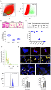
(A) FACS sorting of GFP-labelled AAV CGRP8–37 transduced IFP- MSC (n = 5). Cell sorting of transduced cells resulted in the purification of an aCGRP IFP-MSC subpopulation. (B) aCGRP IFP-MSC showed similar fibroblast-like morphology, but lower clonogenic capacity (72 ± 42 CFU-Fs) to non-transduced IFP-MSC. (C) aCGRP IFP-MSC showed high expression levels of common MSC-defining markers (CD73, CD90, CD105, CD146, CD10, HLA-DR). CD10 expression was similarly high (>95%) whereas CD146 showed reduced expression compared to non-transduced IFP-MSC. (D) aCGRP IFP-MSC sEVs showed high purity and <200 nm sizes. CD63+-selected sEVs showed high positivity for CD9 marker. (E) Immunostaining of HEK 293 cells after incubation with aCGRP IFP-MSC sEVs. Top: Detection of AAV protein in HEK cells incubated with different concentration range of sEVs, Middle: Detection of CGRP protein in HEK cells incubated with different concentration range of sEVs, and bottom: Lower magnification of HEK cells after incubation with sEVs from recombinant MSC (left) and control, sEVs from no recombinant MSC. Plots: Top: Integrated density of AAV signal. aCGRP IFP-MSC sEVs were used at 30 μg/mL (30), 50 μg/mL (50) and 80 μg/mL (80) concentrations. * p < 0.05, ** p < 0.01, and bottom: Integrated density of CGRP signal. * p < 0.05, ** p < 0.01, *** p < 0.001.
Figure 3
Figure 3
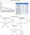
(A) 147 distinct miRNA were present in aCGRP IFP-MSC sEVs (n = 2). Nineteen highly present miRNAs are included in black-bordered box. (B) miRNAs present in aCGRP IFP-MSC sEVs were involved in the regulation of numerous genes and pathways. Predominantly, miRNAs present in sEVs are involved in the regulation of TGF-β/Wnt/FGFR pathways. Putative miRNA interactomes were generated using a miRNet centric network visual analytics platform. The miRNA target gene data were collected from well-annotated database miRTarBase v8.0 and miRNA-gene interactome network refining was performed with 2.0 betweenness cut-off. Values (with 34 cycles cut-off point) were represented in a topology miRNA-gene interactome network using force atlas layout and hypergeometric test algorithm.
Figure 4
Figure 4
(A) In aCGRP IFP-MSC sEVs (n = 2), 19 miRNAs cargos were highly present. These distinct miRNAs regulate genes involved in the production of cytokines, the recruitment of monocytes, and cartilage homeostasis. From these miRNAs, 7 miRNAs were associated in previous studies with significant anti-inflammatory/immunomodulatory effects in vitro and in vivo. Putative miRNA interactomes were generated using a miRNet centric network visual analytics platform. The miRNA target gene data were collected from well-annotated database miRTarBase v8.0 and miRNA-gene interactome network refining was performed with 2.0 betweenness cut-off. Values (with 34 cycles cut-off point) were represented in a topology miRNA-gene interactome network using force atlas layout and hypergeometric test algorithm. (B) In silico analysis revealed a functional correlation of identified miRNAs in aCGRP IFP-MSC sEVs with genes involved in M2 macrophage polarization, immunomodulatory and pain signaling, and cartilage homeostasis. The miRDB online database for prediction of functional miRNA targets has been used to correlate highly expressed target genes in macrophages with specific miRNAs identified by aCGRP IFP-MSC sEV miRNA profiling. MirTarget prediction scores are in the range of 0–100% probability, and candidate transcripts with scores ≥ 50% are presented as predicted miRNA targets in miRDB.
Figure 5
Figure 5
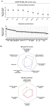
(A,B) Multiple immunomodulatory and reparative molecules secreted as a cargo of aCGRP IFP-MSC sEVs (n = 2). sEVs showed presence of key immunomodulatory molecules including TIMP-2, IL-8, MCP-1, IL-6, ICAM-1, sTNF-RI, MIP-1β, IL-10, and IP-10. In parallel, key reparative molecules including HGF, VEGF, EGFR, IGFBP-1, βFGF, and IGFBP-6 showed presence in aCGRP IFP-MSC sEVs. These proteins are listed in descending order based on their presence levels within sEVs. The miRDB online database for prediction of functional miRNA targets has been used to correlate highly expressed target genes in macrophages with specific miRNAs identified by aCGRP IFP-MSC sEV miRNA profiling. In terms of biological processes, various categories were highly affected and presented as % of proteins involved in a category to the total proteins detected. aCGRP IFP-MSC sEVs protein cargo have effects on PI3K-Akt signaling pathway (59%), MAPK signaling pathway (55%), Ras signaling pathway (52%), Rap1 signaling pathway (43%), cytokine-cytokine receptor interaction (24%), and Jak-STAT signaling pathway (19%). MirTarget prediction scores are in the range of 0–100% probability, and candidate transcripts with scores ≥ 50% are presented as predicted miRNA targets in miRDB.
Figure 6
Figure 6
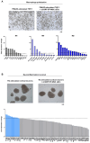
(A) PMA/IO-stimulated THP-1 showed similar morphology with and without aCGRP IFP-MSC sEVs treatment (n = 2). Upon exposure to aCGRP IFP-MSC sEVs, PMA/IO-stimulated THP-1 gene expression analysis showed a strong shift towards M0/M2 macrophage polarization. Notably, exposure to aCGRP IFP-MSC sEVs induced significant expression of key M2-polarization markers such as CD200R1, BMP7, IRF4, IL10, and IL12A. (B) TIC-stimulated cortical neurons showed similar morphology with and without aCGRP IFP-MSC sEVs treatment (n = 2). Upon exposure to aCGRP IFP-MSC sEVs, their molecular profiling indicated an overall reduced neuroinflammatory profile upon exposure to aCGRP IFP-MSC sEVs. From 84 genes, only 14 (SLC6A2, IL1B, PTGS1, EDN1, SCN11A, CCR2, TLR2, GDNF, CD4, OPRD1, ACE, ALOX5, PTGES, CCL12) showed increased expression (>2-fold) compared to TIC-stimulated cortical neurons alone. Blue bars indicate the >2-fold expressed genes. Interestingly, 4 major genes (MAPK8, CD200, MAPK1, PTGES3) involved in neuropathic pain were highly down-regulated (>2-fold) upon exposure to aCGRP IFP-MSC sEVs.

References

    1. Saxler G., Löer F., Skumavc M., Pförtner J., Hanesch U. Localization of SP-and CGRP-immunopositive nerve fibers in the hip joint of patients with painful osteoarthritis and of patients with painless failed total hip arthroplasties. Eur. J. Pain. 2007;11:67. doi: 10.1016/j.ejpain.2005.12.011. - DOI - PubMed
    1. Saito T., Koshino T. Distribution of Neuropeptides in Synovium of the Knee with Osteoarthritis. Clin. Orthop. Relat. Res. 2000;376:172–182. doi: 10.1097/00003086-200007000-00024. - DOI - PubMed
    1. Fernihough J., Gentry C., Bevan S., Winter J. Regulation of calcitonin gene-related peptide and TRPV1 in a rat model of osteoarthritis. Neurosci. Lett. 2005;388:75–80. doi: 10.1016/j.neulet.2005.06.044. - DOI - PubMed
    1. Ferreira-Gomes J., Adães S., Sarkander J., Castro-Lopes J.M. Phenotypic alterations of neurons that innervate osteoarthritic joints in rats. Arthritis Rheum. 2010;62:3677–3685. doi: 10.1002/art.27713. - DOI - PubMed
    1. Hirsch S., Corradini L., Just S., Arndt K., Doods H. The CGRP receptor antagonist BIBN4096BS peripherally alleviates inflammatory pain in rats. Pain. 2013;154:700–707. doi: 10.1016/j.pain.2013.01.002. - DOI - PubMed

Publication types