. 2024 Mar 21;13(6):558.
doi: 10.3390/cells13060558.

Transgene-Free Cynomolgus Monkey iPSCs Generated under Chemically Defined Conditions

Affiliations

Transgene-Free Cynomolgus Monkey iPSCs Generated under Chemically Defined Conditions

Yuliia Tereshchenko et al. Cells. .

Abstract

Non-human primates (NHPs) are pivotal animal models for translating novel cell replacement therapies into clinical applications, including validating the safety and efficacy of induced pluripotent stem cell (iPSC)-derived products. Preclinical development and the testing of cell-based therapies ideally comprise xenogeneic (human stem cells into NHPs) and allogenic (NHP stem cells into NHPs) transplantation studies. For the allogeneic approach, it is necessary to generate NHP-iPSCs with generally equivalent quality to the human counterparts that will be used later on in patients. Here, we report the generation and characterization of transgene- and feeder-free cynomolgus monkey (Macaca fascicularis) iPSCs (Cyno-iPSCs). These novel cell lines have been generated according to a previously developed protocol for the generation of rhesus macaque, baboon, and human iPSC lines. Beyond their generation, we demonstrate the potential of the novel Cyno-iPSCs to differentiate into two clinically relevant cell types, i.e., cardiomyocytes and neurons. Overall, we provide a resource of novel iPSCs from the most frequently used NHP species in the regulatory testing of biologics and classical pharmaceutics to expand our panel of iPSC lines from NHP species with high relevance in preclinical testing and translational research.

Keywords: cardiac differentiation; iPSC; macaque; neuronal differentiation; non-human primate; regeneration; stem cell.


Conflict of interest statement

The authors Y.T., N.E., I.R.-P. and R.B., employed by the German Primate Center-Leibniz Institute for Primate Research, and the authors D.R. and E.G.-R., employed by Charles River Laboratories declare no conflict of interest.

Figures

Figure 1
Figure 1
Cynomolgus macaque iPSC morphology and characterization. (a) Morphology of the four iPSC lines generated in this study and maintained in universal primate pluripotent stem cell (UPPS) medium. iPSC#1.1 and iPSC#1.2 were from Cyno#1, while iPSC#2.1 and iPSC#2.2 were from Cyno#2. Cynomolgus macaque iPSC colonies present human primed iPSC-like morphology, including clear borders and compact colonies consisting of cells that show a high nucleus-to-cytoplasm ratio. Scale bars 100 µm. (b) Two out of four iPSC cell lines are transgene-free at passage ~15, as shown by PCR using primers specific for different regions of the episomal reprogramming plasmids. (c) Pluripotency marker expression, as shown by immunofluorescence. Note that NANOG and SALL4 are not encoded by the reprogramming vectors. Scale bars 20 µm.
Figure 2
Figure 2
Embryoid body formation assay. The four macaque cell lines form cell aggregates and differentiate into representative cell types of the three embryonic germ layers. Staining was conducted for the markers β-tubulin III (ectoderm), alpha-smooth muscle actin (SMA, mesoderm), and α-fetoprotein (AFP, endoderm) in embryoid body outgrowths generated with DPZ_Cyno#1.1, DPZ_Cyno#1.2, DPZ_Cyno#2.1, and DPZ_Cyno#2.2. Scale bars 100 µm and 20 µm.
Figure 3
Figure 3
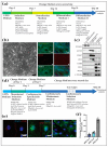
Neuron and cardiomyocyte differentiation protocols were applied to the Cyno-iPSCs. (a) Directed neuronal differentiation of Cyno-iPSCs. (b) Bright-field and immunofluorescence images of neuronal cells. The Cyno-iPSC-derived neurons expressed beta-III-tubulin and showed the characteristic morphology (shown for iPSC#2.1). (c) Protein abundance analysis was conducted on undifferentiated iPSCs and their differentiated counterparts, including cardiomyocyte- and neuron-like cells (Cyno_iPSC#1.1 and 2.1). The analysis encompassed one pluripotency marker (Nanog) and three markers specific to cardiomyocytes (ACTN1, MYH7, and cTNT), as well as three neuronal-specific markers (TUBB3, NEFL, and MAP2). Alfa-tubulin expression is used as a housekeeping marker. (d) Cardiac differentiation of Cyno-iPSCs. (e) Cyno-iPSC cardiomyocytes express cardiac-specific proteins, as shown by immunofluorescence staining (shown for Cyno_iPSC#2.1). The immunofluorescence of cardiac proteins shows the typical sarcomeric structures in the Cyno-iPSC-derived cardiomyocytes: sarcomeric α-actinin, cardiac troponin T (cTNT), and connexin 43 (Cx43). Scale bars, 20 µm. (f) Additionally, the efficiency of differentiation was evaluated by assessing the percentage of alfa-actinin-positive cells in the post-differentiation, pre-selection cell populations (Cyno_iPSC#1.1, #1.2, and #2.1) by fluorescence-activated cell sorting (FACS).

References

    1. Takahashi K., Yamanaka S. Induction of Pluripotent Stem Cells from Mouse Embryonic and Adult Fibroblast Cultures by Defined Factors. Cell. 2006;126:663–676. doi: 10.1016/j.cell.2006.07.024. - DOI - PubMed
    1. Takahashi K., Tanabe K., Ohnuki M., Narita M., Ichisaka T., Tomoda K. Induction of Pluripotent Stem Cells from Adult Human Fibroblasts by Defined Factors. Cell. 2007;131:861–872. doi: 10.1016/j.cell.2007.11.019. - DOI - PubMed
    1. Deinsberger J., Reisinger D., Weber B. Global trends in clinical trials involving pluripotent stem cells: A systematic multi-database analysis. NPJ Regen. Med. 2020;5:15. doi: 10.1038/s41536-020-00100-4. - DOI - PMC - PubMed
    1. Qi Y., Zhang X.-J., Renier N., Wu Z., Atkin T., Sun Z., Ozair M.Z., Tchieu J., Zimmer B., Fattahi F., et al. Combined small-molecule inhibition accelerates the derivation of functional, early-born, cortical neurons from human pluripotent stem cells. Nat. Biotechnol. 2017;35:154–163. doi: 10.1038/nbt.3777. - DOI - PMC - PubMed
    1. Johnsen D.O., Johnson D.K., Whitney R.A. Nonhuman Primates in Biomedical Research. 2nd ed. Elsevier; Amsterdam, The Netherlands: 2012.

Publication types

LinkOut - more resources