. 2024 Feb 21;11(3):200.
doi: 10.3390/bioengineering11030200.

Engineered Dual Antioxidant Enzyme Complexes Targeting ICAM-1 on Brain Endothelium Reduce Brain Injury-Associated Neuroinflammation

Affiliations

Engineered Dual Antioxidant Enzyme Complexes Targeting ICAM-1 on Brain Endothelium Reduce Brain Injury-Associated Neuroinflammation

Brian M Leonard et al. Bioengineering (Basel). .

Abstract

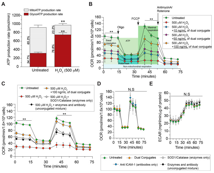
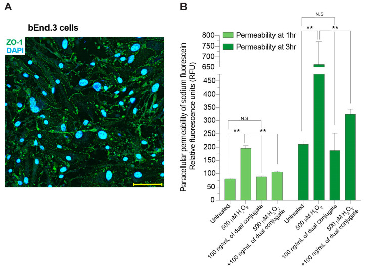
The neuroinflammatory cascade triggered by traumatic brain injury (TBI) represents a clinically important point for therapeutic intervention. Neuroinflammation generates oxidative stress in the form of high-energy reactive oxygen and nitrogen species, which are key mediators of TBI pathology. The role of the blood-brain barrier (BBB) is essential for proper neuronal function and is vulnerable to oxidative stress. Results herein explore the notion that attenuating oxidative stress at the vasculature after TBI may result in improved BBB integrity and neuroprotection. Utilizing amino-chemistry, a biological construct (designated "dual conjugate" for short) was generated by covalently binding two antioxidant enzymes (superoxide dismutase 1 (SOD-1) and catalase (CAT)) to antibodies specific for ICAM-1. Bioengineering of the conjugate preserved its targeting and enzymatic functions, as evaluated by real-time bioenergetic measurements (via the Seahorse-XF platform), in brain endothelial cells exposed to increasing concentrations of hydrogen peroxide or a superoxide anion donor. Results showed that the dual conjugate effectively mitigated the mitochondrial stress due to oxidative damage. Furthermore, dual conjugate administration also improved BBB and endothelial protection under oxidative insult in an in vitro model of TBI utilizing a software-controlled stretching device that induces a 20% in mechanical strain on the endothelial cells. Additionally, the dual conjugate was also effective in reducing indices of neuroinflammation in a controlled cortical impact (CCI)-TBI animal model. Thus, these studies provide proof of concept that targeted dual antioxidant biologicals may offer a means to regulate oxidative stress-associated cellular damage during neurotrauma.

Keywords: blood–brain barrier; brain vasculature; catalase; nanomedicine; neuroinflammation; superoxide dismutase 1; traumatic brain injury.


Conflict of interest statement

The authors declare no conflicts of interest. The funders had no role in the design of the study; in the collection, analyses, or interpretation of data; in the writing of the manuscript; or in the decision to publish the results.

Figures

Figure 1
Figure 1
Development of engineered dual antioxidant enzyme–antibody conjugate complexes. (A) Complexes are generated by “click” chemistry. First, protected sulfhydryls were introduced to the ICAM-1 antibody using N-succinimidyl-S-acetylthioacetate (SATA). Additionally, maleimide groups were added onto the antioxidant enzymes catalase and SOD-1 by the heterobifunctional cross-linker succinimidyl 4-[N-maleimidomethyl]cyclohexane-1-carboxylate (SMCC). Sulfhydryls were deprotected and a conjugation reaction was initiated at a ratio of 2:1:1 (antibody:catalase:SOD1). Unreacted components were removed using Zeba Spin Desalting purification columns. (B) Molecular weights of conjugate complexes were analyzed by SDS-PAGE. Comparisons were made with anti-ICAM-1 antibody only (lane 1), catalase only (lane 2), SOD-1 only (lane 3), unconjugated mixture (lane 4), and conjugate complex (lane 5).
Figure 2
Figure 2
Dual conjugates prevent compromised mitochondrial respiration in brain endothelial cells during oxidative stress insult. Cells were seeded at 16,000 per well and grown until confluence. The cellular oxygen consumption rate (OCR) bioenergetic profile under the indicated conditions was measured in real time using the Seahorse XFe96 Extracellular Flux Analyzer. (A) The effect of the experimental dose of H2O2 (500 μM) on cellular ATP production source, showing a dramatic shift to glycolytic metabolism following insult; (B) dose-dependent assay of dual conjugate (100 ng/mL, 50 ng/mL, 10 ng/mL) in response to 500 μM of H2O2; (C) same schema as panel, B except 100 ng/mL of dual conjugate is compared to equal doses of control groups: free SOD-1 and catalase or an unconjugated mixture of SOD-1, catalase, and anti-ICAM-1; (D) OCR and (E) extracellular acidification rate (ECAR) measurements to determine any basal toxicity of dual conjugates. Vehicle, dual conjugates, or previously mentioned control groups in addition to an anti-ICAM-1 group alone were exposed to cells for 1 h. For (A), means ± SEM, n = 6, ** p < 0.001. For (BE), two-way ANOVA with Dunnett’s test for multiple comparisons was used. N.S = not significant. Baseline and stress-testing phase (FCCP injection) OCR values for (B) (p < 0.0001) and (C) (p < 0.0001) were significantly higher for both the untreated and 100 ng/mL dual-conjugate group compared to different conjugate doses (B) or controls (C).
Figure 3
Figure 3
Engineered dual conjugates attenuate H2O2-induced increases in brain endothelial cell permeability. (A) b.End3 cells were examined by immunofluorescence staining for the expression of the tight-junction protein zonula occludens 1 (ZO-1). ZO-1 (green) was localized at the cell borders and DAPI (blue) stained the cell nuclei. Scale bar equals 10 microns. (B) bEnd.3 cells were grown on transwell membranes until confluence. Cells were then treated with 500 μM H2O2 with or without 100 ng/mL of the dual conjugates. Permeability was assessed at 1 h and 3 h using a 3 kDa fluorescent weight dextran that was applied to the top of the well and measured from the bottom chamber. Relative fluorescence units were determined using a SpectraMax M5e. Treatment with the conjugates reduced permeability changes from exposure to H2O2. ** p < 0.001, n = 3 replicates each condition. N.S = not significant.
Figure 4
Figure 4
Dual conjugates prevent mechanical stretch-induced damage to brain endothelial cells. Cells were seeded at a density of 75,000 per stretch membrane and grown to confluence. (A) Workflow used to develop this model and schema used in the rest of the panels; (B) fluorescent image showing microtears in the endothelial cell monolayer following a stretch with 20% strain (bottom image) compared with a sham group (top image); scale bar 200 microns; (C) initial tests to determine which membrane surface, flat or with nanogrooves orthogonal to the direction of stretch, induced more damage to endothelial cells; (D) comparison of OCR between sham, stretch-injured, and injury + treatment groups; (E) same experiment and cells as (D), measurement of ECAR values. Statistical significance is omitted for clarity. For (CE), two-way ANOVA with Tukey’s test for multiple comparisons was used. Baseline and stress-testing phase (FCCP injection) OCR values for (C,D) are significantly different for all groups (** p < 0.001).
Figure 5
Figure 5
Dual conjugates reduce superoxide production following free radical insult from H2O2, SIN-1, or KO2. Cells were seeded at 55,000 per well (24-well plate) and grown to confluence. (A) Comparison images for groups exposed to 500 μM H2O2 (other treatment groups not shown). The lower-left image shows cells treated with dual conjugates had greatly reduced concentrations of superoxide (red) compared with the insult only or insult + unconjugated mixture images on the right side. Scale bar 200 microns. (B) Fluorescence intensity of MitoSOX red superoxide indicator comparison between all experimental groups, including cells treated with SIN-1 (a superoxide and peroxynitrite producer) and KO2 (a potent superoxide producer). For (B), two-way ANOVA with Tukey’s test for multiple comparisons was used; **** p < 0.0001.
Figure 6
Figure 6
Internalization of dual-conjugate constructs within brain endothelial cells. Optical slices from confocal imaging. Z-stacks identified the top, midpoint and bottom aspects of the cells. Brain endothelial cells (bEnd.3) were stimulated with 50 ng/mL of bacterial LPS for 4 h and then fixed and immunostained with anti-ICAM-1 (green), anti-bovine SOD-1 (red) and nuclear counterstained (DAPI). Labels indicate internalized dual conjugates. Scale bar = 10 microns.
Figure 7
Figure 7
Representative images and quantitative evaluation of post-TBI neuropathological indices for neuronal cell death, astrocyte and microglial activation in animals treated with or without dual conjugates. Mice were placed in the following groups prior to brain harvesting and immunohistochemistry: naïve (craniotomy only but without impact), moderate CCI-TBI and moderate CC-TBI + 100 ng/kg of dual conjugate administered i.v. (A) NeuN staining identifies viable neurons within the cerebral cortex and lack of DAB+ staining for NeuN demonstrates the substantial neuronal loss at 48 h following CCI-TBI. The combination of mechanical strain, oxidative stress and inflammatory responses inevitably results in cell death. However, a rescue of viable neurons can be observed with the presence of dual conjugate administered soon after the TBI was induced. Astrocyte activation was determined by GFAP expression. CCI-TBI shows significant increase at 48 h following CCI-TBI, which is attenuated with the presence of the dual conjugate. Similarly, evaluation of activated microglial by phenotype and augmentation of expression of IBA-1 is clearly seen in the TBI animal. Dual conjugates also reduced the degree of microgliosis. Scale bar 50 microns (B) Bar graphs show the imaging quantification of the neuropathological indices from the groups outlined above. Data are presented as means ± SEM. For (BD), one-way ANOVA with Dunnett’s test for multiple comparisons was used. ** p < 0.001, N.S = not significant.

References

    1. Coronado V.G., Xu L., Basavaraju S.V., McGuire L.C., Wald M.M., Faul M.D., Hemphill J.D. Surveillance for traumatic brain injury-related deaths—United States, 1997–2007. MMWR Surveill Summ. 2011;60:1–32. - PubMed
    1. Satz P., Zaucha K., McCleary C., Light R., Asarnow R., Becker D. Mild head injury in children and adolescents: A review of studies (1970–1995) Psychol. Bull. 1997;122:107–131. doi: 10.1037/0033-2909.122.2.107. - DOI - PubMed
    1. Langlois J.A., Rutland-Brown W., Wald M.M. The epidemiology and impact of traumatic brain injury: A brief overview. J. Head Trauma Rehabil. 2006;21:375–378. doi: 10.1097/00001199-200609000-00001. - DOI - PubMed
    1. Prevention CfDCa . Surveillance Report of Traumatic Brain Injury-Related Deaths by Age Group, Sex, and Mechanism of Injury—United States, 2018 and 2019. Centers for Disease Control and Prevention, US Department of Health and Human Services; Atlanta, GA, USA: 2022.
    1. Rabinowitz A.R., Levin H.S. Cognitive sequelae of traumatic brain injury. Psychiatr. Clin. N. Am. 2014;37:1–11. doi: 10.1016/j.psc.2013.11.004. - DOI - PMC - PubMed

LinkOut - more resources