

Comparative Analysis of Bone Regeneration According to Particle Type and Barrier Membrane for Octacalcium Phosphate Grafted into Rabbit Calvarial Defects
- PMID: 38534489
- PMCID: PMC10968633
- DOI: 10.3390/bioengineering11030215
Comparative Analysis of Bone Regeneration According to Particle Type and Barrier Membrane for Octacalcium Phosphate Grafted into Rabbit Calvarial Defects
Abstract
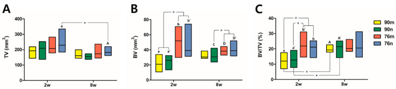
This animal study was aimed to evaluate the efficacy of new bone formation and volume maintenance according to the particle type and the collagen membrane function for grafted octacalcium phosphate (OCP) in rabbit calvarial defects. The synthetic bone substitutes were prepared in powder form with 90% OCP and granular form with 76% OCP, respectively. The calvarial defects were divided into four groups according to the particle type and the membrane application. All specimens were acquired 2 weeks (n = 5) and 8 weeks (n = 5) after surgery. According to the micro-CT results, the new bone volume increased at 2 weeks in the 76% OCP groups compared to the 90% OCP groups, and the bone volume ratio was significantly lower in the 90% OCP group after 2 weeks. The histomorphometric analysis results indicated that the new bone area and its ratio in all experimental groups were increased at 8 weeks except for the group with 90% OCP without a membrane. Furthermore, the residual bone graft area and its ratio in the 90% OCP groups were decreased at 8 weeks. In conclusion, all types of OCP could be applied as biocompatible bone graft materials regardless of its density and membrane application. Neither the OCP concentration nor the membrane application had a significant effect on new bone formation in the defect area, but the higher the OCP concentration, the less graft volume maintenance was needed.
Keywords: bone regeneration; collagen membrane; concentration; octacalcium phosphate.
Conflict of interest statement
The authors declare no conflicts of interest.
Figures


Similar articles
-
Octacalcium Phosphate/Gelatin Composite (OCP/Gel) Enhances Bone Repair in a Critical-sized Transcortical Femoral Defect Rat Model.Clin Orthop Relat Res. 2022 Oct 1;480(10):2043-2055. doi: 10.1097/CORR.0000000000002257. Epub 2022 May 30. Clin Orthop Relat Res. 2022. PMID: 35638896 Free PMC article.
-
Efficacy of Octacalcium Phosphate and Octacalcium Phosphate/Gelatin Composite on the Repair of Critical-Sized Calvarial Defects in Rats.J Dent (Tehran). 2018 Mar;15(2):86-96. J Dent (Tehran). 2018. PMID: 29971126 Free PMC article.
-
Acceleration of bone formation by octacalcium phosphate composite in a rat tibia critical-sized defect.J Orthop Translat. 2022 Oct 12;37:100-112. doi: 10.1016/j.jot.2022.09.007. eCollection 2022 Nov. J Orthop Translat. 2022. PMID: 36262961 Free PMC article.
-
Impact of Octacalcium Phosphate/Gelatin (OCP/Gel) Composite on Bone Repair in Refractory Bone Defects.Tohoku J Exp Med. 2023 Jul 14;260(3):245-252. doi: 10.1620/tjem.2023.J040. Epub 2023 May 18. Tohoku J Exp Med. 2023. PMID: 37197945 Review.
-
The material design of octacalcium phosphate bone substitute: increased dissolution and osteogenecity.Acta Biomater. 2023 Mar 1;158:1-11. doi: 10.1016/j.actbio.2022.12.046. Epub 2022 Dec 27. Acta Biomater. 2023. PMID: 36581004 Review.
Cited by
-
Bioglasses Versus Bioactive Calcium Phosphate Derivatives as Advanced Ceramics in Tissue Engineering: Comparative and Comprehensive Study, Current Trends, and Innovative Solutions.J Funct Biomater. 2025 May 3;16(5):161. doi: 10.3390/jfb16050161. J Funct Biomater. 2025. PMID: 40422826 Free PMC article. Review.
References
-
- Kim J.-S., Jang T.-S., Kim S.-Y., Lee W.-P. Octacalcium Phosphate Bone Substitute (Bontree®): From Basic Research to Clinical Case Study. Appl. Sci. 2021;11:7921. doi: 10.3390/app11177921. - DOI
Grants and funding
LinkOut - more resources
Full Text Sources
