. 2024 Mar 20;14(6):653.
doi: 10.3390/diagnostics14060653.

Evaluation of Whole Brain Intravoxel Incoherent Motion (IVIM) Imaging

Affiliations

Evaluation of Whole Brain Intravoxel Incoherent Motion (IVIM) Imaging

Kamil Lipiński et al. Diagnostics (Basel). .

Abstract

Intravoxel Incoherent Motion (IVIM) imaging provides non-invasive perfusion measurements, eliminating the need for contrast agents. This work explores the feasibility of IVIM imaging in whole brain perfusion studies, where an isotropic 1 mm voxel is widely accepted as a standard. This study follows the validity of a time-limited, precise, segmentation-ready whole-brain IVIM protocol suitable for clinical reality. To assess the influence of SNR on the estimation of S0, f, D*, and D IVIM parameters, a series of measurements and simulations were performed in MATLAB for the following three estimation techniques: segmented grid search, segmented curve fitting, and one-step curve fitting, utilizing known "ground truth" and noised data. Scanner-specific SNR was estimated based on a healthy subject IVIM MRI study in a 3T scanner. Measurements were conducted for 25.6 × 25.6 × 14.4 cm FOV with a 256 × 256 in-plane resolution and 72 slices, resulting in 1 × 1 × 2 mm voxel size. Simulations were performed for 36 SNR levels around the measured SNR value. For a single voxel grid, the search algorithm mean relative error Ŝ0, f^, D^*, and D^ of at the expected SNR level were 5.00%, 81.91%, 76.31%, and 18.34%, respectively. Analysis has shown that high-resolution IVIM imaging is possible, although there is significant variation in both accuracy and precision, depending on SNR and the chosen estimation method.

Keywords: DWI; IVIM; MRI; SNR; brain; perfusion.


Conflict of interest statement

The authors declare no conflicts of interest.

Figures

Figure 1
Figure 1
A bi-exponential IVIM model illustration. The orange curve represents the prediction from over-threshold b-values. The blue curve represents the actual data. The difference between the prediction from high b-values and the actual data is attributed to the influence of blood circulation and is referred to as pseudo-diffusion [8]. It is seen that b-values where scanning is sensitive for fast components lie in the range of approx. 0–250.
Figure 2
Figure 2
(a) Histogram of SNR (left). (b) Heatmap of SNR shown as axial slices 20 mm from each other (right).
Figure 3
Figure 3
Gray matter coverage on DWI scan. The yellow shape represents the coverage of the gray matter map with a probability > 80% overlayed on b0 image, regridded to IVIM DWI resolution.
Figure 4
Figure 4
RMSE of parameter fitting for three methods of single voxel estimation.
Figure 5
Figure 5
RMSE of parameter fitting for three methods. Estimation was made for clustered voxels and each estimation for the signal was averaged from eight voxels.
Figure 6
Figure 6
RMSE of parameter fitting for three methods. Estimation made for clustered voxels, each estimation for signal averaged from 27 voxels.
Figure 7
Figure 7
RMSE of parameter fitting for three methods. Estimation was made for clustered voxels and each estimation for the signal was averaged from 64 voxels.
Figure 8
Figure 8
Brodmann areas atlas overlayed on DWI scans. Red and green shape represents the coverage of the Brodmann atlas template, regridded to IVIM DWI resolution.
Figure 9
Figure 9
Comparison of (a) diffusion, (b) pseudo-diffusion parameter estimation in mm2/s, and (c) blood fraction parameter estimation; calculated on the signal from Brodmann areas.
Figure 10
Figure 10
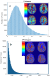
Histogram of estimate value (x-axis) on white and grey matter (sum of >70% TPM maps) and heatmap of voxels using grid search calculation method.: (a) f and (b) D*.

References

    1. Essig M., Shiroishi M.S., Nguyen T.B., Saake M., Provenzale J.M., Enterline D., Anzalone N., Dörfler A., Rovira À., Wintermark M., et al. Perfusion MRI: The Five Most Frequently Asked Technical Questions. AJR Am. J. Roentgenol. 2013;200:24–34. doi: 10.2214/AJR.12.9543. - DOI - PMC - PubMed
    1. Wahsner J., Gale E.M., Rodríguez-Rodríguez A., Caravan P. Chemistry of MRI Contrast Agents: Current Challenges and New Frontiers. Chem. Rev. 2019;119:957–1057. doi: 10.1021/acs.chemrev.8b00363. - DOI - PMC - PubMed
    1. Le Bihan D., Breton E., Lallemand D., Grenier P., Cabanis E., Laval-Jeantet M. MR Imaging of Intravoxel Incoherent Motions: Application to Diffusion and Perfusion in Neurologic Disorders. Radiology. 1986;161:401–407. doi: 10.1148/radiology.161.2.3763909. - DOI - PubMed
    1. Einstein A. On the Movement of Small Particles Suspended in Stationary Liquids Required by the Molecular-Kinetic Theory of Heat. Ann. Phys. 1905;322:549–560. doi: 10.1002/andp.19053220806. - DOI
    1. Brown R. XXVII. A Brief Account of Microscopical Observations Made in the Months of June, July and August 1827, on the Particles Contained in the Pollen of Plants; and on the General Existence of Active Molecules in Organic and Inorganic Bodies. Philos. Mag. 1828;4:161–173. doi: 10.1080/14786442808674769. - DOI

LinkOut - more resources