Review
. 2024 Mar 21;11(3):95.
doi: 10.3390/jcdd11030095.

Cardiac Allograft Vasculopathy: Challenges and Advances in Invasive and Non-Invasive Diagnostic Modalities

Affiliations
Review

Cardiac Allograft Vasculopathy: Challenges and Advances in Invasive and Non-Invasive Diagnostic Modalities

Moaz A Kamel et al. J Cardiovasc Dev Dis. .

Abstract

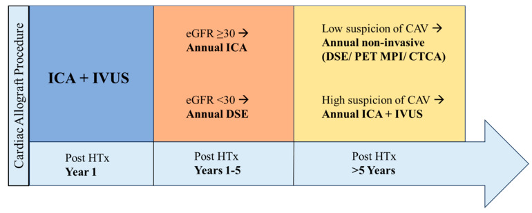
Cardiac allograft vasculopathy (CAV) is a distinct form of coronary artery disease that represents a major cause of death beyond the first year after heart transplantation. The pathophysiology of CAV is still not completely elucidated; it involves progressive circumferential wall thickening of both the epicardial and intramyocardial coronary arteries. Coronary angiography is still considered the gold-standard test for the diagnosis of CAV, and intravascular ultrasound (IVUS) can detect early intimal thickening with improved sensitivity. However, these tests are invasive and are unable to visualize and evaluate coronary microcirculation. Increasing evidence for non-invasive surveillance techniques assessing both epicardial and microvascular components of CAV may help improve early detection. These include computed tomography coronary angiography (CTCA), single-photon emission computed tomography (SPECT), positron emission tomography (PET), and vasodilator stress myocardial contrast echocardiography perfusion imaging. This review summarizes the current state of diagnostic modalities and their utility and prognostic value for CAV and also evaluates emerging tools that may improve the early detection of this complex disease.

Keywords: angiography; cardiac allograft vasculopathy; echocardiography; nuclear imaging.


Conflict of interest statement

The authors declare no conflicts of interest.

Figures

Figure 1
Figure 1
A 53-year-old male patient who developed severe CAV two years after HTx. (A) Successful intervention of the middle left anterior descending artery (green circle) with one drug-eluting stent (preintervention stenosis was 90%). (B) Successful intervention of the middle right coronary artery (green circle) with one drug-eluting stent (preintervention stenosis was 70%).
Figure 2
Figure 2
A 62-year-old male patient who developed CAV 10 years after heart transplant. (A) Conventional ICA showing no significant stenosis; middle left anterior descending artery is 10% obstructed by diffuse disease and middle right coronary artery is 20% obstructed by diffuse disease. (B) IVUS was performed in mid left anterior descending artery and confirmed the presence of significant atherosclerosis. CAV, cardiac allograft vasculopathy; ICA, invasive coronary angiography; IVUS, intravascular ultrasound.
Figure 3
Figure 3
(A) Cardiac magnetic resonance imaging (CMR) showing clear thinning (white arrows) of the anterolateral, inferolateral, and inferior mid-cavity, with subendocardial delayed enhancement in these regions suggesting a vascular etiology. The hypertrophy of the septum is profound but is presumably compensatory due to the extensive loss of lateral wall myocardium. While this CMR finding could be multifactorial, invasive coronary angiography showed a severe obstruction (white arrow) in the proximal circumflex artery (B).
Figure 4
Figure 4
Moderate to large reversible perfusion defect (white arrows) in the anterior and inferolateral wall showed using positron emission tomography (A) and computed tomography cardiac perfusion test (B). Coronary allograft vasculopathy was confirmed with invasive coronary angiography; this study showed a severe obstruction (white circle) in the mid circumflex artery (C).
Figure 5
Figure 5
CAV surveillance over time based on expert opinion. CAV, cardiac allograft vasculopathy; HTx, heart transplant; ICA, invasive coronary angiography; IVUS, intravascular ultrasound; DSE, dobutamine stress echocardiography; PET, positron emission tomography; MPI, myocardial perfusion imaging; CTCA, computed tomography coronary angiography.

References

    1. Sciaccaluga C., Ghionzoli N., Mandoli G.E., Sisti N., D’Ascenzi F., Focardi M., Bernazzali S., Vergaro G., Emdin M., Valente S., et al. The role of non-invasive imaging modalities in cardiac allograft vasculopathy: An updated focus on current evidences. Heart Fail. Rev. 2022;27:1235–1246. doi: 10.1007/s10741-021-10155-0. - DOI - PMC - PubMed
    1. Mehra M.R. The scourge and enigmatic journey of cardiac allograft vasculopathy. J. Heart Lung Transplant. 2017;36:1291–1293. doi: 10.1016/j.healun.2017.10.010. - DOI - PubMed
    1. Khush K.K., Cherikh W.S., Chambers D.C., Harhay M.O., Hayes D., Jr., Hsich E., Meiser B., Potena L., Robinson A., Rossano J.W., et al. The International Thoracic Organ Transplant Registry of the International Society for Heart and Lung Transplantation: Thirty-sixth adult heart transplantation report—2019; focus theme: Donor and recipient size match. J. Heart Lung Transplant. 2019;38:1056–1066. doi: 10.1016/j.healun.2019.08.004. - DOI - PMC - PubMed
    1. Abrahimi P., Liu R., Pober J.S. Blood Vessels in Allotransplantation. Am. J. Transplant. 2015;15:1748–1754. doi: 10.1111/ajt.13242. - DOI - PubMed
    1. Chih S., Chong A.Y., Mielniczuk L.M., Bhatt D.L., Beanlands R.S. Allograft Vasculopathy: The Achilles’ Heel of Heart Transplantation. J. Am. Coll. Cardiol. 2016;68:80–91. doi: 10.1016/j.jacc.2016.04.033. - DOI - PubMed

LinkOut - more resources