Review
. 2024 Mar 6;22(3):126.
doi: 10.3390/md22030126.

Current Status of Indole-Derived Marine Natural Products: Synthetic Approaches and Therapeutic Applications

Affiliations
Review

Current Status of Indole-Derived Marine Natural Products: Synthetic Approaches and Therapeutic Applications

Sergio Fernández et al. Mar Drugs. .

Abstract

Indole is a versatile pharmacophore widely distributed in bioactive natural products. This privileged scaffold has been found in a variety of molecules isolated from marine organisms such as algae and sponges. Among these, indole alkaloids represent one of the biggest, most promising family of compounds, having shown a wide range of pharmacological properties including anti-inflammatory, antiviral, and anticancer activities. The aim of this review is to show the current scenario of marine indole alkaloid derivatives, covering not only the most common chemical structures but also their promising therapeutic applications as well as the new general synthetic routes developed during the last years.

Keywords: biological activity; indole alkaloids; marine resources; synthetic strategies; therapeutic application.


Conflict of interest statement

The authors declare no conflicts of interest.

Figures

Figure 1
Figure 1
Example of marine drugs accepted by the FDA.
Figure 2
Figure 2
Structure/activity relationships of indole derivatives.
Figure 3
Figure 3
Structure of some marine indole alkaloids with anticancer activity.
Figure 4
Figure 4
Structures of C3-acyclic substituted SIAs 15.
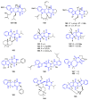
Figure 5
Figure 5
Structures of C3-carbaldehyde/carboxy-substituted SIAs 610.
Figure 6
Figure 6
Biogenetic route to obtain Anthranoside C (11).
Figure 7
Figure 7
Structures of prenylated SIAs 1417.
Figure 8
Figure 8
Biogenesis of isomers 18 and 19.
Figure 9
Figure 9
Synthetic approaches in the preparation of functionalized SIAs.
Figure 10
Figure 10
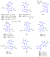
Trachycladindole A–G (2632), aplysinopsins (33), and their derivatives (3440).
Figure 11
Figure 11
Trachycladindole hypothetical biosynthesis by A. Hentz.
Figure 12
Figure 12
Stanovnik and Svete’s synthesis of Aplysinopsin derivate 39.
Figure 13
Figure 13
Structure of Meridianins A–G (4147).
Figure 14
Figure 14
Jing and Yang synthesis of Meridianin D (44).
Figure 15
Figure 15
Fresneda and Molina’s synthesis of Meridianins C (43) and D (44).
Figure 16
Figure 16
Structure of bis-indoles 5053 and tris-indole 54.
Figure 17
Figure 17
Structure of bromodeoxytopsentin (55) and dibromodeoxytopsentin (56).
Figure 18
Figure 18
Structure of eusynstelamides A–B and D–F (5761).
Figure 19
Figure 19
Structure of Hamacanthins A–B (6263).
Figure 20
Figure 20
Janosik et al. [64] synthesis of Rhopaladin C (53).
Figure 21
Figure 21
Basic structures of Simple Diketopiperazines.
Figure 22
Figure 22
Chemical structures of simple DKPs 6468 with C3-methylene bridge.
Figure 23
Figure 23
Chemical structures of simple DKPs 6982 with C3-methylene bridge.
Figure 24
Figure 24
Chemical structures of simple DKPs 83102 with C3-ethylidene bridge.
Figure 25
Figure 25
Chemical structures of bis-indol DKPs 103105.
Figure 26
Figure 26
Possible disconnections of a 2,5-diketopiperazine ring.
Figure 27
Figure 27
Aza-Wittig cyclization to synthesize DKPs 107.
Figure 28
Figure 28
Synthesis and biosynthesis of Brevianamide F (64).
Figure 29
Figure 29
Synthesis of Neochenulin A (94).
Figure 30
Figure 30
Synthesis of Aspergilazine A (103).
Figure 31
Figure 31
Structures of compounds 113118.
Figure 32
Figure 32
Structures of compounds 119129.
Figure 33
Figure 33
Example of synthesis of breviamides 132135.
Figure 34
Figure 34
Synthesis of dimethyhydropyranoindole nucleus.
Figure 35
Figure 35
Structures of compounds 136146.
Figure 36
Figure 36
Structures of compounds 147165.
Figure 37
Figure 37
Synthesis of Indole diketopiperazine alkaloids.
Figure 38
Figure 38
Structures of compounds 173 and 174.
Figure 39
Figure 39
Classical synthesis of Flustramine C (178).
Figure 40
Figure 40
Synthesis of the flustramines analogs 179.
Figure 41
Figure 41
Synthetic routes of tricyclic HPI.
Figure 42
Figure 42
Structures of compounds 182185.
Figure 43
Figure 43
Structures of compounds 186203.
Figure 44
Figure 44
Structures of compounds 204206.
Figure 45
Figure 45
Structures of compounds 207213.
Figure 46
Figure 46
Structures of compounds 214216.
Figure 47
Figure 47
Representative commercialized β-carboline drugs.
Figure 48
Figure 48
Structure of Norharmane 217.
Figure 49
Figure 49
C1-substituted βC compounds 217237.
Figure 50
Figure 50
C1-substituted βC compounds 238245.
Figure 51
Figure 51
C1-substituted βC compounds 246248.
Figure 52
Figure 52
C1-substituted βC compounds 249251.
Figure 53
Figure 53
C1-substituted βC compounds 252260.
Figure 54
Figure 54
C1-substituted βC compounds 261268.
Figure 55
Figure 55
C1-substituted βC compounds 269270.
Figure 56
Figure 56
C1-substituted βC compounds 271277.
Figure 57
Figure 57
Chemical structure of Shishijimicin A–C (278280).
Figure 58
Figure 58
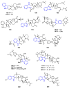
Chemical structures of Manzamines 281291.
Figure 59
Figure 59
Chemical structures of Manzamines 292297.
Figure 60
Figure 60
Chemical structures of Manzamines 298306.
Figure 61
Figure 61
Chemical structures of Manzamines 307312.
Figure 62
Figure 62
N2-substituted βC compounds 313317.
Figure 63
Figure 63
C3-substituted βC compounds 318325.
Figure 64
Figure 64
Chemical structures of Hyrtioerectine B (326).
Figure 65
Figure 65
Annelated C1-N2 βC compounds 327339.
Figure 66
Figure 66
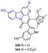
Annelated βC compounds 340341.
Figure 67
Figure 67
Naturally occurring marine βC 1,1-dimers 342344.
Figure 68
Figure 68
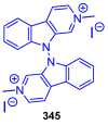
Structure of marine βC 9,9-dimer 345.
Figure 69
Figure 69
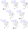
Structure of manzamine hybrid dimers 346348.
Figure 70
Figure 70
Structure of Kauluamine (349).
Figure 71
Figure 71
Most employed synthetic routes for synthesizing βCs.
Figure 72
Figure 72
Other classical general synthetic routes towards the synthesis of βCs.
Figure 73
Figure 73
Representative modern approaches towards the synthesis of βCs.

Similar articles

Cited by

References

    1. Karthikeyan A., Joseph A., Nair B.G. Promising Bioactive Compounds from the Marine Environment and Their Potential Effects on Various Diseases. J. Genet. Eng. Biotechnol. 2022;20:14–52. doi: 10.1186/s43141-021-00290-4. - DOI - PMC - PubMed
    1. Carroll A.R., Copp B.R., Davis R.A., Keyzers R.A., Prinsep M.R. Marine Natural Products. Nat. Prod. Rep. 2023;40:275–325. doi: 10.1039/D2NP00083K. - DOI - PubMed
    1. Zhang L., An R., Wang J., Sun N., Zhang S., Hu J., Kuai J. Exploring Novel Bioactive Compounds from Marine Microbes. Curr. Opin. Microbiol. 2005;8:276–281. doi: 10.1016/j.mib.2005.04.008. - DOI - PubMed
    1. Malve H. Exploring the Ocean for New Drug Developments: Marine Pharmacology. J. Pharm. Bioall. Sci. 2016;8:83–91. doi: 10.4103/0975-7406.171700. - DOI - PMC - PubMed
    1. Xu Z., Eichler B., Klausner E.A., Duffy-Matzner J., Zheng W. Lead/Drug Discovery from Natural Resources. Molecules. 2022;27:8280. doi: 10.3390/molecules27238280. - DOI - PMC - PubMed

LinkOut - more resources