Review
. 2024 Mar 5;8(1):10.
doi: 10.3390/vision8010010.

Angioid Streaks Remain a Challenge in Diagnosis, Management, and Treatment

Affiliations
Review

Angioid Streaks Remain a Challenge in Diagnosis, Management, and Treatment

Georgios Tsokolas et al. Vision (Basel). .

Abstract

Aim: Angioid streaks (ASs) are a rare retinal condition and compromise visual acuity when complicated with choroidal neovascularization (CNV). They represent crack-like dehiscences at the level of the Bruch's membrane. This objective narrative review aims to provide an overview of pathophysiology, current treatment modalities, and future perspectives on this condition. Materials and Methods: A literature search was performed using "PubMed", "Web of Science", "Scopus", "ScienceDirect", "Google Scholar", "medRxiv", and "bioRxiv." Results: ASs may be idiopathic, but they are also associated with systemic conditions, such as pseudoxanthoma elasticum, hereditary hemoglobinopathies, or Paget's disease. Currently, the main treatment is the use of anti-vascular endothelial growth factors (anti-VEGF) to treat secondary CNV, which is the major complication observed in this condition. If CNV is detected and treated promptly, patients with ASs have a good chance of maintaining functional vision. Other treatment modalities have been tried but have shown limited benefit and, therefore, have not managed to be more widely accepted. Conclusion: In summary, although there is no definitive cure yet, the use of anti-VEGF treatment for secondary CNV has provided the opportunity to maintain functional vision in individuals with AS, provided that CNV is detected and treated early.

Keywords: Bruch’s membrane; Paget’s disease; aflibercept; angioid streaks; bevacizumab; brolucizumab; faricimab; hemoglobinopathies; peau d’orange; pseudoxanthoma elasticum; ranibizumab.


Conflict of interest statement

The authors declare no conflicts of interest.

Figures

Figure 1
Figure 1
Near-infrared reflectance (NIR) imaging and 488 nm fundus autofluorescence in pseudoxanthoma elasticum. Angioid streaks and peau d’orange are best and most reliably visible on NIR reflectance imaging (B,E,J), correlating well with findings on funduscopy (A,D,G,K). Peau d’orange is usually not discernible on 488 nm fundus autofluorescence images (C,F). Angioid streaks may present with reduced autofluorescence (C,F) but may as well remain undetected on autofluorescence imaging (H,I). Note the reticular drusen on NIR reflectance and 488 nm autofluorescence, which are sometimes associated with pseudoxanthoma elasticum (H,I) [5].
Figure 2
Figure 2
Pattern dystrophy-like changes and atrophy in pseudoxanthoma elasticum on 488 nm fundus autofluorescence imaging. Atrophic lesions and pattern dystrophy-like changes are typical features of advanced pseudoxanthoma elasticum. Compared to funduscopic images (A,C,E,G), these lesions are best visible on 488 nm fundus autofluorescence images (B,D,F,H). Pattern dystrophy-like lesions encompass different patterns of increased autofluorescence. Depending on the stage of pattern dystrophy changes, atrophy of the retinal pigment epithelium with reduced autofluorescence may be present (B,D,F) [5].
Figure 3
Figure 3
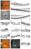
Features of pseudoxanthoma elasticum on optical coherence tomography (OCT). Calcification of Bruch’s membrane may be seen on OCT images. This is best illustrated within areas of peau d’orange, the transition zone between the calcified and un-calcified Bruch membranes (AE). The horizontal arrow in (A,B) indicates the placement of the OCT scan in (C). Magnifications (2.5×) of characteristic details in (C) are shown in (D,E). Area * corresponds to (D) and area # to (E). Areas of increased reflectivity within the outer zone of the RPE–Bruch’s membrane complex (arrow heads in (D)) correlate with the whitish opaque fundus reflex on color images (A) and the increased signal on near-infrared reflectance images (B). Areas of lower reflectivity ((E), arrows in (D)) correlate to the normal fundus reflex. Angioid streaks correlate to breaks within the thickened and hyperreflective Bruch’s membrane ((FH), arrows). Fibrovascular tissue may grow through such breaks (I,J). A typical complication of angioid streaks is the development of choroidal neovascularization leading to retinal exsudation (K). Eventually, atrophy of the retinal pigment epithelium is associated with atrophic changes in the photoreceptor layer with (L) or without (M) cystoid retinal lesions. In some patients there may be persistent subretinal fluid independent of choroidal neovascularization (N,Q). If longstanding, a vitelliform lesion may present with deposition of yellowish hyperautofluorescent material at the bottom of the lesion (OQ). The green arrow in (P) indicates the placement of the OCT scan in (Q) [5].
Figure 4
Figure 4
Multimodal imaging findings of choroidal neovascularization (CNV). Angioid streaks are observed on the red-free image (a); however, no hemorrhage is evident. The overlay of false-colored optical coherence tomography angiography (OCT-A) over the color photograph (b) outlines the area of choroidal neovascularization (CNV). En Face OCT-A (c) demonstrates two areas of CNV (red and blue arrows) that demonstrate a tangled morphology of vascular networks. Sites of CNV closely correlate to sites of angioid streaks ((d); arrowheads) when OCT-A is compared to face reflectance images. Structural OCT scans (I,II) confirm a mixed type 1 (I) and type 2 (II) neovascular lesion that arises in proximity to sites of Bruch’s membrane disruption [47].
Figure 5
Figure 5
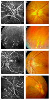
Features of pseudoxanthoma elasticum on fluorescein angiography. Angioid streaks typically show variable staining on fluorescein angiography (A,B). Comet tail lesions appear as hyperfluorescent spots with their tail toward the optic disk (C,D). Choroidal neovascularizations are mostly classic membranes. Sometimes, their detection may be difficult due to adjacent staining of angioid streaks (EH) [5].
Figure 6
Figure 6
Comparison of clinical features on late phase fluorescein angiography, early and late phase indocyanine green angiography (ICG-A), and funduscopy. Late-phase fluorescein angiography shows variable staining of angioid streaks (A,E,I), which corresponds well with findings on funduscopy (B,F,J). A characteristic finding on late-phase ICG-A is a centrally reduced fluorescence with a spotted transition zone to normal peripheral fluorescence (C,G,K). Angioid streaks are well visible within the dark, non-fluorescent area. Note that there is no correlation between color images (B,F,J) or early ICG-A frames (D,H,L). Comet tail lesions (J) are usually hyperfluorescent on late-phase fluorescein angiograms (I) and hypofluorescent on ICG late-phase angiograms (K) [5].

References

    1. Kanski J. Clinical Ophthalmology: A Systematic Approach. 8th ed. Elsevier; Amsterdam, The Netherlands: 2015.
    1. Doyne R.W. Choroidal and retinal changes. The results of blows on the eyes. Trans. Ophthalmol. Soc. UK. 1889;9:128.
    1. Chatziralli I., Saitakis G., Dimitriou E., Chatzirallis A., Stoungioti S., Theodossiadis G., Theodossiadis P. Angioid streaks: A comprehensive review from pathophysiology to treatment. Retina. 2018;39:1–11. doi: 10.1097/IAE.0000000000002327. - DOI - PubMed
    1. Plange O. Über streifenförmige Pigmentbildung mit sekundären Veränderungen der Netzhaut infolge von Hämorrhagien. Arch. Augenheilkd. 1891;23:78–90.
    1. Gliem M., De Zaeytijd J., Finger R.P., Holz F.G., Leroy B.P., Issa P.C. An update on the ocular phenotype in patients with pseudoxanthoma elasticum. Front. Genet. 2013;4:4–14. doi: 10.3389/fgene.2013.00014. - DOI - PMC - PubMed