. 2024 Jun 3;223(6):e202305038.
doi: 10.1083/jcb.202305038. Epub 2024 Mar 27.

The GTPase activating protein Gyp7 regulates Rab7/Ypt7 activity on late endosomes

Affiliations

The GTPase activating protein Gyp7 regulates Rab7/Ypt7 activity on late endosomes

Nadia Füllbrunn et al. J Cell Biol. .

Abstract

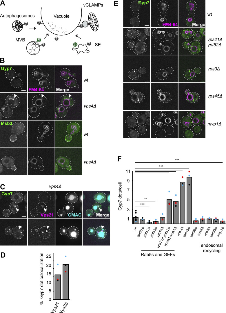
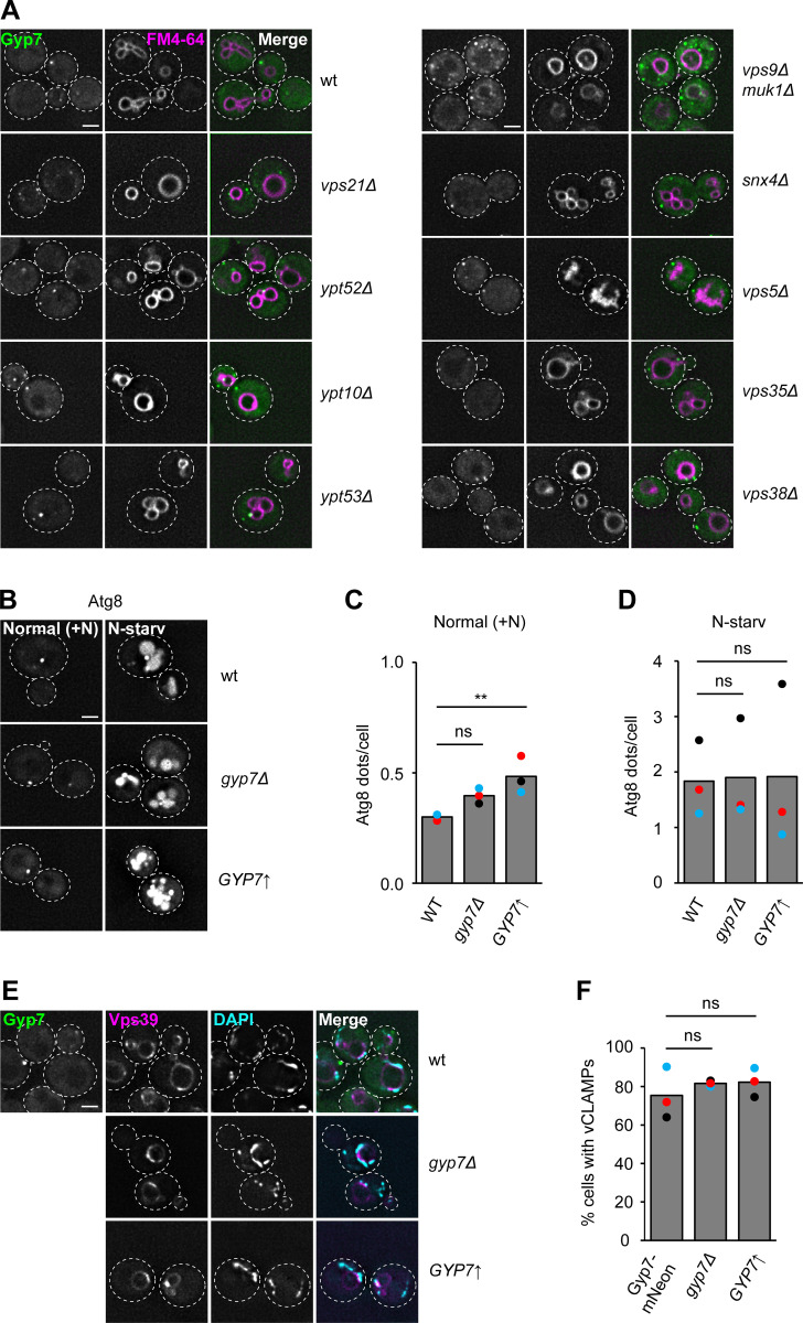
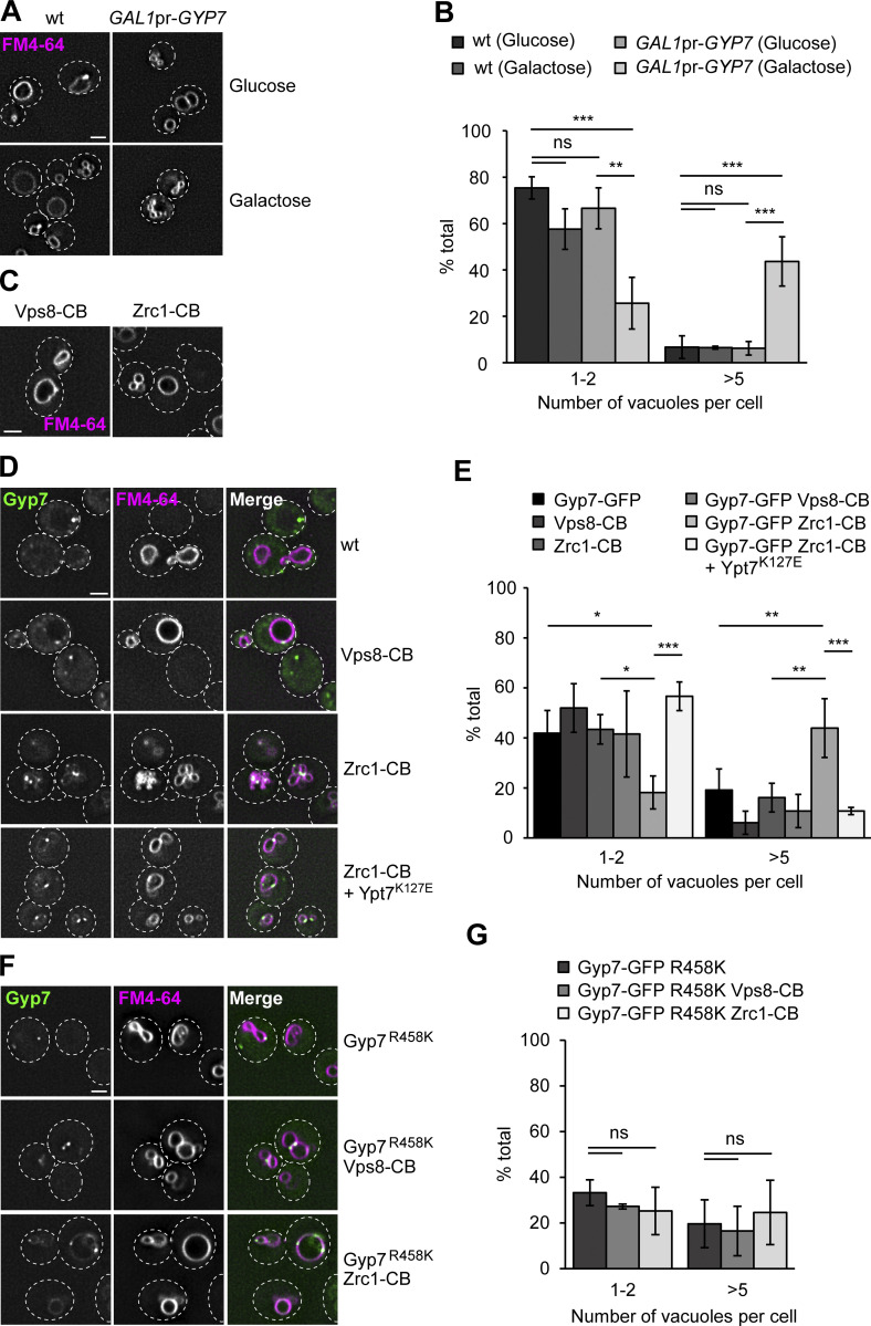
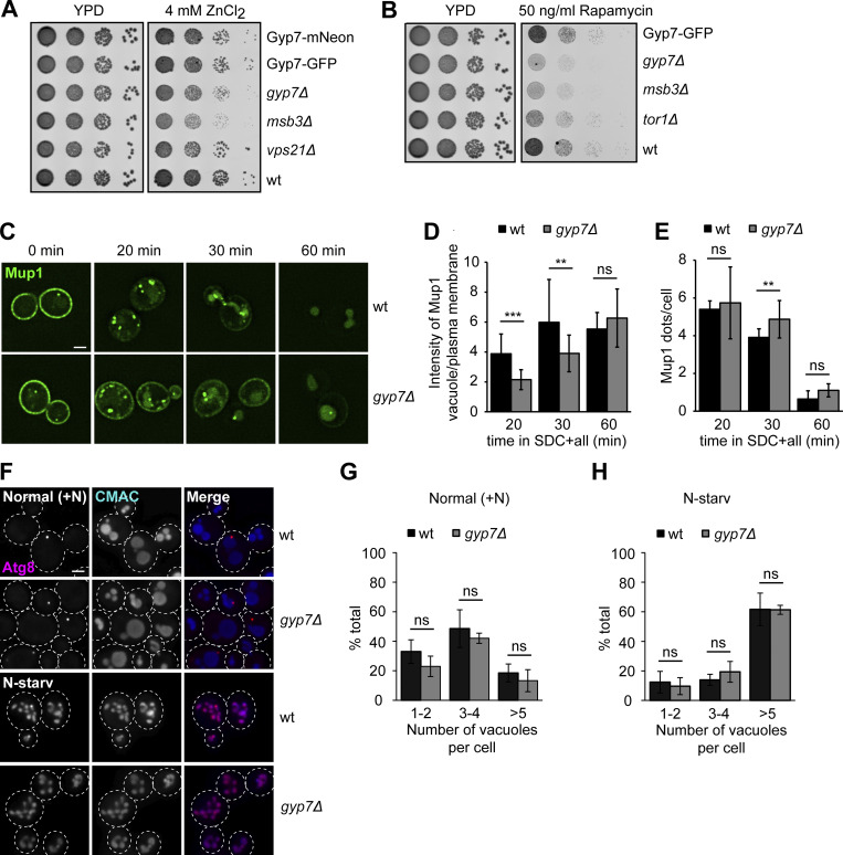
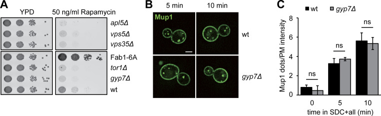
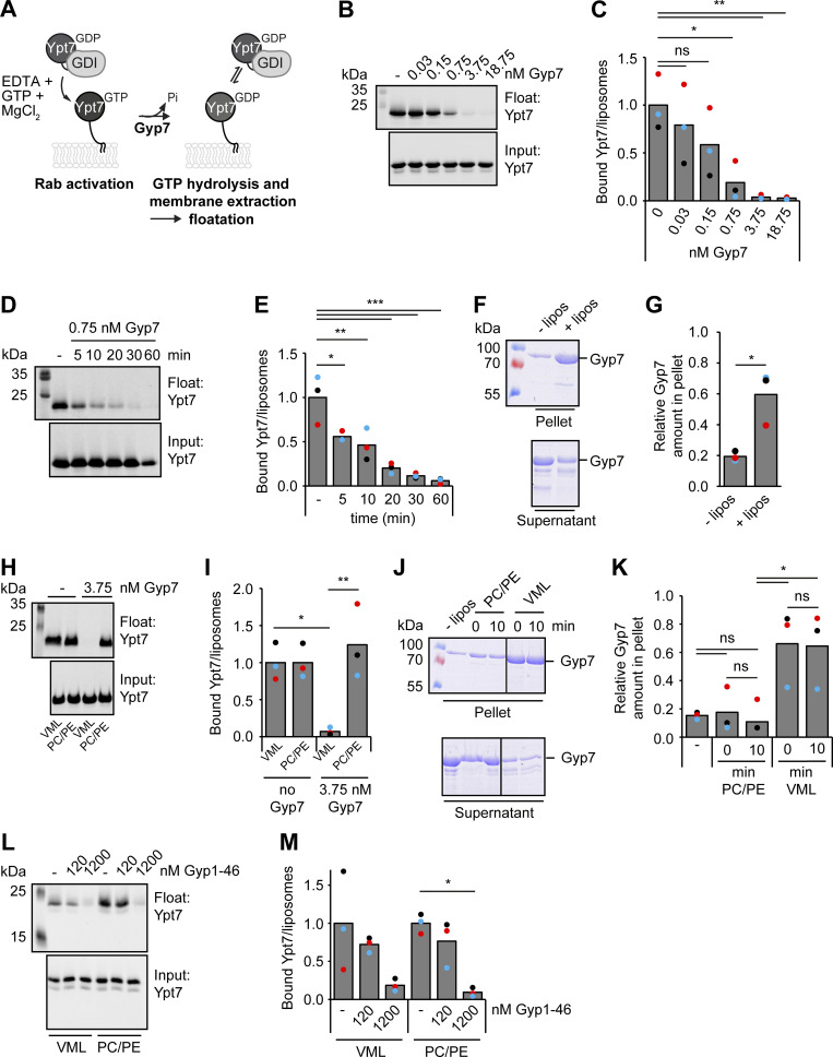
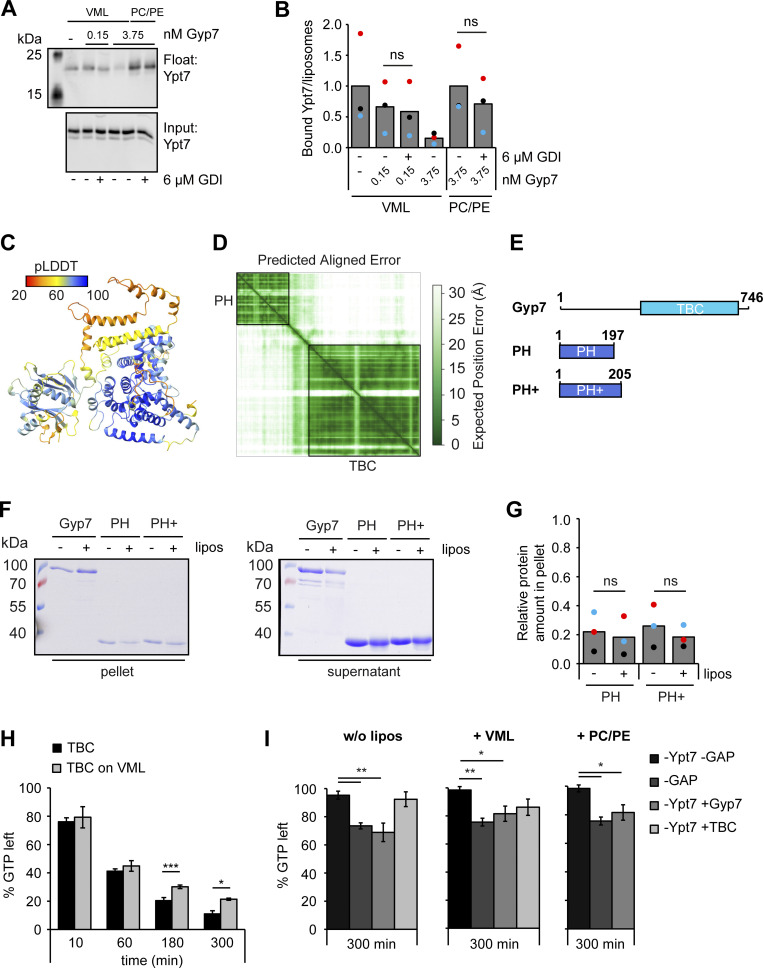
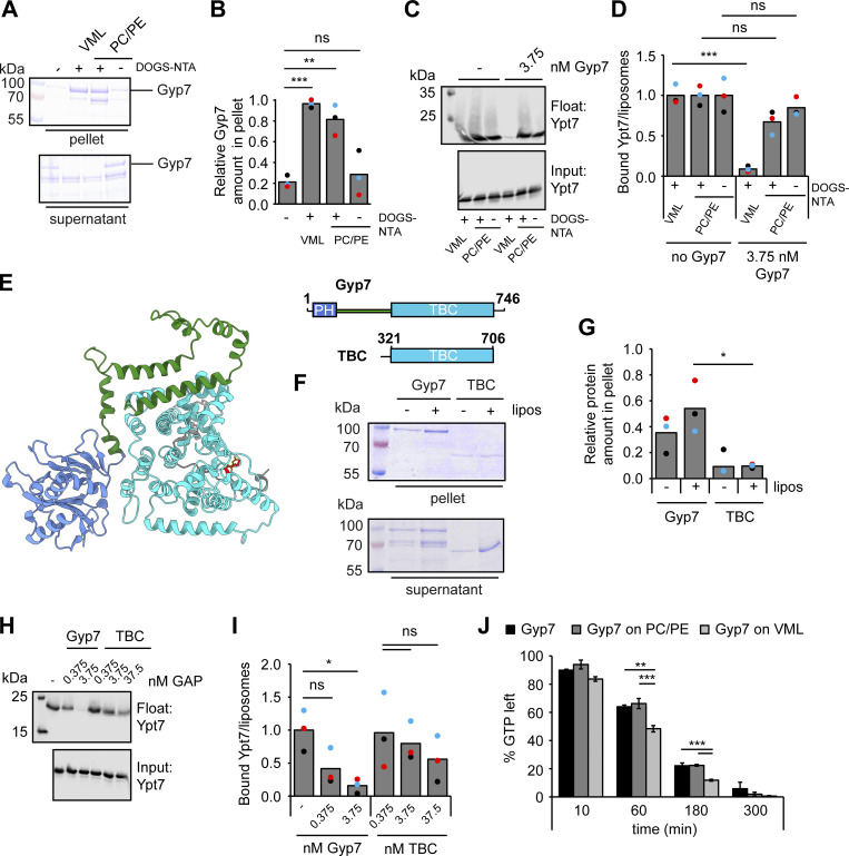
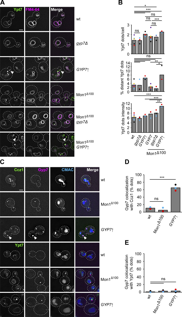
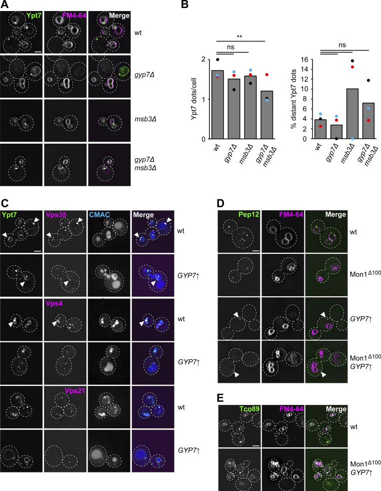
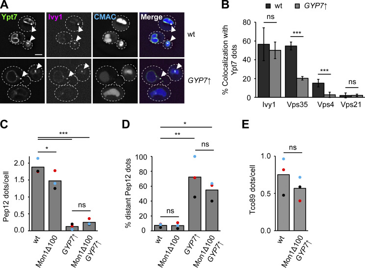
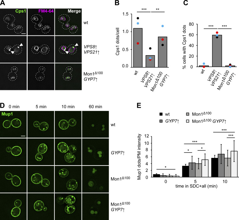
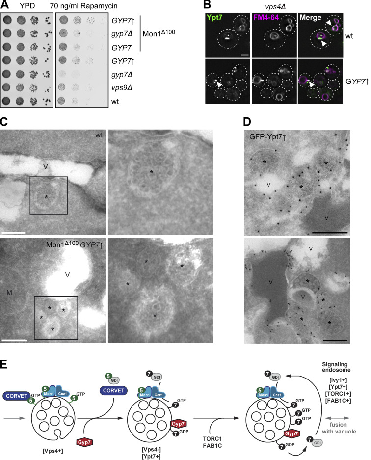
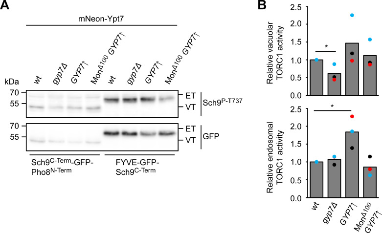
Organelles of the endomembrane system contain Rab GTPases as identity markers. Their localization is determined by guanine nucleotide exchange factors (GEFs) and GTPase activating proteins (GAPs). It remains largely unclear how these regulators are specifically targeted to organelles and how their activity is regulated. Here, we focus on the GAP Gyp7, which acts on the Rab7-like Ypt7 protein in yeast, and surprisingly observe the protein exclusively in puncta proximal to the vacuole. Mistargeting of Gyp7 to the vacuole strongly affects vacuole morphology, suggesting that endosomal localization is needed for function. In agreement, efficient endolysosomal transport requires Gyp7. In vitro assays reveal that Gyp7 requires a distinct lipid environment for membrane binding and activity. Overexpression of Gyp7 concentrates Ypt7 in late endosomes and results in resistance to rapamycin, an inhibitor of the target of rapamycin complex 1 (TORC1), suggesting that these late endosomes are signaling endosomes. We postulate that Gyp7 is part of regulatory machinery involved in late endosome function.


Conflict of interest statement

Disclosures: The authors declare no competing interests exist.

Figures

Figure 1.
Figure 1.
Gyp7 localization depends on a functional endosomal system. (A) Overview of Ypt7 function in fusion and fission reactions at the vacuole. For details, see text. (B) Localization of endogenously expressed Gyp7 and Msb3. Gyp7 and Msb3 were C-terminally tagged with mNeonGreen in wild-type (wt) and vps4Δ cells. Vacuolar membranes were stained with FM4-64 (see Materials and methods). Cells were imaged by fluorescence microscopy. Individual slices are shown. Arrowheads depict Gyp7 accumulations. Dashed lines indicate yeast cell boundaries. Scale bar, 2 μm. (C) Localization of endosomal markers relative to Gyp7. Marker proteins mCherry-Vps21 and Vps35-2xmKate were coexpressed in vps4Δ cells encoding endogenous Gyp7-mNeonGreen. Vacuoles were stained with CMAC (see Materials and methods). Cells were imaged by fluorescence microscopy. Individual slices are shown. Arrowheads depict representative colocalization. Dashed lines indicate yeast cell boundaries. Scale bar, 2 μm. (D) Quantification of Gyp7 puncta colocalizing with endosomal markers in C. Cells (n ≥ 100) from three independent experiments were quantified in Fiji. Bar graphs represent the averages from three experiments and puncta represent the mean of each experiment. (E) Localization of Gyp7 in selected deletion mutants. Gyp7 was tagged with mNeonGreen in wild-type, vps21Δ ypt52Δ, vps3Δ, vps45Δ, and mvp1Δ cells. Vacuolar membranes were stained with FM4-64. Cells were imaged by fluorescence microscopy and individual slices are shown. Dashed lines indicate yeast cell boundaries. Scale bar, 2 μm. (F) Quantification of Gyp7 puncta per cell in E and Fig. S1 A. Cells (n ≥ 100) from three independent experiments were quantified in Fiji. Bar graphs represent the averages from three experiments and puncta represent the mean of each experiment. P value **<0.01, ***<0.001, using ANOVA one-way test.
Figure S1.
Figure S1.
Gyp7 does not affect Ypt7 function in autophagy or vCLAMP formation. (A) Localization of Gyp7 in selected deletion mutants. Gyp7 was tagged with mNeonGreen in wild-type (wt), vps21Δ, ypt52Δ, ypt10Δ, ypt53Δ, vps9Δ muk1Δ, snx4Δ, vps5Δ, vps35Δ, and vps38Δ cells. Vacuolar membranes were stained with FM4-64. Cells were imaged by fluorescence microscopy and individual slices are shown. Dashed lines indicate yeast cell boundaries. Scale bar, 2 μm. (B) Localization of Atg8 upon deletion or overexpression of Gyp7. Gyp7 was either deleted or expressed from the TEF1 promoter in cells encoding the autophagy-specific marker protein mCherry-Atg8. Cells were grown in SDC+all and then shifted to SD-N for 1 h (see Materials and methods). Cells were imaged by fluorescence microscopy and individual slices are shown. Dashed lines indicate yeast cell boundaries. Scale bar, 2 μm. (C) Quantification of Atg8 dots per cell in B during growth. Cells (n ≥ 150) from three independent experiments were quantified in Fiji. Bar graphs represent the averages from three experiments and dots represent the mean of each experiment. P value ns, **<0.01, using ANOVA one-way test. (D) Quantification of Atg8 dots per cell in B during N-starvation. Quantification was performed as in C. P value ns, using ANOVA one-way test. (E) Formation of vCLAMPs upon deletion or overexpression of Gyp7. The vCLAMP-forming protein mCherry-Vps39 was expressed from the TEF1 promoter in cells encoding Gyp7-mNeonGreen, gyp7Δ, and TEF1pr-GYP7 cells. Mitochondria were stained with DAPI (see Materials and methods). Cells were imaged by fluorescence microscopy and individual slices are shown. Dashed lines indicate yeast cell boundaries. Scale bar, 2 μm. (F) Quantification of E. Colocalization of mCherry-Vps39 enrichments and DAPI-stained mitochondria was defined as vCLAMP. Cells with ≥1 vCLAMP were counted as vCLAMP-positive cells. Cells (n ≥ 150) from three independent experiments were quantified in Fiji. Bar graphs represent the averages from three experiments and dots represent the mean of each experiment. P value ns, using ANOVA one-way test.
Figure 2.
Figure 2.
Vacuolar localization of Gyp7 impairs vacuolar function. (A) Vacuole morphology upon galactose-induced overexpression of Gyp7. Gyp7 was expressed from the GAL1 promoter. Wild-type (wt) cells and cells encoding GAL1pr-GYP7 were grown in glucose- or galactose-containing media (see Materials and methods). Vacuolar membranes were stained with FM4-64. Cells were imaged by fluorescence microscopy and individual slices are shown. Dashed lines indicate yeast cell boundaries. Scale bar, 2 μm. (B) Quantification of the number of vacuoles per cell in A. Cells were grouped into three different classes: 1–2 vacuoles, 3–4 vacuoles (not shown), and >5 vacuoles. Cells (n ≥ 100) from three independent experiments were quantified in Fiji. Bar graphs represent the averages and error bars the SD from three experiments. P value ns, **<0.01, ***<0.001 using ANOVA one-way test. (C) Vacuole morphology of cells expressing Vps8- or Zrc1-Chromobody. Vps8 and Zrc1 were C-terminally tagged with a nanobody against GFP (CB). Vacuolar membranes were stained with FM4-64. Cells were imaged by fluorescence microscopy and individual slices are shown. Dashed lines indicate yeast cell boundaries. Scale bar, 2 μm. (D) Vacuole morphology of cells with Gyp7 targeted to endosomes or the vacuole. Vps8 and Zrc1 were C-terminally tagged with CB in cells expressing Gyp7-GFP. Where indicated, a Ypt7 fast-cycling mutant (Ypt7K127E) was expressed from an integrative plasmid. Vacuolar membranes were stained with FM4-64. Cells were imaged by fluorescence microscopy and individual slices are shown. Dashed lines indicate yeast cell boundaries. Scale bar, 2 μm. (E) Quantification of the number of vacuoles per cell in C and D. Cells were classified as in B. Cells (n ≥ 150) from three independent experiments were quantified in Fiji. Bar graphs represent the averages and error bars the SD from three experiments. P value *<0.05, **<0.01 and ***<0.001, using ANOVA one-way test. (F) Vacuole morphology of cells expressing Gyp7R458K, the catalytic dead mutant of Gyp7. The mutation was introduced into cells expressing Gyp7-GFP. Where indicated, Vps8 and Zrc1 were C-terminally tagged with a CB. Vacuolar membranes were stained with FM4-64. Cells were imaged by fluorescence microscopy and individual slices are shown. Dashed lines indicate yeast cell boundaries. Scale bar, 2 μm. (G) Quantification of the number of vacuoles per cell in F. Cells were classified as in B. Cells (n ≥ 130) from three independent experiments were quantified in Fiji. Bar graphs represent the averages and error bars the SD from three experiments. P value ns, using ANOVA one-way test.
Figure 3.
Figure 3.
Gyp7 is required for endosomal physiology and efficient endocytosis. (A) Growth assay on ZnCl2-containing plates. Indicated yeast strains were grown to the same OD600 in YPD media and serial dilutions were spotted onto agar plates containing YPD or YPD supplemented with 4 mM ZnCl2 (see Materials and methods). Plates were incubated at 30°C for several days before imaging. Images are representative for three independent experiments. (B) Growth assay on rapamycin-containing plates. Indicated yeast strains were spotted onto agar plates containing YPD or YPD supplemented with 50 ng/ml rapamycin as in A. Plates were incubated at 30°C for several days before imaging. Images are representative of three independent experiments. (C) Endocytosis of Mup1 in wild-type (wt) and gyp7Δ cells. Cells were grown to logarithmic phase in SDC-MET media, analyzed by fluorescence microscopy, and then shifted to SDC+all media. Cells were imaged at indicated time points by fluorescence microscopy. Individual slices are shown. Scale bar, 2 μm. (D) Quantification of the vacuole to plasma membrane fluorescence intensity ratio of Mup1 in C. The maximal fluorescence intensity of Mup1-GFP signal in the vacuolar lumen was divided by the maximal intensity of Mup1 at the plasma membrane. For each time point, cells (n ≥ 100) from three independent experiments were quantified in Fiji. Bar graphs represent the averages and error bars the SD from three experiments. P value ns, **<0.01, ***<0.001, using two-sided Student’s t test. (E) Quantification of Mup1-GFP puncta per cell in C. For each time point, cells (n ≥ 100) from three independent experiments were quantified in Fiji. Bar graphs represent the averages and error bars the SD from three experiments. P value ns, **<0.01, using two-sided Student’s t test. (F) Vacuole morphology of wild-type and gyp7Δ cells in growth and starvation conditions. Cells were grown in SDC+all and then shifted to SD-N for 2 h, where indicated (see Materials and methods). Cells were imaged by fluorescence microscopy and individual slices are shown. Dashed lines indicate yeast cell boundaries. Scale bar, 2 μm. (G) Quantification of the number of vacuoles per cell in F during growth. Cells were grouped into three different classes: 1–2 vacuoles, 3–4 vacuoles, and >5 vacuoles. Cells (n ≥ 150) from three independent experiments were quantified in Fiji. Bar graphs represent the averages from three experiments and puncta represent the mean of each experiment. P value ns, using ANOVA one-way test. (H) Quantification of the number of vacuoles per cell in F during nitrogen starvation. Cells were grouped as described in G. Cells (n ≥ 150) from three independent experiments were quantified in Fiji. Bar graphs represent the averages from three experiments and puncta represent the mean of each experiment. P value ns, using ANOVA one-way test.
Figure S2.
Figure S2.
Gyp7 function is required for normal TORC1 activity. (A) Growth assay on rapamycin-containing plates. Indicated yeast strains were grown to the same OD600 in YPD media and serial dilutions were spotted onto agar plates containing YPD or YPD supplemented with 50 ng/ml rapamycin. Plates were incubated at 30°C for several days before imaging. Images are representative of three independent experiments. (B) Endocytosis of Mup1 in wild-type (wt) and gyp7Δ cells as in Fig. 3 C. Cells were imaged at indicated time points by fluorescence microscopy. Individual slices are shown. Scale bar, 2 μm. (C) Quantification of the number of dots to plasma membrane (PM) intensity of Mup1 ratio in B and Fig. 3 C. For each cell, the number of Mup1 dots was divided by the maximum fluorescence intensity of Mup1-GFP signal at the plasma membrane. For each time point, cells (n ≥ 100) from three independent experiments were quantified in Fiji. Bar graphs represent the averages and error bars the SD from three experiments. P value ns, using two-sided Student’s t test.
Figure 4.
Figure 4.
Gyp7 requires a distinct membrane environment for efficient GAP activity. (A) Overview of the GDI extraction assay. 250 μM liposomes with VML composition are preloaded with 0.6 μM Ypt7-GDI complex in the presence of 3.75 mM EDTA and 125 μM GTP. The nucleotide binding is stabilized by addition of 7.5 mM MgCl2. Incubation with the GAP Gyp7 triggers GTP hydrolysis. GDI extracts inactivated Ypt7 from liposomal membranes. Liposomes with bound Ypt7 are floated in a sucrose gradient and separated from unbound protein. Floated membrane fractions and inputs were analyzed by western blotting (see Materials and methods). (B) Ypt7 inactivation increases with the concentration of Gyp7. Assay was performed as in A. Reactions were incubated with different amounts of Gyp7 for 1 h. Control reaction contained no Gyp7. 40% of the float was analyzed together with 3% input by western blotting using an anti-Ypt7 antibody. (C) Quantification of bound Ypt7 to liposomes in B. Band intensity of Ypt7 signal in float was measured in Fiji and compared with input. Reactions containing Gyp7 were normalized to the average value of the control reaction. Bar graphs represent the averages from three independent experiments and puncta represent the mean of each experiment. P value ns, *<0.05, **<0.01, using ANOVA one-way test. (D) Kinetics of Gyp7 activity toward Ypt7-GTP. Assay was performed as in A. Reactions were incubated with 0.75 nM Gyp7 for different time points. Control reaction contained no Gyp7. 40% of the float was analyzed together with 3% input by western blotting using an anti-Ypt7 antibody. (E) Quantification of bound Ypt7 to liposomes in D. Quantification was performed as in C. P value *<0.05, **<0.01, ***<0.001 using ANOVA one-way test. (F) Membrane association of Gyp7. 715 μM liposomes with VML composition were incubated with 715 nM Gyp7 for 10 min. Membranes were separated from supernatant by centrifugation at 100,000 g and both fractions were analyzed by SDS-PAGE and Coomassie staining. Control reaction contained no liposomes (see Materials and methods). (G) Quantification of the relative Gyp7 amount in the pellet in F. Band intensity of Gyp7 signal in the pellet was measured in Fiji and compared with Gyp7 signal in the supernatant. Bar graphs represent the averages from three independent experiments and puncta represent the mean of each experiment. P value *<0.05, using two-sided Student’s t test. (H) Comparison of Gyp7 activity on liposomes with VML composition and PC/PE liposomes. The assay was performed as in A. 3.75 nM Gyp7 was added to reactions containing liposomes with VML composition or PC/PE liposomes for 10 min. Control reactions contained respective liposomes and no Gyp7. 40% of the float was analyzed together with 3% input by western blotting using an anti-Ypt7 antibody. (I) Quantification of bound Ypt7 to liposomes in H. Quantification was performed as in C. Reactions containing Gyp7 were normalized to the average value of the respective control reaction. P value *<0.05, **<0.01, using ANOVA one-way test. (J) Association of Gyp7 with liposomes of VML composition and PC/PE liposomes. 715 nM Gyp7 was incubated with 715 μM liposomes for 0 and 10 min. Membrane association was analyzed as in F. (K) Quantification of the relative Gyp7 amount in the pellet in J. Quantification was performed as in G. P value ns, *<0.05, using ANOVA one-way test. (L) Comparison of Gyp1-46 activity on liposomes with VML composition and PC/PE liposomes. Assay was performed as in A, except for the addition of Gyp1-46 instead of Gyp7 to reactions. Reactions were incubated with different amounts of Gyp1-46 for 10 min. Control reactions contained respective liposomes and no GAP. 40% of the float was analyzed together with 3% input by western blotting using an anti-Ypt7 antibody. (M) Quantification of bound Ypt7 to liposomes in L. Quantification was performed as in C. Reactions containing Gyp1-46 were normalized to the average value of the respective control reaction. P value *<0.05, using ANOVA one-way test. Source data are available for this figure: SourceData F4.
Figure S3.
Figure S3.
The N-terminal PH domain of Gyp7 does not bind membranes. (A) 250 μM PC/PE liposomes or liposomes of VML composition were preloaded with 0.6 μM Ypt7-GDI complex in the presence of 3.75 mM EDTA and 125 μM GTP. Nucleotide binding was stabilized by addition of 7.5 mM MgCl2. Reactions were incubated with different amounts of Gyp7 and, where indicated, with 6 μM GDI for 10 min. Liposomes were floated in a sucrose gradient. Control reaction contained respective liposomes of VML composition and no Gyp7. 40% of the float was analyzed together with 3% input by western blotting using an anti-Ypt7 antibody. (B) Quantification of bound Ypt7 to liposomes in A. Band intensity of Ypt7 signal in float was measured in Fiji and compared with input. Reactions containing Gyp7 were normalized to the average value of the control reaction. Bar graphs represent the averages from three independent experiments and dots represent the mean of each experiment. P value ns, using ANOVA one-way test. (C) AlphaFold2 structure prediction of Gyp7 color-coded according to the pLDDT values. (D) Plot of the Predicted Aligned Error of C with the PH and TBC domain of Gyp7 labeled. (E) Comparison of full-length Gyp7 and its PH domain. Two PH domain constructs (PH = aa 1–197; PH+ = aa 1–205) contain the N-terminal region of Gyp7 (aa 1–746). (F) Membrane association of the PH domain compared to full-length Gyp7. 715 nM protein was incubated with 715 μM liposomes of VML composition for 10 min. Membranes were separated from supernatant by centrifugation at 100,000 g and both fractions were analyzed by SDS-PAGE and Coomassie staining. Control reactions contained no liposomes. (G) Quantification of the relative protein amount in the pellet in F. Band intensity of protein signal in the pellet was measured in Fiji and compared with the protein signal in the supernatant. Bar graphs represent the averages from three independent experiments and dots represent the mean of each experiment. P value ns, using ANOVA one-way test. (H) Comparison of the Gyp7 TBC domain activity toward soluble Ypt7-GTP in solution and on membranes. 5 μM Ypt7 was incubated with 5 μM GAP and 50 μM GTP in the presence of 1 mM DTT, 20 mM EDTA, and 5 mM MgCl2. Where indicated, reactions contained 1 mM liposomes with VML composition. Control reactions contained no Ypt7, no GAP, or neither Ypt7 nor GAP (see Fig. S3 I). Reactions were stopped after 0, 10, 60, 180, and 300 min by snap-freezing and boiling at 95°C. Samples were applied to a HPLC system and the absorbance of GDP and GTP was monitored at 254 nm. Peaks were analyzed with OpenChrom and for each time point the percentage of GDP and GTP in the samples was determined. The percentage of GTP left at each time point was normalized to the respective percentage of GTP at t = 0 min. Normalized % GTP left plotted against the time in min. Bar graphs represent the averages and error bars the SD from three independent experiments. P value *<0.05, ***<0.001, using ANOVA one-way test. (I) No major GTP hydrolysis in control reactions of Fig. 5 J and H after 300 min. Control reactions contained no Ypt7, no GAP, or neither Ypt7 nor GAP. For each time point the percentage of GDP and GTP in the reactions was determined. The percentage of GTP left of each sample at t = 300 min was normalized to the respective percentage of GTP at t = 0 min. Normalized % GTP left plotted against the time in min. Bar graphs represent the averages and error bars the SD from three independent experiments. P value *<0.05, **<0.01, using ANOVA one-way test. Source data are available for this figure: SourceData FS3.
Figure 5.
Figure 5.
Gyp7 is activated by a distinct membrane environment. (A) Membrane association of Gyp7 with DOGS-NTA containing liposomes. 715 nM Gyp7 was incubated with 715 μM liposomes (VML + DOGS-NTA, PC/PE + DOGS-NTA, PC/PE) for 10 min. Membranes were separated from supernatant by centrifugation at 100,000 g and both fractions were analyzed by SDS-PAGE and Coomassie staining. Control reaction contained no liposomes. (B) Quantification of the relative Gyp7 amount in the pellet in A. Band intensity of Gyp7 signal in the pellet was measured in Fiji and compared with Gyp7 signal in the supernatant. Bar graphs represent the averages from three independent experiments and puncta represent the mean of each experiment. P value ns, **<0.01, ***<0.001, using ANOVA one-way test. (C) Comparison of Gyp7 activity on DOGS-NTA containing liposomes. 250 μM liposomes were preloaded with 0.6 μM Ypt7:GDI complex in the presence of 3.75 mM EDTA and 125 μM GTP. Nucleotide binding was stabilized by addition of 7.5 mM MgCl2. Reactions were incubated with 3.75 μM Gyp7 for 10 min. Liposomes were floated in a sucrose gradient. Control reactions contained no Gyp7. 40% of the float was analyzed together with 3% input by western blotting using an anti-Ypt7 antibody. (D) Quantification of bound Ypt7 to liposomes in C. Band intensity of Ypt7 signal in float was measured in Fiji and compared to input. Reactions containing Gyp7 were normalized to the average value of the respective control reaction. Bar graphs represent the averages from three independent experiments and puncta represent the mean of each experiment. P value ns, ***<0.001, using ANOVA one-way test. (E) AlphaFold2 structure prediction of Gyp7. The N-terminal PH domain is colored blue and the C-terminal TBC domain is colored cyan with the catalytic Arg (R458) and Glu (Q531) residues shown red in stick representation. A middle domain, which is modeled with low predicted local distance difference test (pLDDT) confidence scores (Fig. S3, C and D), is colored green. (F) Membrane association of the TBC domain compared to full-length Gyp7. Gyp7 and the TBC domain were incubated with liposomes of VML composition as in A. Control reactions contained no liposomes. (G) Quantification of the relative amount of Gyp7 in the pellet in F. Quantification performed as in B. P value *<0.05, using ANOVA one-way test. (H) Comparison of Gyp7 and TBC domain activities on liposomes with VML composition. Assay was performed as in C. Pre-loaded liposomes were incubated with different amounts of Gyp7 or the TBC domain for 10 min. (I) Quantification of bound Ypt7 to liposomes in H. Quantification was performed as in D. Reactions containing GAP were normalized to the average value of the control reaction. P value ns, *<0.05, using ANOVA one-way test. (J) Comparison of Gyp7 activity toward soluble Ypt7-GTP in solution and on membranes. 5 μM Ypt7 was incubated with 5 μM GAP and 50 μM GTP in the presence of 1 mM DTT, 20 mM EDTA, and 5 mM MgCl2. Where indicated, reactions contained 1 mM liposomes with VML composition or PC/PE liposomes. Control reactions contained no Ypt7, no GAP, or neither Ypt7 nor GAP (see Fig. S3 I). Reactions were stopped after 0, 10, 60, 180, and 300 min by snap-freezing and boiling at 95°C. Samples were applied to a HPLC system and the absorbance of GDP and GTP was monitored at 254 nm. Peaks were analyzed with OpenChrom and for each time point the percentage of GDP and GTP in the samples was determined. The percentage of GTP left at each time point was normalized to the respective percentage of GTP at t = 0 min. Normalized % GTP left plotted against the time in min. Bar graphs represent the averages and error bars the SD from three independent experiments. P value **<0.01, ***<0.001, using ANOVA one-way test. Source data are available for this figure: SourceData F5.
Figure 6.
Figure 6.
Gyp7 and Mon1–Ccz1 shift Ypt7 from the vacuole to dot-like structures. (A) The localization of Ypt7 depends on the expression level or activity of Gyp7 and Mon1–Ccz1. Endogenous mNeon-Ypt7 was expressed from an integrative plasmid in ypt7Δ cells. Where indicated, 100 amino acids at the N-terminus of Mon1 were deleted (Mon1Δ100). Gyp7 was either deleted or expressed from the TEF1 promoter in mNeon-Ypt7 expressing cells with wild-type (wt) Mon1 or Mon1Δ100. Vacuolar membranes were stained with FM4-64. Cells were imaged by fluorescence microscopy. Individual slices are shown. Arrowheads depict Ypt7 accumulations not proximal to the vacuole. Dashed lines indicate yeast cell boundaries. Scale bar, 2 μm. (B) Quantification of the total number of Ypt7 puncta per cell, the percentage of distant Ypt7 puncta, and the fluorescence intensity of Ypt7 puncta in A. The number of distant Ypt7 puncta (not at the vacuole) was divided by the total number of Ypt7 puncta per cell. The maximum fluorescence intensity of mNeon-Ypt7 puncta was normalized to the maximum fluorescence intensity of mNeon-Ypt7 at the vacuolar membrane. Cells (n ≥ 100) from three independent experiments were quantified in Fiji. Bar graphs represent the averages from three experiments and puncta represent the mean of each experiment. P value ns, *<0.05, **<0.01, ***<0.001, using ANOVA one-way test. (C) Localization of Gyp7 relative to Ypt7 and Mon1–Ccz1. Gyp7 was C-terminally tagged with 2xmKate in the Mon1100 strain, in TEF1pr-GYP7 or wild-type cells encoding endogenous Ccz1-mNeon (top) or mNeon-Ypt7 (bottom). Vacuoles were stained with CMAC. Cells were imaged by fluorescence microscopy. Individual slices are shown. Arrowheads depict representative colocalization. Dashed lines indicate yeast cell boundaries. Scale bar, 2 μm. (D) Quantification of Gyp7 puncta colocalizing with Ccz1 puncta in C. Cells (n ≥ 100) from three independent experiments were quantified in Fiji. Bar graphs represent the averages from three experiments and puncta represent the mean of each experiment. P value ns, ***<0.001, using ANOVA one-way test. (E) Quantification of Gyp7 puncta colocalizing with Ypt7 puncta in C. Cells (n ≥ 100) from three independent experiments were quantified in Fiji. Bar graphs represent the averages from three experiments and puncta represent the mean of each experiment. P value ns, using ANOVA one-way test.
Figure S4.
Figure S4.
Confined Ypt7 dots correspond to signaling endosomes. (A) Localization of mNeon-Ypt7 in wild-type (wt) cells and cells with Gyp7 and/or Msb3 deleted. Vacuolar membranes were stained with FM4-64. Cells were imaged by fluorescence microscopy. Individual slices are shown. Dashed lines indicate yeast cell boundaries. Scale bar, 2 μm. (B) Quantification of the total number of Ypt7 dots per cell and the percentage of distant Ypt7 dots in A. The number of distant Ypt7 dots (not at the vacuole) was divided by the total number of Ypt7 dots per cell. Cells (n ≥ 150) from three independent experiments were quantified in Fiji. Bar graphs represent the averages from three experiments and dots represent the mean of each experiment. P value ns, **<0.01, using ANOVA one-way test. (C) Localization of mNeon-Ypt7 dots relative to endosomal marker proteins. Endosomal markers Vps35-mKate, Vps4-3xHA-mCherry, and mCherry-Vps21 were co-expressed in TEF1pr-GYP7 or wild-type cells encoding endogenous mNeon-Ypt7. Vacuoles were stained with CMAC. Cells were imaged by fluorescence microscopy. Individual slices are shown. Arrowheads depict representative colocalization. Dashed lines indicate yeast cell boundaries. Scale bar, 2 μm. (D) Localization of GFP-Pep12 in wild-type cells and cells expressing Gyp7 from the TEF1 promoter and/or Mon1Δ100-Ccz1. Vacuolar membranes were stained with FM4-64. Cells were imaged by fluorescence microscopy. Individual slices are shown. Arrowheads depict distant Pep12 dots. Dashed lines indicate yeast cell boundaries. Scale bar, 2 μm. (E) Localization of Tco89-mNeon in wild-type and TEF1pr-GYP7 cells. Vacuolar membranes were stained with FM4-64. Cells were imaged by fluorescence microscopy. Individual slices are shown. Dashed lines indicate yeast cell boundaries. Scale bar, 2 μm.
Figure 7.
Figure 7.
Ypt7-positive puncta correspond to signaling endosomes. (A) Localization of mNeon-Ypt7 puncta relative to the endosomal marker Ivy1. Ivy1-mKate was expressed in TEF1pr-GYP7 or wild-type (wt) cells encoding endogenous mNeon-Ypt7. Vacuoles were stained with CMAC. Cells were imaged by fluorescence microscopy. Individual slices are shown. Arrowheads depict representative colocalization. Dashed lines indicate yeast cell boundaries. Scale bar, 2 μm. (B) Quantification of Ypt7 colocalizing with endosomal markers in A and Fig. S4 C. Cells (n ≥ 100) from three independent experiments were quantified in Fiji. Bar graphs represent the averages and error bars the SD from the three experiments. P value ns, ***<0.001, using two-sided Student’s t test. (C) Quantification of the number of Pep12 puncta per cell in Fig. S4 D. Cells (n ≥ 150) from three independent experiments were quantified in Fiji. Bar graphs represent the averages from three experiments and puncta represent the mean of each experiment. P value *<0.05, ***<0.001, using ANOVA one-way test. (D) Quantification of the percentage of distant Pep12 puncta in Fig. S4 D. The number of distant Pep12 puncta (not at the vacuole) was divided by the total number of Pep12 puncta per cell. Cells (n ≥ 150) from three independent experiments were quantified in Fiji. Bar graphs represent the averages from three experiments and puncta represent the mean of each experiment. P value *<0.05, **<0.01, using ANOVA one-way test. (E) Quantification of the number of Tco89 puncta per cell in Fig. S4 E. Cells (n ≥ 150) from three independent experiments were quantified in Fiji. Bar graphs represent the averages from three experiments and puncta represent the mean of each experiment. P value ns, using two-sided Student’s t test.
Figure 8.
Figure 8.
Enhanced Ypt7 cycling affects endocytic trafficking. (A) Localization of Cps1 in wild-type (wt), TEF1pr-VPS8 ADHpr-VPS21, and Mon1Δ100-Ccz1 TEF1pr-GYP7 cells. Vacuolar membranes were stained with FM4-64. Cells were imaged by fluorescence microscopy. Individual slices are shown. Arrowheads depict Cps1 accumulations next to the vacuole. Dashed lines indicate yeast cell boundaries. Scale bar, 2 μm. (B) Quantification of the number of Cps1 puncta per cell in A. Cells (n ≥ 140) from three independent experiments were quantified in Fiji. Bar graphs represent the averages from three experiments and puncta represent the mean of each experiment. P value **<0.01, ***<0.001, using ANOVA one-way test. (C) Quantification of the percentage of cells with Cps1 accumulations in A. The number of cells with Cps1 accumulations at the vacuole was divided by the total number of cells. Cells (n ≥ 140) from three independent experiments were quantified in Fiji. Bar graphs represent the averages from three experiments and puncta represent the mean of each experiment. P value ***<0.001, using ANOVA one-way test. (D) Endocytosis of Mup1 in cells with altered expression or activity of Gyp7 and Mon1–Ccz1. Cells were grown to logarithmic phase in SDC-MET media, analyzed by fluorescence microscopy, and then shifted to SDC+all media. Cells were imaged at indicated time points by fluorescence microscopy. Individual slices are shown. Scale bar, 2 μm. This is the same assay shown in Fig. 3 C with different mutants and time points analyzed. (E) Quantification of the number of puncta to plasma membrane fluorescence intensity of Mup1 ratio in D. For each cell, the number of Mup1 puncta was divided by the maximum fluorescence intensity of Mup1-GFP signal at the plasma membrane (PM). For each time point, cells (n ≥ 100) from three independent experiments were quantified in Fiji. Bar graphs represent the averages and error bars the SD from three experiments. P value *<0.05, ***<0.001, using ANOVA one-way test.
Figure 9.
Figure 9.
Ypt7 functions on mature endosomes. (A) Growth assay on rapamycin-containing plates. Indicated yeast strains were grown to the same OD600 in YPD media and serial dilutions were spotted onto agar plates containing YPD or YPD supplemented with 70 ng/ml rapamycin. Plates were incubated at 37°C for several days before imaging. Images are representative of three independent experiments. (B) Ypt7 accumulates in the Class E compartment. Endogenous mNeon-Ypt7 was expressed from an integrative plasmid in ypt7Δ vps4Δ cells. Where indicated, Gyp7 was expressed from the TEF1 promoter. Vacuolar membranes were stained with FM4-64. Cells were imaged by fluorescence microscopy. Individual slices are shown. Arrowheads depict Ypt7 accumulations in the Class E compartment. Dashed lines indicate yeast cell boundaries. Scale bar, 2 μm. (C) Electron microscopy analysis of cells expressing mNeon-Ypt7 in wild-type (wt) and Mon1∆100-Ccz1 TEF1pr-GYP7 cells (see Materials and methods). M, mitochondria; V, vacuole; asterisk, multivesicular body. Scale bars, 200 nm. (D) IEM analysis of cells expressing TEF1pr-GFP-YPT7. Ypt7 was detected by using anti-GFP antibodies and protein A–conjugated gold (see Materials and methods). Asterisk, multivesicular body; V, vacuole. Scale bars, 200 nm. (E) Working model of Gyp7 function on MVBs. MVBs form with the help of ESCRTs on Vps21/Rab5-positive endosomes (left), which carry yet inactive Mon1–Ccz1. Maturation of endosomes includes recruitment of Gyp7 and loss of Rab5 and its effector CORVET. Some of these late endosomes also acquire TORC1 and the Fab1 complex, thus turn into signaling endosomes. This may affect Gyp7 and Mon1–Ccz1 activity and thus control the available Ypt7 pool.
Figure S5.
Figure S5.
Gyp7 affects endosomal and vacuolar TORC1 activity. (A) mNeon-Ypt7 background cells with either wild-type (wt) expression levels of Gyp7, Gyp7 deleted, Gyp7 overexpressed from the TEF1 promoter or both overexpressed Gyp7 combined with truncation of Mon1 (Mon1∆100) were transformed with either vacuolar (VT, Sch9C-term-GFP-Pho8N-term) or endosomal (ET, FYVE-GFP-Sch9C-term) TORC1 activity reporters and grown exponentially in SDC+all. After SDS-PAGE analysis of the corresponding extracted proteins, the expression of the ET/VT reporters was detected by immunoblotting using anti-GFP antibodies, and their phosphorylation levels by using phospho-specific anti-Sch9-pThr737 antibodies were analyzed likewise. (B) Quantifications of the ET/VT assay in A, expressed as the ratios of Sch9-pThr737/GFP signals and normalized with wild-type cells. Bar graphs represent the averages from three independent experiments, and dots represent the mean of each experiment. P value *<0.05, using a two-sided Student’s t test. Source data are available for this figure: SourceData FS5.

References

    1. Albert, S., Will E., and Gallwitz D.. 1999. Identification of the catalytic domains and their functionally critical arginine residues of two yeast GTPase-activating proteins specific for Ypt/Rab transport GTPases. EMBO J. 18:5216–5225. 10.1093/emboj/18.19.5216 - DOI - PMC - PubMed
    1. Araki, M., Yoshimoto K., Ohta M., Katada T., and Kontani K.. 2021. Development of a versatile HPLC-based method to evaluate the activation status of small GTPases. J. Biol. Chem. 297:101428. 10.1016/j.jbc.2021.101428 - DOI - PMC - PubMed
    1. Arlt, H., Auffarth K., Kurre R., Lisse D., Piehler J., and Ungermann C.. 2015. Spatiotemporal dynamics of membrane remodeling and fusion proteins during endocytic transport. Mol. Biol. Cell. 26:1357–1370. 10.1091/mbc.e14-08-1318 - DOI - PMC - PubMed
    1. Babst, M., Wendland B., Estepa E.J., and Emr S.D.. 1998. The Vps4p AAA ATPase regulates membrane association of a Vps protein complex required for normal endosome function. EMBO J. 17:2982–2993. 10.1093/emboj/17.11.2982 - DOI - PMC - PubMed
    1. Balderhaar, H.J., Arlt H., Ostrowicz C., Bröcker C., Sündermann F., Brandt R., Babst M., and Ungermann C.. 2010. The Rab GTPase Ypt7 is linked to retromer-mediated receptor recycling and fusion at the yeast late endosome. J. Cell Sci. 123:4085–4094. 10.1242/jcs.071977 - DOI - PubMed

Publication types

MeSH terms

Substances