. 2024 May 1;35(5):ar66.
doi: 10.1091/mbc.E23-05-0204. Epub 2024 Mar 27.

After wounding, a G-protein coupled receptor promotes the restoration of tension in epithelial cells

Affiliations

After wounding, a G-protein coupled receptor promotes the restoration of tension in epithelial cells

Ivy S Han et al. Mol Biol Cell. .

Abstract

The maintenance of epithelial barrier function involves cellular tension, with cells pulling on their neighbors to maintain epithelial integrity. Wounding interrupts cellular tension, which may serve as an early signal to initiate epithelial repair. To characterize how wounds alter cellular tension we used a laser-recoil assay to map cortical tension around wounds in the epithelial monolayer of the Drosophila pupal notum. Within a minute of wounding, there was widespread loss of cortical tension along both radial and tangential directions. This tension loss was similar to levels observed with Rok inactivation. Tension was subsequently restored around the wound, first in distal cells and then in proximal cells, reaching the wound margin ∼10 min after wounding. Restoring tension required the GPCR Mthl10 and the IP3 receptor, indicating the importance of this calcium signaling pathway known to be activated by cellular damage. Tension restoration correlated with an inward-moving contractile wave that has been previously reported; however, the contractile wave itself was not affected by Mthl10 knockdown. These results indicate that cells may transiently increase tension and contract in the absence of Mthl10 signaling, but that pathway is critical for fully resetting baseline epithelial tension after it is disrupted by wounding.


Figures

FIGURE 1:
FIGURE 1:
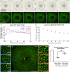
Tension is lost around wounds. (A) D melanogaster pupa (left) with pupal case removed (right) to expose notum (outlined by black rectangle). (B) Map of distances where tension was measured. Wound is designated as 0 µm (yellow X). Tension was measured at ∼70, 110, and 210 µm on either side of the wound (yellow lightning bolts). Map is overlaid on an image of the notum with pnr domain labeled by red nuclei. Closest distance (∼70 µm) corresponds to the edge of nuclear membrane damage visible in wounded samples. (C) Two types of borders measured: ML borders, which are radial to the wound at this location, and AP borders, which are tangential to the wound at this location. (D) Example of laser-induced recoil for a ML cell border. TCJs of ablated cell border indicated by magenta arrows. See also: Supplemental Movie 1. (D′) Diagram illustrating how laser-induced recoil reports cortical tension: low tension corresponds to low recoil velocity; high tension corresponds to high recoil velocity. (E) In unwounded control samples, tension was fairly constant at all locations tested for both ML and AP borders. n = 10 to 16 for each bin. (F) In unwounded pnr > Rok RNAi samples, significantly lower tension was detected in both ML and AP borders. Reduced tension was confined to the domain expressing Rok RNAi. n = 3 to 7 for each bin. (G) In wounded control samples, tension was reduced in a gradient for both ML and AP borders, with lower tension closer to the wound. Measurements taken 1–10 min postwound. n = 9 to 22 for each bin. Graph bars represent mean ± SEM. *p < 0.05, **p < 0.01, ***p < 0.001, ****p < 0.0001 by one-way ANOVA with multiple comparisons. Scale bars = 50 µm (B and C), or 10 µm (D).
FIGURE 2:
FIGURE 2:
Tension is restored over time. (A) At ∼70 µm from the wound, cortical tension is reduced by half by 1–2 min after wounding and restored to prewound levels within 15–18 min. Data collected 2–8 min after wounding shows a mix of tension levels (light gray, from Figure 1G); n = 6, 7, 30, and 9. (B) Data from panel A showing average recoil displacements versus time. (C) At ∼135 µm from the wound, cortical tension is reduced by half by 30 s after wounding and restored to prewound levels within 5–6 min; n = 10, 11, and 6. (D) Data from panel C showing average recoil displacements versus time. Graph bars represent mean ± SEM. *p < 0.05, **p < 0.01, ****p < 0.0001 by one-way ANOVA with multiple comparisons. Shaded regions in B and D represent ± one SD. All measurements conducted on ML borders.
FIGURE 3:
FIGURE 3:
Tension is restored as the contractile wave moves toward the wound. (A) A wave of cell contractions travels inward over 12 min after wounding and can be visualized in Ecad-GFP-labeled pupae by measuring displacement fields between successive frames and calculating the local area strain (one of n = 5 shown). Negative values correspond to contractions. See also Supplemental Movie 2. (B) The contractile wave can also be visualized as a wave of apical MyoII-GFP accumulation traveling inward after wounding (one of n = 7 shown). See also Supplemental Movie 3. Note that A and B show different wounds. (C) Peaks of radial MyoII-GFP intensity from B move closer to the wound over time, tracking progression of the contractile wave. (D) The average location of the wave over time, as determined from data similar to panel C averaged over n = 7 wounds. (E-E′′) Using Ecad-GFP to monitor the contractile wave, tension was measured before (blue) and after (red) the contractile wave passed through a cell ∼70 µm from the wound. Yellow arrowheads in E′ and E′′ show contracting cells. See also Supplemental Movie 4. (F) Tension at ∼70 µm from the wound is restored as the wave passes through. Graph bars represent mean ± SEM. n = 3 and 3; **p < 0.01 by paired sample t test. Scale bars = 50 µm (A, B, and E), or 10 µm (E′ and E′′).
FIGURE 4:
FIGURE 4:
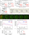
The restoration of tension after wounding requires the GPCR Mthl10. (A) In unwounded flies, knockdown of mthl10 within the pnr domain had no effect on cortical tension for both ML and AP borders; n = 3 to 7 per bin. (B) The wound-induced loss of tension persists after wounding in the pnr domain where mthl10 was knocked down, indicating that Mthl10 is required for the restoration of tension; n = 3 to 9 per bin. (C) IP3R is downstream of Mthl10 and is also required for the restoration of tension after wounding, indicating that Mthl10 signaling restores tension through IP3R. Measurements in B-C taken 1–10 min postwound; n = 10 to 14 per bin. (D–G) The contractile wave does not require mthl10. Wave symmetry was not disturbed when mthl10 was knocked down in the pnr domain, as visualized with area strain maps calculated for displacement fields between successive Ecad-GFP images (D; n = 4; see also Supplemental Movie 5) or with MyoII-GFP (E; n = 7, see also Supplemental Movie 6). Radial profile analysis (F and F′) showed no significant difference between control and pnr domains in pnr > mthl10 RNAi samples compared with control samples (G; n = 6 and 8). (H) The contractile wave exists without mthl10, but it requires mthl10 to restore tension after wounding. Tension is not restored at ∼70 µm even 15 min postwound in pnr > mthl10RNAi samples. Graph bars represent mean ± SEM; n = 5 and 5; *p < 0.05, ***p < 0.001, ****p < 0.0001 by one-way ANOVA with multiple comparisons (A, B, and C), unpaired t test (G), or paired sample t test (H). Scale bars = 50 µm.
FIGURE 5:
FIGURE 5:
Mthl10 is also required for tension restoration around scanned wounds. (A and B) In scan-wounded control samples, there is no evident wave of contraction (n = 5; see also Supplemental Movie 7) or myosin accumulation (n = 5; see also Supplemental Movie 8). Different wounds are shown in A and B. (C) Within ∼2–10 min of completing a scanned wound, postwound tension is fairly uniform at all locations tested; n = 4 to 8 per bin. (D) In contrast, for scan-wounded pnr > mthl10 RNAi samples, there is reduced tension in the knockdown domain, indicating that scanned wounds also require Mthl10 for tension restoration; n = 9 to 10 per bin. All measurements were taken on ML cell–cell borders. Graph bars represent mean ± SEM. ***p < 0.001, ****p < 0.0001 by one-way ANOVA with multiple comparisons. Scale bars = 50 µm.
FIGURE 6:
FIGURE 6:
Schematic of how Mthl10-driven calcium signaling and the wound-induced contractile wave coordinate to restore tension around epithelial wounds.

Update of

Similar articles

Cited by

References

    1. Alisafaei F, Chen X, Leahy T, Janmey PA, Shenoy VB (2021). Long-range mechanical signaling in biological systems. Soft Matter 17, 241–253. - PMC - PubMed
    1. Antunes M, Pereira T, Cordeiro JV, Almeida L, Jacinto A (2013). Coordinated waves of actomyosin flow and apical cell constriction immediately after wounding. J Cell Biol 202, 365–379. - PMC - PubMed
    1. Bonnet I, Marcq P, Bosveld F, Fetler L, Bellaiche Y, Graner F (2012). Mechanical state, material properties and continuous description of an epithelial tissue. J R Soc Interface 9, 2614–2623. - PMC - PubMed
    1. Brand AH, Perrimon N (1993). Targeted gene expression as a means of altering cell fates and generating dominant phenotypes. Development 118, 401–415. - PubMed
    1. Cao J, Wang J, Jackman CP, Cox AH, Trembley MA, Balowski JJ, Cox BD, De Simone A, Dickson AL, Di Talia S, et al. (2017). Tension creates an endoreplication wavefront that leads regeneration of epicardial tissue. Dev Cell 42, 600–615.e604. - PMC - PubMed

Substances

LinkOut - more resources