

Arterial spin labelling magnetic resonance imaging and perfusion patterns in neurocognitive and other mental disorders: a systematic review
- PMID: 38536448
- PMCID: PMC11150205
- DOI: 10.1007/s00234-024-03323-0
Arterial spin labelling magnetic resonance imaging and perfusion patterns in neurocognitive and other mental disorders: a systematic review
Abstract
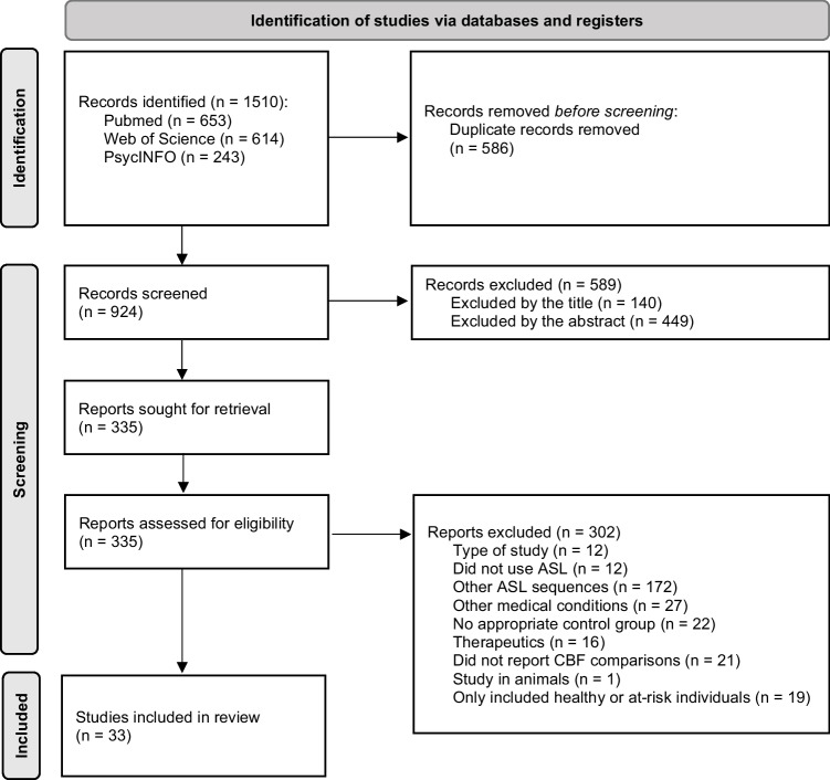
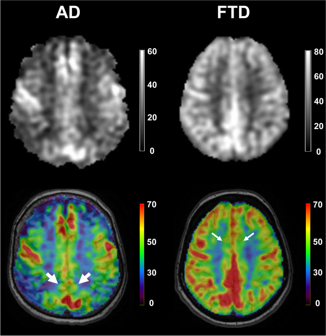
We reviewed 33 original research studies assessing brain perfusion, using consensus guidelines from a "white paper" issued by the International Society for Magnetic Resonance in Medicine Perfusion Study Group and the European Cooperation in Science and Technology Action BM1103 ("Arterial Spin Labelling Initiative in Dementia"; https://www.cost.eu/actions/BM1103/ ). The studies were published between 2011 and 2023 and included participants with subjective cognitive decline plus; neurocognitive disorders, including mild cognitive impairment (MCI), Alzheimer's disease (AD), frontotemporal lobar degeneration (FTLD), dementia with Lewy bodies (DLB) and vascular cognitive impairment (VCI); as well as schizophrenia spectrum disorders, bipolar and major depressive disorders, autism spectrum disorder, attention-deficit/hyperactivity disorder, panic disorder and alcohol use disorder. Hypoperfusion associated with cognitive impairment was the major finding across the spectrum of cognitive decline. Regional hyperperfusion also was reported in MCI, AD, frontotemporal dementia phenocopy syndrome and VCI. Hypoperfused structures found to aid in diagnosing AD included the precunei and adjacent posterior cingulate cortices. Hypoperfused structures found to better diagnose patients with FTLD were the anterior cingulate cortices and frontal regions. Hypoperfusion in patients with DLB was found to relatively spare the temporal lobes, even after correction for partial volume effects. Hyperperfusion in the temporal cortices and hypoperfusion in the prefrontal and anterior cingulate cortices were found in patients with schizophrenia, most of whom were on medication and at the chronic stage of illness. Infratentorial structures were found to be abnormally perfused in patients with bipolar or major depressive disorders. Brain perfusion abnormalities were helpful in diagnosing most neurocognitive disorders. Abnormalities reported in VCI and the remaining mental disorders were heterogeneous and not generalisable.
Keywords: Arterial spin labeling; Cerebral blood flow; Cognitive impairment; Mental disorders; Perfusion.
© 2024. The Author(s).
Conflict of interest statement
None.
Figures
References
Publication types
MeSH terms
Substances
LinkOut - more resources
Full Text Sources
Medical
