

ESR essentials: MRI of the knee-practice recommendations by ESSR
- PMID: 38536461
- PMCID: PMC11399221
- DOI: 10.1007/s00330-024-10706-7
ESR essentials: MRI of the knee-practice recommendations by ESSR
Abstract
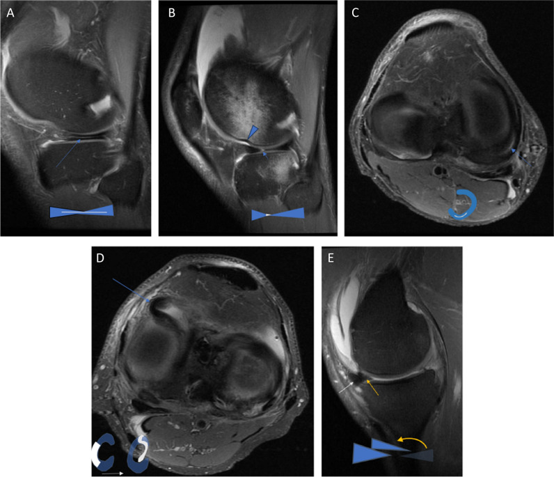
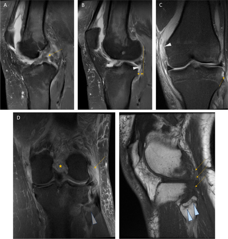
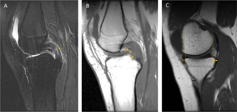
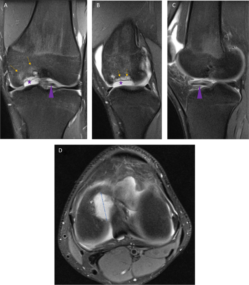
Many studies and systematic reviews have been published about MRI of the knee and its structures, discussing detailed anatomy, imaging findings, and correlations between imaging and clinical findings. This paper includes evidence-based recommendations for a general radiologist regarding choice of imaging sequences and reporting basic MRI examinations of the knee. We recommend using clinicians' terminology when it is applicable to the imaging findings, for example, when reporting meniscal, ligament and tendon, or cartilage pathology. The intent is to standardise reporting language and to make reports less equivocal. The aim of the paper is to improve the usefulness of the MRI report by understanding the strengths and limitations of the MRI exam with regard to clinical correlation. We hope the implementation of these recommendations into radiological practice will increase diagnostic accuracy and consistency by avoiding pitfalls and reducing overcalling of pathology on MRI of the knee. CLINICAL RELEVANCE STATEMENT: The recommendations presented here are meant to aid general radiologists in planning and assessing studies to evaluate acute and chronic knee findings by advocating the use of unequivocal terminology and discussing the strengths and limitations of MRI examination of the knee. KEY POINTS: • On MRI, the knee should be examined and assessed in three orthogonal imaging planes. • The basic general protocol must yield T2-weighted fluid-sensitive and T1-weighted images. • The radiological assessment should include evaluation of ligamentous structures, cartilage, bony structures and bone marrow, soft tissues, bursae, alignment, and incidental findings.
Keywords: Cruciate ligaments; Evidence-based practice; Knee; Magnetic resonance imaging; Menisci.
© 2024. The Author(s).
Conflict of interest statement
The authors of this manuscript declare no relationships with any companies, whose products or services may be related to the subject matter of the article.
Figures
Similar articles
-
[Comparison of the Arthroscopic Finding in the Knee Joint and the MRI - Retrospective Study].Acta Chir Orthop Traumatol Cech. 2017;84(4):285-291. Acta Chir Orthop Traumatol Cech. 2017. PMID: 28933331 Czech.
-
Magnetic resonance imaging of the knee: An overview and update of conventional and state of the art imaging.J Magn Reson Imaging. 2017 May;45(5):1257-1275. doi: 10.1002/jmri.25620. Epub 2017 Feb 17. J Magn Reson Imaging. 2017. PMID: 28211591 Review.
-
The cost-effectiveness of magnetic resonance imaging for investigation of the knee joint.Health Technol Assess. 2001;5(27):1-95. doi: 10.3310/hta5270. Health Technol Assess. 2001. PMID: 11532240 Review.
-
Usefulness of simultaneous acquisition of spatial harmonics technique for MRI of the knee.AJR Am J Roentgenol. 2004 Jun;182(6):1411-5. doi: 10.2214/ajr.182.6.1821411. AJR Am J Roentgenol. 2004. PMID: 15149984
-
Evaluation of acute knee pain in primary care.Ann Intern Med. 2003 Oct 7;139(7):575-88. doi: 10.7326/0003-4819-139-7-200310070-00010. Ann Intern Med. 2003. PMID: 14530229 Review.
Cited by
-
Tibial tuberosity-trochlea groove distance is dependent on flexion angle and intra-articular version in different magnetic resonance imaging recording techniques.J Exp Orthop. 2025 Jun 5;12(2):e70300. doi: 10.1002/jeo2.70300. eCollection 2025 Apr. J Exp Orthop. 2025. PMID: 40476014 Free PMC article.
-
Lower extremity MRI: are their requests always appropriate in France?Eur Radiol. 2025 Aug;35(8):4692-4698. doi: 10.1007/s00330-025-11402-w. Epub 2025 Feb 4. Eur Radiol. 2025. PMID: 39903237 Free PMC article.
-
Improving tissue contrast visualization in two-point Dixon MRI using dark-fat processing: application in clinical knee imaging.Skeletal Radiol. 2025 Aug;54(8):1739-1748. doi: 10.1007/s00256-025-04925-2. Epub 2025 Apr 12. Skeletal Radiol. 2025. PMID: 40220143
-
Medical slice transformer for improved diagnosis and explainability on 3D medical images with DINOv2.Sci Rep. 2025 Jul 4;15(1):23979. doi: 10.1038/s41598-025-09041-8. Sci Rep. 2025. PMID: 40615608 Free PMC article.
-
Atypically Displaced Meniscal Tears: An Educational Review with Focus on MRI and Arthroscopy.Clin Pract. 2025 Jun 12;15(6):109. doi: 10.3390/clinpract15060109. Clin Pract. 2025. PMID: 40558227 Free PMC article. Review.
References
-
- Kassarjian A, Fritz L, Afonso P et al (2016) Guidelines for MR imaging of sports injuries. Eur Soc Skelet Radiol. Available via https://essr.org/content-essr/uploads/2016/10/ESSR-MRI-Protocols-Knee.pdf. Accessed 15 Nov 2023
Publication types
MeSH terms
LinkOut - more resources
Full Text Sources
Medical
Miscellaneous
