. 2024 Oct;34(10):6940-6952.
doi: 10.1007/s00330-024-10624-8. Epub 2024 Mar 27.

Automated graded prognostic assessment for patients with hepatocellular carcinoma using machine learning

Affiliations

Automated graded prognostic assessment for patients with hepatocellular carcinoma using machine learning

Moritz Gross et al. Eur Radiol. 2024 Oct.

Abstract

Background: Accurate mortality risk quantification is crucial for the management of hepatocellular carcinoma (HCC); however, most scoring systems are subjective.

Purpose: To develop and independently validate a machine learning mortality risk quantification method for HCC patients using standard-of-care clinical data and liver radiomics on baseline magnetic resonance imaging (MRI).

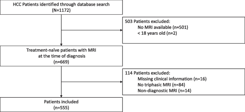
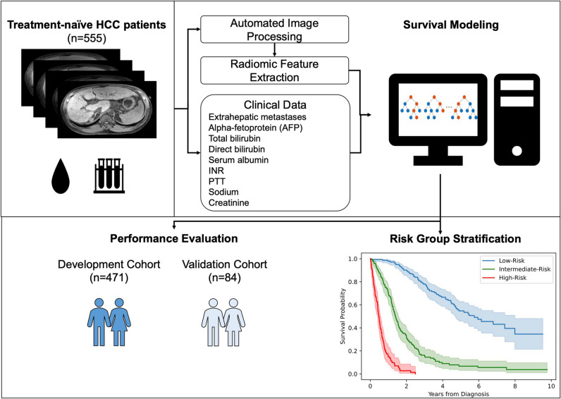
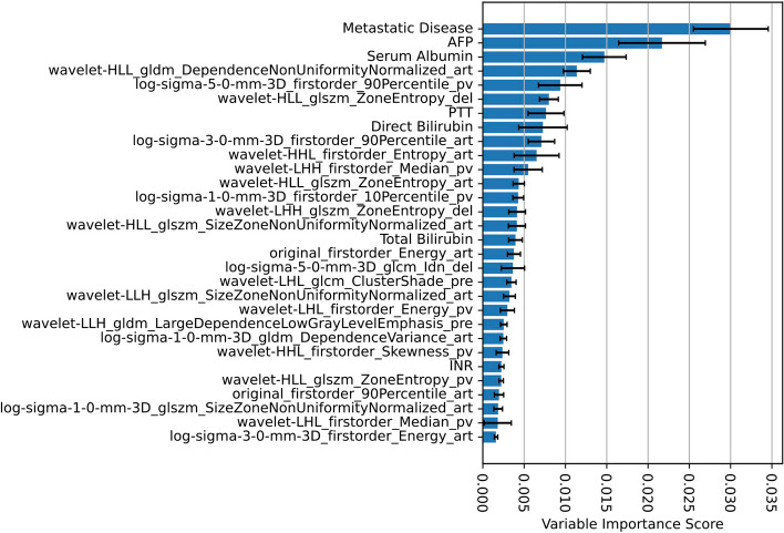
Methods: This retrospective study included all patients with multiphasic contrast-enhanced MRI at the time of diagnosis treated at our institution. Patients were censored at their last date of follow-up, end-of-observation, or liver transplantation date. The data were randomly sampled into independent cohorts, with 85% for development and 15% for independent validation. An automated liver segmentation framework was adopted for radiomic feature extraction. A random survival forest combined clinical and radiomic variables to predict overall survival (OS), and performance was evaluated using Harrell's C-index.

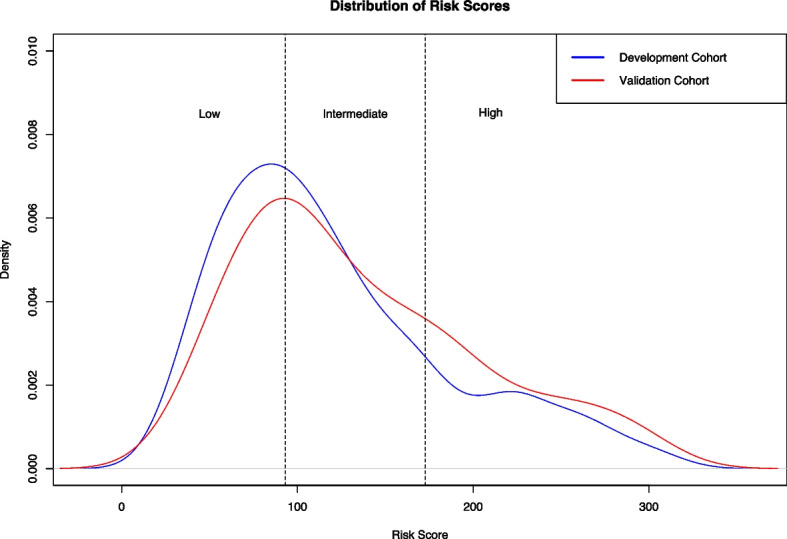
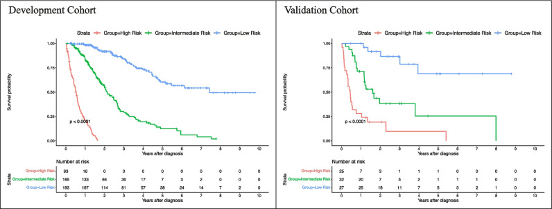
Results: A total of 555 treatment-naïve HCC patients (mean age, 63.8 years ± 8.9 [standard deviation]; 118 females) with MRI at the time of diagnosis were included, of which 287 (51.7%) died after a median time of 14.40 (interquartile range, 22.23) months, and had median followed up of 32.47 (interquartile range, 61.5) months. The developed risk prediction framework required 1.11 min on average and yielded C-indices of 0.8503 and 0.8234 in the development and independent validation cohorts, respectively, outperforming conventional clinical staging systems. Predicted risk scores were significantly associated with OS (p < .00001 in both cohorts).

Conclusions: Machine learning reliably, rapidly, and reproducibly predicts mortality risk in patients with hepatocellular carcinoma from data routinely acquired in clinical practice.

Clinical relevance statement: Precision mortality risk prediction using routinely available standard-of-care clinical data and automated MRI radiomic features could enable personalized follow-up strategies, guide management decisions, and improve clinical workflow efficiency in tumor boards.

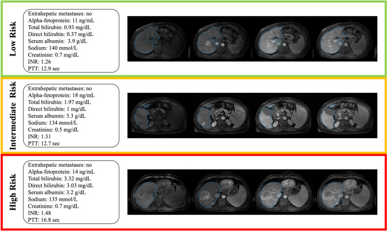
Key points: • Machine learning enables hepatocellular carcinoma mortality risk prediction using standard-of-care clinical data and automated radiomic features from multiphasic contrast-enhanced MRI. • Automated mortality risk prediction achieved state-of-the-art performances for mortality risk quantification and outperformed conventional clinical staging systems. • Patients were stratified into low, intermediate, and high-risk groups with significantly different survival times, generalizable to an independent evaluation cohort.

Keywords: Hepatocellular carcinoma; Machine learning; Magnetic resonance imaging; Medical image processing; Risk assessment.


Conflict of interest statement

The authors of this manuscript declare no relationships with any companies whose products or services may be related to the subject matter of the article.

Figures

Fig. 1
Fig. 1
Flowchart of patient inclusion and exclusion. From an institutional database with 1172 patients, 555 patients (118 females, 437 males, 63.8 ± 8.9 years) with imaging- or histopathologically proven treatment-naïve hepatocellular carcinoma and baseline multiphasic contrast-enhanced magnetic resonance imaging at the time of diagnosis were included in the study
Fig. 2
Fig. 2
Model development. An automated liver segmentation framework was adopted for radiomic feature extraction after automated image co-registration. To predict overall survival, a random survival forest was fit from a combination of clinical and radiomic variables. Model performance was evaluated using Harrell’s C-index and the area under the time-dependent receiver operating characteristic curve (AUC). Patients were stratified into low-, intermediate-, and high-risk groups based on their predicted risk scores
Fig. 3
Fig. 3
Variable importance scores. The bar chart shows the mean variable importance score (error bars show standard deviation) of each included variable of the final risk prediction model obtained by 10 permutations of random shuffling. Naming convention of radiomic features: The prefix specifies the image type (original image or filter-derived MR image (“log”: Laplacian of Gaussian) with extraction parameters); the suffix specifies the MR contrast phase (“_pre”: pre-contrast phase, “_art”: late arterial phase, “_pv”: portal venous phase, “_del”: delayed phase). Equations for the calculation of each radiomic feature are available in ref. [22]. (AFP: Alpha-fetoprotein; INR: international normalized ratio; PTT: partial thromboplastin time)
Fig. 4
Fig. 4
Distribution of predicted risk scores in the development- and independent validation cohort. Based on the risk score predictions of the survival model in the development cohort, we derived two cutoff points (93.08 and 172.73) to stratify patients into low-, intermediate-, and high-risk groups. We applied the same cutoff points for stratification in the independent validation cohort. For plotting, a Gaussian smoothing kernel was used
Fig. 5
Fig. 5
Example cases. Standard-of-care clinical data and axial pre-contrast-, late arterial, portal venous-, and delayed-phase MRI with corresponding automated liver segmentations overlaid in blue. Low-risk group: A 59-year-old male patient presenting with a focal 4.5 cm lesion in the right liver lobe. The patient was censored after 8.75 years. Intermediate-risk group: A 54-year-old male patient presenting with a focal 3.1 cm lesion in the right liver lobe. The patient died 15 months after diagnosis. High-risk group: A 54-year-old male patient presenting with a focal 5.7 cm lesion in the right liver lobe. The patient died 6.7 months after diagnosis
Fig. 6
Fig. 6
Kaplan-Meier curves of the proposed risk groups in the development- and validation cohort

References

    1. Sung H, Ferlay J, Siegel RL et al (2021) Global Cancer Statistics 2020: GLOBOCAN estimates of incidence and mortality worldwide for 36 cancers in 185 countries. CA Cancer J Clin 71:209–249 10.3322/caac.21660 - DOI - PubMed
    1. Jemal A, Bray F, Center MM, Ferlay J, Ward E, Forman D (2011) Global cancer statistics. CA Cancer J Clin 61:69–90 10.3322/caac.20107 - DOI - PubMed
    1. Siegel RL, Miller KD, Jemal A (2019) Cancer statistics, 2019. CA Cancer J Clin 69:7–34 10.3322/caac.21551 - DOI - PubMed
    1. White DL, Thrift AP, Kanwal F, Davila J, El-Serag HB (2017) Incidence of hepatocellular carcinoma in All 50 United States, From 2000 Through 2012. Gastroenterology 152:812-820.e815 10.1053/j.gastro.2016.11.020 - DOI - PMC - PubMed
    1. Perz JF, Armstrong GL, Farrington LA, Hutin YJ, Bell BP (2006) The contributions of hepatitis B virus and hepatitis C virus infections to cirrhosis and primary liver cancer worldwide. J Hepatol 45:529–538 10.1016/j.jhep.2006.05.013 - DOI - PubMed

LinkOut - more resources