Review
. 2024 Oct;34(10):838-853.
doi: 10.1016/j.tcb.2024.02.009. Epub 2024 Mar 27.

Blebology: principles of bleb-based migration

Affiliations
Review

Blebology: principles of bleb-based migration

Juan Manuel García-Arcos et al. Trends Cell Biol. 2024 Oct.

Abstract

Bleb-based migration, a conserved cell motility mode, has a crucial role in both physiological and pathological processes. Unlike the well-elucidated mechanisms of lamellipodium-based mesenchymal migration, the dynamics of bleb-based migration remain less understood. In this review, we highlight in a systematic way the establishment of front-rear polarity, bleb formation and extension, and the distinct regimes of bleb dynamics. We emphasize new evidence proposing a regulatory role of plasma membrane-cortex interactions in blebbing behavior and discuss the generation of force and its transmission during migration. Our analysis aims to deepen the understanding of the physical and molecular mechanisms of bleb-based migration, shedding light on its implications and significance for health and disease.

Keywords: actomyosin; amoeboid migration; bleb migration; cell polarity; small GTPase.


Conflict of interest statement

Declaration of interests The authors declare that they have no known competing financial interests or personal relationships that could have appeared to influence this review.

Figures

Figure 1:
Figure 1:. The Abercrombie steps of bleb-based migration.
Top left panel, “Front-Rear polarity establishment”: the cell establishes a gradient of front rear-polarity markers guided by self-polarization mechanisms or a gradient of extracellular cues (e.g.: chemotactic gradient), this increases the probability of bleb formation at the leading edge. Top right panel, “Leading-edge extension”: bleb formation at the leading edge combined with cortical retrograde flows deplete cortical materials from the leading edge promoting bleb extension. Bottom right panel: “Friction/adhesion with substrate”: cells interact with the substrate in different ways, i) specific cell-cell adhesions through cadherins, specific weak or nascent focal adhesions by integrins, non-specific interactions between the cell surface and the surrounding environment, or by steric hindrance due to substrate topography. Bottom left panel: “De-adhesion/translocation”: blebbing cells generally translocate fast due to the absence of strong adhesions or the complete absence of adhesions. The ECM (extracellular matrix) is generally not degraded by proteolysis, which forces cells to deform on their way. In some cases, blebs can detach from the cell and migrate on their own, forming cytoplasts.
Figure 2:
Figure 2:. Possible front-rear polarity markers in bleb-based migration.
Biochemical markers characteristic of the cell front (magenta) and the cell rear (blue) in migrating blebbing cells. First column: membrane-proximal cortical actin (MPA) is enriched at the rear of migrating cells [68], and depleted locally before protrusion initiation [67]. Second column: PI3K can enrich the cell front with PIP3 and PTEN the cell rear with PIP2, contributing to the maintenance of cell polarity in blebbing cells [26,120].Third column: Rac has been reported to be active at the cell front to confine bleb formation to the leading edge [46,64], while ERM proteins at the cell rear can be phosphorylated, which restricts membrane blebbing [46,121]. Fourth column: Due to the combined effect of bleb formation and cell-scale cortical flows, actomyosin is depleted from the cell front and depleted at the cell rear. This might change depending on cell morphology.
Figure 3:
Figure 3:. Force transmission mechanisms during bleb migration.
Black arrows represent the active friction forces contributing to cell migration. Left column: Melanoma cells in collagen form transient focal adhesion at the base of blebs (depicted in green), where integrin mediates force transmission from bleb retraction to the extracellular matrix. Additionally, blebs were shown to push collagen fibers forward and interdigitate between collagen fibers. This process of pushing-pulling could eventually wear off the collagen matrix (depicted in light blue) in the absence of metalloproteases [26]. Center column: The force transmission in zebrafish PGCs occurs at the level of the cell body, and not the bleb. This is mediated by e-cadherins (depicted in light purple), beta-catenins (dark purple), and ezrin (orange) which transmit forces from the retrograde flow to surrounding cells [46]. Right column: Confined cancer cells in microfluidic devices use unspecific electrostatic interactions between transmembrane or surface proteins (depicted in grey) and the surroundings (blue) to transmit forces from the retrograde flow to the non-adhesive PDMS walls, possibly through ezrin (orange) or other proteins [33].
Figure I:
Figure I:. Examples of cell morphologies and their classification of the mesenchymal (blue) versus amoeboid (magenta) continuum.
Cells on the left-most side display stereotypical shapes in the mesenchymal mode of migration dominated by actin polymerization-driven protrusions like lamellipodia, ruffles, and filopodia. Cells on the right-most side represent cell shapes during amoeboid migration dominated by pressure-driven protrusions like blebs and pseudopodia. The drawing shows the continuum in the morphology that can be found in many cells where they can exhibit different protrusions within the same cell.
Figure II:
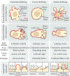
Figure II:. Different regimes of actin-membrane dynamics at the leading edge of blebbing cells.
The membrane is drawn in black and actin in red. Arrows represent the direction of membrane displacement. Top row: non-polarized cells under different blebbing regimes. Middle row: Polarized cells under different blebbing regimes. Bottom row: Single bleb dynamics in transient, circus, and stable blebs. The membrane is drawn in black, actin in red and myosin II in green. Black arrows represent the direction of membrane displacement. Transient blebs have a short cycle of expansion, cortex formation, and bleb retraction. Circus blebs display an asymmetry in the retraction and localization of actomyosin, “traveling” through the cell surface tangentially. Stable blebs display a “stable phase” where cortical flow emerges and an actomyosin gradient is established.

References

    1. Krause M and Gautreau A (2014) Steering cell migration: lamellipodium dynamics and the regulation of directional persistence. Nat Rev Mol Cell Biol 15, 577–590 - PubMed
    1. Gardel ML et al. (2010) Mechanical Integration of Actin and Adhesion Dynamics in Cell Migration. Annu. Rev. Cell Dev. Biol. 26, 315–333 - PMC - PubMed
    1. Bailly M and Condeelis J (2002) Cell motility: insights from the backstage. Nat Cell Biol 4, E292–E294 - PubMed
    1. Bodor DL et al. (2020) Of Cell Shapes and Motion: The Physical Basis of Animal Cell Migration. Developmental Cell 52, 550–562 - PubMed
    1. Friedl P and Wolf K (2010) Plasticity of cell migration: a multiscale tuning model. Journal of Cell Biology 188, 11–19 - PMC - PubMed

LinkOut - more resources