. 2024 Mar 27;15(1):2687.
doi: 10.1038/s41467-024-46737-3.

Uncovering structural themes across cilia microtubule inner proteins with implications for human cilia function

Affiliations

Uncovering structural themes across cilia microtubule inner proteins with implications for human cilia function

Jens S Andersen et al. Nat Commun. .

Abstract

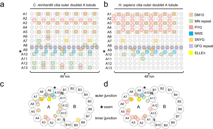
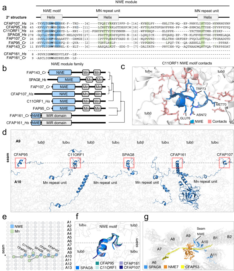
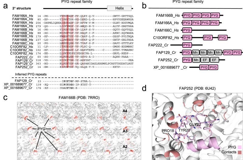
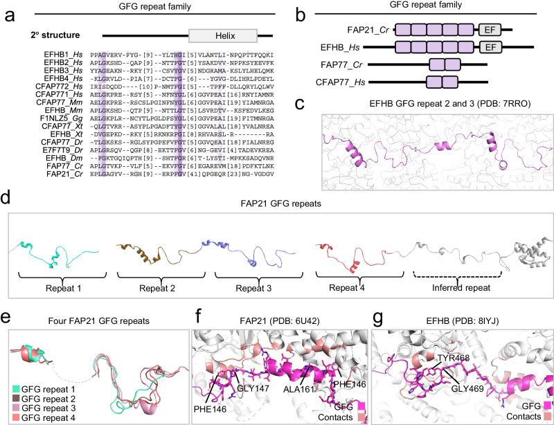
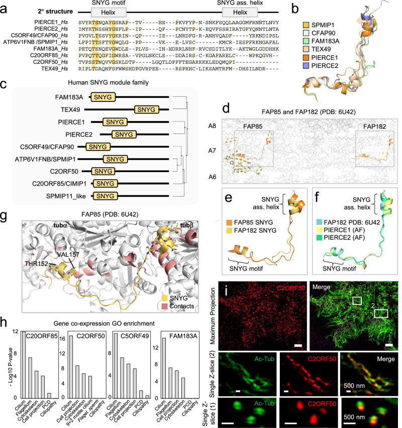
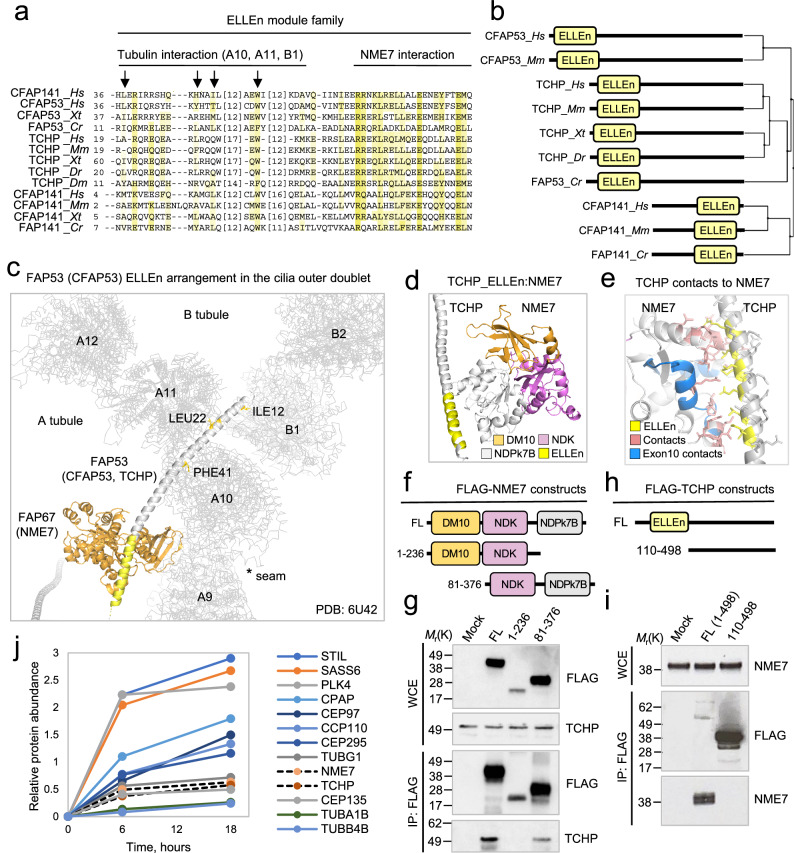
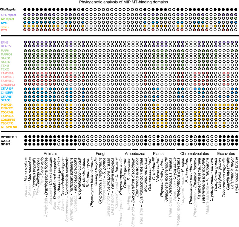
Centrosomes and cilia are microtubule-based superstructures vital for cell division, signaling, and motility. The once thought hollow lumen of their microtubule core structures was recently found to hold a rich meshwork of microtubule inner proteins (MIPs). To address the outstanding question of how distinct MIPs evolved to recognize microtubule inner surfaces, we applied computational sequence analyses, structure predictions, and experimental validation to uncover evolutionarily conserved microtubule- and MIP-binding modules named NWE, SNYG, and ELLEn, and PYG and GFG-repeat by their signature motifs. These modules intermix with MT-binding DM10-modules and Mn-repeats in 24 Chlamydomonas and 33 human proteins. The modules molecular characteristics provided keys to identify elusive cross-species homologs, hitherto unknown human MIP candidates, and functional properties for seven protein subfamilies, including the microtubule seam-binding NWE and ELLEn families. Our work defines structural innovations that underpin centriole and axoneme assembly and demonstrates that MIPs co-evolved with centrosomes and cilia.


Conflict of interest statement

The authors declare no competing interests.

Figures

Fig. 1
Fig. 1. Schematic representation of the positions of MIP modules inside the motile cilia outer doublet of C. reinhardtii and H. sapiens based on cryo-EM structures of ciliary doublet microtubules.
a, b Distribution of MIPs along the axoneme at periodically defined positions. c, d Lateral distribution of MIPs at the protofilaments of the A- and B-tubules. The position of the seam and of the outer and inner junctions between the A- and B-tubules are indicated.
Fig. 2
Fig. 2. New members of the DM10 domain containing protein family.
a Multiple sequence alignment of the DM10 domain from the human and C. reinhardtii proteins identified by iterative HMM-to-HMM searches. Proteins are designated by their UniProt identifiers. Coloring schemes as per ClustalX parameters with modifications. Predicted regions corresponding to β-sheets and based on RIB72 DM10 domain, are shown above the alignment. b Schematic representation of protein architectures of the DM10 domain family. Phylogenetic relationships were calculated using average distances and percent identity (PID) between DM10 modules used in the alignment. c Superimposition of C. reinhardtii RIB72 DM10 domain structure with the AlphaFold2-predicted DM10 domain structure of CAPS2. d Gene co-expression analysis of CAPS2 across non-cancerous ciliated Head and Neck Squamous cell Carcinoma (HNSC), Kidney renal clear cell carcinoma (KICH), Lung Adenocarcinoma (LUAD), Lung Squamous cell Carcinoma (LUSC), and Uterine Corpus Endometrial Carcinoma (UCEC) tissues. PCC = Pearson correlation coefficient. e Gene ontology (GO) enrichment analysis of CAPS2 co-expressed genes identified in (d). Enrichment P-values were obtained using the PANTHER Classification System for the slim cellular component devised by the PANTHER software. f RNA-seq ratios obtained from NIH3T3 fibroblast extracts. Ratios calculated as transcripts per million (TPM) of serum starved cells (24 h) over cycling cells. Source data are provided as a Source Data file. g Immunoblot of cell extracts from hTERT-RPE1 cells grown with or without serum. Proteins were probed with indicated antibodies. The shown immunoblot is representative of two independent experiments. h Immunofluorescence microscopy micrographs of motile cilia on human bronchial epithelial cells imaged by structured illumination microscopy. Cells were probed with the indicated antibodies. The images are representative of two independent experiments. Source data are provided as a Source Data file. Scale bar: 5 μm in the top row, 1 μm in the second row.
Fig. 3
Fig. 3. Identification of Mn repeats in eukaryotic MIPs.
a Multiple sequence alignment of the Mn repeat units from human proteins identified by iterative HMM-to-HMM searches. The expanded alignment with C. reinhardtii Mn repeat proteins is shown in supplementary Fig. S2c. Proteins are designated by their UniProt identifiers. Coloring schemes as per ClustalX parameters with modifications. Shown in green are positions where hydrophilicity and aromatic residues are conserved and participate in the short α-helix of individual Mn repeat unit. Predicted regions corresponding to α-helices and based on FAP363 Mn repeats, are shown above the alignment. b Portions of the elongated Mn repeat proteins SAXO1, SAXO2, and TEX26 as predicted by comparative modeling as well as deep learning by AlphaFold2. c Module architecture of proteins with known and predicted Mn repeats and inferred Mn repeat. d Superimposed Mn repeat units of human CFAP95, CFAP107, and SPAG8as assessed by Pymol. e Examples of Mn repeat unit contact sites with the MT lattice in mammals. The Mn unit is shown in green, and MT contact side chains are shown in red.
Fig. 4
Fig. 4. Identification of a universal seam-binding module in MIPs across eukaryotes.
a Multiple sequence alignment of the NWE module from the five human and four C. reinhardtii proteins identified by iterative HMM-to-HMM searches. Except for CFAP161, the NWE module consists of the co-evolved triad of an N-terminal NWE motif adjoined to two Mn repeat units. In FAP161 and CFAP161 the Mn repeats are absent. The amino acid numbers of the Mn repeat units are indicated. Coloring schemes as per ClustalX parameters with modifications. Positions comprising the NWE motif are highlighted in blue. The Mn repeat units are colored green as in Fig. 3. Predicted regions corresponding to α-helices are shown above the alignment. b Module architecture of predicted NWE module proteins in humans and C. reinhardtii. Phylogenetic relationships were calculated using average distances and percent identity (PID) between NWE modules used in the alignment. c The NWE motif in C11ORF1 as found in complex with the MTs inner surface MTs in the H. sapiens cilia outer doublet. d NWE motifs and Mn repeats across the cilia outer doublet seam in H. sapiens. e Distribution of the NWE motifs and Mn repeats along the axoneme at periodically defined positions. f Structural superimposition of the NWE motifs at the seam. g The NWE motif of SPAG8 binds MTs at the A-tubule seam region of the outer doublet. NME7 associates with the ELLEn module of CFAP53 (see Fig. 8).
Fig. 5
Fig. 5. Identification of MT inner surface binding PYG repeats in MIPs.
a Multiple sequence alignment of the PYG repeats in four human and three C. reinhardtii proteins identified by HMM-to-HMM searches. Only the outermost N-terminal PYG repeat unit for each protein is shown. Proteins are designated by their UniProt identifiers. Coloring schemes as per ClustalW parameters with modifications. b Module architecture of predicted PYG repeat proteins in human and C. reinhardtii. c PYG repeats of human FAM166B shown in the outer doublet lumen MT interface. d Structure of the PYG repeat in FAP252 where contacts between the MT lattice and the PYG repeat is shown for the conserved residues.
Fig. 6
Fig. 6. Identification of MT inner surface binding GFG repeats in MIPs.
a Multiple sequence alignment of the GFG repeat in two human and two C. reinhardtii proteins identified by iterative HMM-to-HMM searches. Proteins are designated by their UniProt identifiers. Coloring schemes as per ClustalW parameters with modifications. b Module architecture and evolutionary relationships of the GFG repeat family members in C. reinhardtii and humans. c The GFG repeat 2 in human EFHB. d Module architecture of C. reinhardtii FAP21 (human EFHB) showing the GFG repeats along the length of FAP21. In FAP21, four GFG repeats were identified by sequence alignment and a fifth was inferred from visual inspection of the FAP21 tertiary structure in the C. reinhardtii outer doublet. e Superimposition of four GFG repeats from FAP21. The highly divergent central portion of each GFG repeat (dotted line) is omitted to allow superimposition. f One GFG repeat unit of FAP21 in contact with the MT lattice. Contact sites are highlighted in red. Conserved aromatic and glycine residues are shown (g) One GFG repeat unit of EFHB in contact with the MT lattice. Contact sites are highlighted in red. Conserved aromatic and glycine residues are shown.
Fig. 7
Fig. 7. Identification of SNYG modules in C. reinhardtii and mammalian MIPs.
a Multiple sequence alignment of the SNYG module in eight human proteins identified by iterative HMM-to-HMM searches. The SNYG module consists of two α-helices, one representing the conserved Mn repeat-like motif bearing a highly conserved glycine, and the other helix here named the SNYG motif associated helix (SNYG ass. helix). Proteins are designated by their UniProt identifiers. Coloring schemes as per ClustalW parameters with modifications. Predicted regions corresponding to α-helices are shown above the alignment. b Superimposition of experimentally determined SNYG modules from mammalian PIERCE1, PIERCE2, TEX49, FAM183A, C5ORF49 (CFAP90), and ATP6V1FNB (SPMIP1). Structures were derived from PDB: 8OTZ. c Module architecture of predicted SNYG module proteins in human. d The SNYG module in FAP85 and FAP182 (shown in boxes) in complex with the tubulin lattice of the C. reinhardtii cilia outer doublet A-tubule (PDB: 6U42). Protofilament number shown right. e Superimposition of SNYG module structures from FAP85 and FAP182. f Superimpositions of FAP182 SNYG module structures with AlphaFold2-predicted SNYG modules of PIERCE1 and PIERCE2. g The FAP85 SNYG modules in complex with tubA and tubB. Contact sites between tubulins and conserved threonine or hydrophobic residues are highlighted in red. h GO enrichment of gene co-expression analysis (top 6) of the four SNYG family members C20ORF85, C2ORF50, C5ORF49, and FAM183A. Enrichment P-values were obtained using the PANTHER Classification System for the slim cellular component devised by the PANTHER software. i Immunofluorescence microscopy micrographs of motile cilia on cultured human bronchial epithelial cells imaged by structured illumination microscopy. Cells were stained with indicated antibodies. The images are representative of two independent experiments. Scale bar: 5 μm in the top row, 500 nm in the second row.
Fig. 8
Fig. 8. The ELLEn module binds FAP67 and NME7 in the cilia outer doublet lumen.
a Multiple sequence alignment of the ELLEn module across species. Proteins are designated by their UniProt identifiers. Species represented are Homo sapiens (Hs), Mus musculus (Mm), Xenopus tropicalis (Xt), Danio rerio (Dr), Drosophila melanogaster (Dm), and C. reinhardtii (Cr). Coloring schemes as per ClustalW parameters with modifications. b Module architecture and evolutionary relationships of the ELLEn module family members across species. Phylogenetic relationships are calculated using average distances and percent identity (PID) between ELLEn modules used in the alignment. c Associations of the ELLEn module in FAP53 with FAP67 (NME7 in human). d, e AlphaFold2 prediction of the human complex and the contacts between the ELLEn module of TCHP and NME7. f Schematic representations of FLAG-NME7 constructs. g FLAG pulldown analysis and immunoblot of FLAG-NME7 constructs expressed in HEK293T cells. Proteins were probed with the indicated antibodies. The immunoblots are representative of two independent experiments. h Schematic representations of FLAG-TCHP constructs. i FLAG pulldown analysis and immunoblot of FLAG-TCHP constructs expressed in HEK293T cells. Proteins were probed with the indicated antibodies. The immunoblots are representative of three independent experiments. j Quantitative analysis of centrosome-associated proteins during centriole biogenesis. An array of representative centrosome proteins implicated in centriole duplication are shown in solid lines indicating temporal dynamics of centrosome protein recruitments. NME7 and TCHP are indicated with dashed lines. The quantitative analysis is representative of experiments performed in two replicates. Source data are provided as a Source Data file.
Fig. 9
Fig. 9. Co-evolution of MIP modules with centrosomes and cilia.
Coulson plot demonstrating occurrence and absence (or loss) of identified MIP modules in 52 eukaryotic genomes. Rows show individual proteins or groups of proteins divided into the five module families GFG repeat (purple), Mn repeat (green), NWE module (blue), SNYG module (yellow), and PYG repeat (pink). The presence of a centrosome or cilium is shown for each organism in the top row.

References

    1. Ma M, et al. Structure of the decorated ciliary doublet microtubule. Cell. 2019;179:909–922.e12. doi: 10.1016/j.cell.2019.09.030. - DOI - PMC - PubMed
    1. Gui, M. et al. De novo identification of mammalian ciliary motility proteins using cryo-EM. Cell184, 5791–5806.e19 (2021). - PMC - PubMed
    1. Wang X, et al. Cryo-EM structure of cortical microtubules from human parasite Toxoplasma gondii identifies their microtubule inner proteins. Nat. Commun. 2021;12:3065. doi: 10.1038/s41467-021-23351-1. - DOI - PMC - PubMed
    1. Gui M, Wang X, Dutcher SK, Brown A, Zhang R. Ciliary central apparatus structure reveals mechanisms of microtubule patterning. Nat. Struct. Mol. Biol. 2022;29:483–492. doi: 10.1038/s41594-022-00770-2. - DOI - PMC - PubMed
    1. Leung MR, et al. The multi-scale architecture of mammalian sperm flagella and implications for ciliary motility. EMBO J. 2021;40:e107410. doi: 10.15252/embj.2020107410. - DOI - PMC - PubMed

Substances

LinkOut - more resources